Impact of dead space closure and lymph vessel ligation during MRM on Post-operative seroma formation: A two institutional randomized study.
•
2 recomendaciones•829 vistas

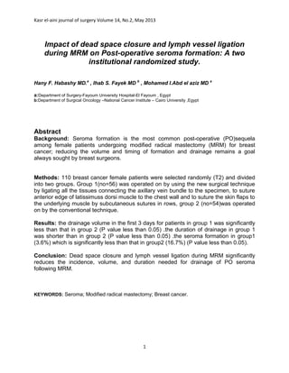
Hany F. Habashy MD.a , Ihab S. Fayek MD b , Mohamed I.Abd el aziz MD a a:Department of Surgery-Fayoum University Hospital-El Fayoum , Egypt. b:Department of Surgical Oncology –National Cancer Institute – Cairo University ,Egypt. Kasr el-aini journal of surgery Volume 14, No.2, May 2013

Recomendados
Recomendados
Más contenido relacionado
La actualidad más candente
La actualidad más candente (19)
Similar a Impact of dead space closure and lymph vessel ligation during MRM on Post-operative seroma formation: A two institutional randomized study.
Similar a Impact of dead space closure and lymph vessel ligation during MRM on Post-operative seroma formation: A two institutional randomized study. (8)
Más de Dr./ Ihab Samy
Más de Dr./ Ihab Samy (20)
Último
Último (20)
Impact of dead space closure and lymph vessel ligation during MRM on Post-operative seroma formation: A two institutional randomized study.
- 1. Kasr el-aini journal of surgery Volume 14, No.2, May 2013 1 Impact of dead space closure and lymph vessel ligation during MRM on Post-operative seroma formation: A two institutional randomized study. Hany F. Habashy MD.a , Ihab S. Fayek MD b , Mohamed I.Abd el aziz MD a a:Department of Surgery-Fayoum University Hospital-El Fayoum , Egypt b:Department of Surgical Oncology –National Cancer Institute – Cairo University ,Egypt Abstract Background: Seroma formation is the most common post-operative (PO)sequela among female patients undergoing modified radical mastectomy (MRM) for breast cancer; reducing the volume and timing of formation and drainage remains a goal always sought by breast surgeons. Methods: 110 breast cancer female patients were selected randomly (T2) and divided into two groups. Group 1(no=56) was operated on by using the new surgical technique by ligating all the tissues connecting the axillary vein bundle to the specimen, to suture anterior edge of latissimuss dorsi muscle to the chest wall and to suture the skin flaps to the underlying muscle by subcutaneous sutures in rows, group 2 (no=54)was operated on by the conventional technique. Results: the drainage volume in the first 3 days for patients in group 1 was significantly less than that in group 2 (P value less than 0.05) .the duration of drainage in group 1 was shorter than in group 2 (P value less than 0.05) .the seroma formation in group1 (3.6%) which is significantly less than that in group2 (16.7%) (P value less than 0.05). Conclusion: Dead space closure and lymph vessel ligation during MRM significantly reduces the incidence, volume, and duration needed for drainage of PO seroma following MRM. KEYWORDS: Seroma; Modified radical mastectomy; Breast cancer.
- 2. Kasr el-aini journal of surgery Volume 14, No.2, May 2013 2 Introduction Seroma is defined as a serous fluid collection that develops under the skin flaps following mastectomy or in the axillary dead space after axillary dissection.(1) Seroma formation is the most frequent post-operative complication after breast cancer surgery. It occurs in most patients after mastectomy and is now increasingly being considered side effect of surgery rather than a complication however, all patients are not clinically symptomatic. (2, 9) Incidence of seroma formation after breast surgery varies between 2.5% and 51%. (3-5) Although seroma is not life threatening, it can lead to significant morbidity (e.g. flap necrosis, wound dehiscence, predisposes to sepsis, prolonged recovery period, multiple physician visits) and may delay adjuvant therapy. (6,7) Fluid collection is ideally managed by repeated needle aspiration to seal the skin flaps against the chest wall. The aetiology of seroma formation is not yet quite clear.(10) Several factors have been investigated as the cause of seroma formation these include age, duration of wound drainage, use of pressure garment, postoperative arm activity, intraoperative lymphatic channel ligation was done or not, preoperative chemotherapy, and use of electro cautery. (3, 8, 19) Studies on the composition of the fluid collected from postmastectomy drainage suggest an inflammatory origin, while others have hypothesized that seroma is most likely to originate from lymph. (11, 12) The optimal way to reduce incidence of seroma formation is unknown. Some evidence exists that placing sutures from the skin flaps to the underlying muscles can obliterate the dead space, thus reducing the seroma formation. (13, 14, 15) This study aiming to reduce the incidence of seroma formation by using an altered surgical technique, is a randomized trial based on the hypothesis that axillary lymph leakage and dead space are major important contributors to seroma formation,and that surgical techniques applied to seal off axillary interrupted lymph vessels and to obliterate the dead space might reduce the incidence of this complication. Methods Patients and surgical technique This study was conducted in the department of surgery at Fayoum university hospital and the National cancer institute - Cairo University.
- 3. Kasr el-aini journal of surgery Volume 14, No.2, May 2013 3 Figure 1: ligation of lymphatic vessels Figure2 (a): suturing the anterior border of LDM to chest wall. Figure 2(b): closure of dead space between LDM and chest wall. Figure3: suturing the skin flap to underlying muscle in rows.
- 4. Kasr el-aini journal of surgery Volume 14, No.2, May 2013 4 From May 2011 to March 2012, 110 patients with late T2 breast cancer who were scheduled as candidates for elective unilateralmodified radical mastectomy without immediate reconstruction were randomly preoperatively divided into 2 groups, group1 and group 2, for which mastectomies were performed using different surgical techniques. Surgery for the 2 groups was performed by the same surgical team. Axillary dissection was done up to level III in all cases. In group 1, in an attempt to seal off axillary lymphatic vessels and obliterate the dead space, we used the technique of lymph vessel ligation and suturing skin flaps to the underlying muscle, and all the tissues connecting axillary vein to the specimen were ligated. Once the anterior surface of the axillary vein was exposed, sharp rather than blunt dissection was performed, and the bleeding vessels and all of the fat pads were ligated just below the axillary vein with 3-0 vicryl sutures. (Fig.1) Next, the thoracodorsal vein, with its accompanying artery and nerve comprising the thoracodorsal neurovascular bundle, was identified, and the small vascular branch along with all the fat pad coursing toward the axillary contents was carefully ligated. Then, the long thoracic nerve, usually seen lying against chest wall, was identified and preserved. The dissection was then completed by freeing the inferior axillary contents from the chest wall, followed by insertion of a suction drain in the axilla. The anterior edge of latissimus dorsi was identified by electrocautery and then stitched to the serratus anterior muscle anterior to the long thoracic nerve which was preserved after removal of the specimen.(Fig.2) The skin flaps were fixed by subcutaneous sutures to the underlying musclebyapproximately 15 vicryl sutures at periodic intervals, placed from the skin flap to the underlying muscle, if moderate to severe dimpling was observed, the stitch was removed and replaced.(Fig.3) In group 2, the control group, the procedure was generally performed in a similar fashion, but axillary dissection was carried out bluntly instead of sharply; the bleeding vessel branches linking axillary vein bundle or thoracodorsal bundle to the specimen were freed and ligated, and the accompanying fat pad was freed bluntly or cut with electrocautery or scalpel but was not ligated. The anterior edge of the latissimus dorsi muscle was not fixed to the chest wall. Skin flaps was not fixed to underlying muscle.
- 5. Kasr el-aini journal of surgery Volume 14, No.2, May 2013 5 In both groups, all resected specimens were examined and the lymph nodes dissected, counted, and assessed histopathologically for metastases. External compression dressing was provided over the axilla for the first 3 postoperative days and the patients were encouraged to do active and passive shoulder exercises starting from day one postoperative. The drain 18 Fr.double arms redivac was inserted. The bottle was changed every 24 hours and the daily drain output was measured and recorded. Drain obstruction was recorded if it occurred an adjustment was needed. The drain was removed when the output was less than 50 mL in 24 hours regardless of time elapsed after surgery. Each patient was seen 1 week after discharge and weekly thereafter or more frequently as needed. The daily drain output in the first 3 postoperative days was measured in each group and compared between the 2groups. The length of drainage in both groups was calculated and compared. The operative time, defined as the time from onset of surgery to the end of wound closure, was calculated. The operative time was compared between the 2 groups. The associated morbidity in the form of seroma formation, hematoma, flap necrosis, and wound infection was recorded. Seroma was defined as a fluid collection via palpation on clinical examination. A blood collection under the skin, removable by aspiration, was considered as hematoma. Flap necrosis was defined as any visible necrosis along the edge of the wound. Results: In all, 110 breast cancer patients were included into the study with 56in group 1 and 54 in group 2. The two groups were not statistically different regarding age, weight, number of axillary lymph nodes resected, and number of cases with positive nodes indicating the success of randomization. (Table 1) The mean time of surgery was about 18 minutes longer for group 1 than that for group 2 (103.41 minutes vs. 85.63 minutes). No patients in either groups received intraoperative blood transfusion. 3 patients had the drain obstructed in group 2, whereas none had the problem in group 1 (P<0.05). The daily drain output in the initial 3 postoperative days in group 1 was significantly less than that in group 2 (P<0.05). Drains were removed earlier in group 1 than those in group 2 (P< 0.05). The incidence of seroma formation for group 1 (3.6%) was significantly less than that for group 2 (16.7%) (P< 0.05). A small hematoma, probably due to the penetration of a suture into a vessel in the muscle tissue, was observed in the first postoperative day in 1 patient in group 1, but no significant difference was found between the 2 groups in terms of hematoma, wound infection, or flap necrosis. No other complications were seen for all patients in the study.(Table 2)
- 6. Kasr el-aini journal of surgery Volume 14, No.2, May 2013 6 Moreover, as is shown inTable 2, the median drainage volume decreased very quickly in group 1 in the initial 3 postoperative days and the individual drainage volume in day 3 was no more than 50 mL/24 h for all patients in group 1; in contrast, the median drainage volume did not decrease so rapidly in group 2, and the individual drainage volume was still greater than 100 mL/24 h after 3 postoperative days in some patients. Table 1: Patients characteristics and surgical intervention. NS= No Statistical Significance. Table 2: Comparison between operating time, drainage volume and complications NS= No Statistical Significance. variables Group1(no=56) Group 2 no=(54) P Value Mean age (year) 50.41 51.30 NS Mean weight (Kg) 71.43 68.17 NS Mean No. of lymph nodes resected 27.34 28.15 NS Variables Group 1(no=56) Group 2 no=(54) P value Operative time (min) 103.41 85.63 Less than 0.05 Drain volume day 1 (ml) 153.65 241.81 Less than 0.05 Drain volume day 2(ml) 78.24 156.32 Less than 0.05 Drain volume day 3(ml) 54.47 122.98 Less than 0.05 Time of drainage(days)removed when less than 50cc/day for 2 successive days 5.81 11.07 Less than 0.05 Obstruction of drain (%) 0 5.36 Less than 0.05 Hematoma (%) 1.8 0 NS Wound infection (%) 0 0 NS Flap necrosis (%) 0 0 NS Seroma formation (%) 3.6 16.7 Less than 0.05
- 7. Kasr el-aini journal of surgery Volume 14, No.2, May 2013 7 Discussion: The pathogenesis of seroma has not been fully understood. (16) However, several studies have indicated that it may originate from lymph. (11, 12) McCaul et al., 2000 investigated the hematologic and biochemical composition of the drain fluid on days 3 and 4 after mastectomy and axillary clearance and found that it was most similar to inflammatory exudates, although lymph and serum undoubtedly made a small contribution.(18)Then the size of the dead space after wound closure and handling tissues during the operation may have been factors that significantly affect fluid collection postoperatively. McCaul et al., 2000 pointed out that the elements of surgical technique may well be responsible for the observed intersurgeon variation in frequency of postmastectomy seroma formation. (18) Historically, Halsted suggested obliteration of the dead space particularly in the axilla to facilitate wound healing.(17) More recently, Chilson et al., 1992 noted that there was a decrease in the incidence of post- mastectomy seroma in their institution and the number of postoperative clinic visits for aspiration of seroma when flap tacking was performed.(19) O'Dwyer et al., 1991 reported that closing the dead space by suturing skin flaps to the underlying muscle combined with early drain removal correlated with a low incidence of seroma formation after mastectomy.(20) In a prospective randomized clinical trial, Coveney et al., 1993 showed significantly less drainage in the group of patients in whom skin flaps were sutured down to the chest wall muscles compared with those with just conventional skin flap closure.(14) In a more recent prospective study, Schuijtvlot et al., 2002 compared the effect of buttress suture with non- buttress suture after breast-conserving surgery and axillary dissection. They showed that the use of buttress suture reduced seroma formation. (13) Following axillary dissection without axillary drain Classe et al., 2006 showed that axillary padding using axillary aponeurosis and local muscle flaps to fill the dead space could be used as an alternative to closed vacuum suction after axillary lymphadenectomy with early discharge. (15) As evidenced above, seroma formation was reduced somehow by sutures placed underneath the skin flap; however, it remains a significant complication. (13, 20) In this randomized study, we ligated all tissue just below the axillary vein and below the thoracodorsal vein in an attempt to seal off the axillary lymph vessels, which were believed to be the source of the lymph leakage. Additionally, suturing the anterior edge of the latissimus dorsi muscle to the chest wall could collapse the lateral dead space between the muscle and the chest wall, therefore avoiding burying the drain tube in the
- 8. Kasr el-aini journal of surgery Volume 14, No.2, May 2013 8 dead space, which sometimes occurs and results in the obstruction of the tube. The use of the sutures in conjunction with negative pressure drains act in concert to obliterate the dead space as we observed. With the combination of those techniques, the incidence of seroma formation was reduced to a very low level 3.6% in the study group. The intensity and duration of the exudates were reduced significantly in the initial 3 postoperative days, and the length of drainage was significantly shortened compared with that in the control group (group 2). Although time to operate was slightly longer than that in group 2, the difference became modest as operating skills improved, and no additional complications such as dysfunction of shoulder movement were observed. Finally, it is important to note that the 3 different surgical interventions—sewing down the skin flaps, ligating lymphatics, and suturing the latissimus dorsi reduced the incidence of seroma significantly. However, it is impossible to determine which of these, or which combination of these, actually produced the observed effect. What is more, in our experience in the study, the seroma formed after the altered interventions, although occurring in only 3.6 %of cases, was more difficult to manage than that formed after conventional procedures. Further studies are needed to determine the pathogenesis of seroma formation and the optimal way to reduce it. Conclusion Ligating transected axillary lymph channels and obliterating the subcutaneous cavity can significantly reduce the incidence of seroma formation after modified radical mastectomy without increasing morbidity as was observed in this randomized clinical study. The daily drain output in the initial 3 postoperative days was significantly reduced and the length of drainage was reduced considerably compared with the control arm (P<0 .05). This operating technique may thus be recommended as an effective approach to reducing incidence of seroma formation after modified radical mastectomy. References: 1- Gonzalez EA, Saltzstein EC, Riedner CS, Nelson BK:Seroma formation following breast cancer surgery.Breast J 2003, 9:385-388. 2- Pogson CJ, Adwani A, Ebbs SR:Seroma following breast cancer surgery.Eur J SurgOncol 2003, 29:711- 717 3- Barwell J, Campbell L, Watkins RM, Teasdale C: How long should suction drains stay in after breast surgery with axillary dissection? Ann R CollSurgEngl 1997, 79:435-437. 4- Woodworth PA, McBoyle MF, Helmer SD, Beamer RL: Seroma formation after breast cancer surgery:
- 9. Kasr el-aini journal of surgery Volume 14, No.2, May 2013 9 incidence and predicting factors.AmSurg 2000, 66:444-450. 5- Brayant M, Baum M:Postoperative seroma following mastectomy and axillary dissection. Br J Surg 1987, 74:1187. 6- Budd DC, Cochran RC, Sturtz DL, Fouty WJ:Surgical morbidity after mastectomy operations. Am J Surg 1978, 135:218-220. 7- Aitkin DR, Minton JP: Complications associated with mastectomy.SurgClin North Am 1983, 63:1331-1352. 8- Dawson I, Stam L, Heslinga JM, Kalsbeck HL:Effect of shoulder immobilization on wound seroma and shoulder dysfunction following modified radical mastectomy: a randomized prospective clinical trial.Br J Surg 1989, 76:311-312. 9- Kuroi K, Shimozuma K, Taguchi T, et al. Evidence-based risk factors for seroma formation in breast surgery. Jpn J Clin Oncol. 2006; 36:197–206. 10- Agrawal A; Ayantunde AA; Cheung KL .Concepts of seroma formation and prevention in breast cancer surgery Aust NZ J Surg, 76 (2006), pp. 1088–1095 11-Watt-Boolsen S, Nielsen VB, Jensen J et al. Postmastectomy seroma .A study of the nature and origin of seroma after mastectomy.Danish Med Bull, 36 (1989), pp. 487–489. 12- Bonnema J, Ligtenstein DA, Wiggers Tet al. The composition of serous fluid after axillary dissection.Eur J Surg, 165 (1999), pp. 9–13. 13- Schuijtvlot M, Sahu AK, Cawthorn SJ.A prospective audit of the use of a buttress suture to reduce seroma formation following axillary node dissection without drains.Breast, 11 (2002), pp. 94–96 14- Coveney EC, O'Dwyer PJ, Geraghty JG et al. Effect of closing dead space on seroma formation after mastectomy—a prospective randomised clinical trial.Eur J SurgOncol, 19 (1993), pp. 143–146 15- Classe JM, Berchery D, Campion L et al. Randomized clinical trial comparing axillary padding with closed suction drainage for the axillary wound after lymphadenectomy for breast cancer.Br J Surg, 93 (2006), pp. 820–824. 16- Kuroi K, Shimozuma K, Taguchi T et al. Pathophysiology of seroma in breast cancer.Breast Cancer, 12 (2005), pp. 288– 293. 17- Halsted WS.Developments in the skin grafting operations for cancer of the breast JAMA, 60 (1913), pp. 416–418. 18- McCaul JA, Aslaam A, Spooner RJ et al. Etiology of seroma formation in patients undergoing surgery for breast cancer.Breast, 9 (2000), pp. 144–148. 19- Chilson TR, Chan FD, Lonser RR et al. Seroma prevention after modified radical mastectomy. Am Surg, 58 (1992), pp. 750– 754. 20- O'Dwyer PJ, O'Higgins NJ, James AG.Effect of closing dead space on incidence of seroma after mastectomy. Surg Gynecol Obstet, 172 (1991), pp. 55–56.