This study evaluated a new method called the Etest GRD for detecting Staphylococcus aureus with reduced susceptibility to glycopeptides (hVISA) by comparing it to the current gold standard population analysis profile area under the curve ratio (PAP-AUC) method. The Etest GRD was 77% sensitive and 98% specific at 24 hours, improving to 93% sensitive and 82% specific at 48 hours compared to PAP-AUC. The Etest GRD is simpler to perform than PAP-AUC and may be feasible for use in clinical microbiology laboratories to help detect hVISA.
Call Girls Rishikesh Just Call 8250077686 Top Class Call Girl Service Available
dkn520.pdf
1. Evaluation of the Etest GRD for the detection of
Staphylococcus aureus with reduced susceptibility to glycopeptides
Steven N. Leonard1–3, Kerri L. Rossi1, Karly L. Newton1 and Michael J. Rybak1,2,4*
1
Anti-Infective Research Laboratory, Eugene Applebaum College of Pharmacy and Health Sciences, Wayne State
University, 259 Mack Ave., Detroit, MI 48201, USA; 2
Detroit Receiving Hospital, 4201 Saint Antoine St.,
Detroit, MI 48201, USA; 3
Northeastern University, School of Pharmacy, 206 Mugar Building, 360 Huntington
Ave., Boston, MA 02115, USA; 4
School of Medicine, Wayne State University, Detroit, MI 48201, USA
Received 18 September 2008; returned 21 October 2008; revised 17 November 2008; accepted 2 December 2008
Objectives: Continued glycopeptide-selective pressure has led to non-susceptible strains of
Staphylococcus aureus including heterogeneously vancomycin-intermediate S. aureus (hVISA). The
gold standard for identification of hVISA is the population analysis profile area under the curve ratio
(PAP-AUC), though this method is time-consuming and labour-intensive. The objective of this study was
to compare a new method for detection of hVISA, the Etest GRD, to PAP-AUC and to macro Etest.
Methods: One hundred clinical hVISA and 50 clinical fully vancomycin-susceptible S. aureus (VSSA),
confirmed by PAP-AUC, were evaluated. Microtitre and Etest MICs were determined by standard testing
procedures on all isolates. Macro Etest was performed according to referenced procedures. The Etest
GRD was tested using a 0.5 McFarland standard on Mueller–Hinton agar 1 5% blood and read at 24 and
48 h. If either the vancomycin or the teicoplanin end of the GRD strip was 8 and the vancomycin Etest
was 4, the isolate was considered hVISA.
Results: Vancomycin MIC50/MIC90 for hVISA and VSSA was 1.5/2 mg/L and 1/1.5 mg/L, respectively, by
Etest and vancomycin MIC50/MIC90 for hVISA and VSSA was 1/2 mg/L for both by microtitre; MIC values
for hVISA being significantly higher (P 0.023). At 24 h, the Etest GRD was 77% sensitive and 98%
specific, and at 48 h, it was 93% sensitive and 82% specific compared with PAP-AUC. Macro Etest was
83% sensitive and 94% specific at 48 h.
Conclusions: Etest GRD was simple to perform and may be feasible for clinical microbiology labora-
tories. This test may be useful for clinical detection of hVISA.
Keywords: hVISA, hGISA, heteroresistance, MRSA
Introduction
The continued emergence of strains of Staphylococcus aureus
with reduced susceptibility to glycopeptides, including heterore-
sistant strains such as heterogeneously vancomycin-intermediate
S. aureus (hVISA), presents a clinical challenge. Infection with
hVISA has been associated with high bacterial load infections,
prolonged fever and bacteraemia, increased length of hospital
stay and vancomycin treatment failure.1 –4
This is particularly
concerning because these organisms generally go undetected in
the clinical laboratory as they are considered vancomycin-
susceptible by traditional MIC testing.2,5
Due to this difficulty in
detection, the prevalence of hVISA is difficult to estimate and
ranges from 2% to 11%.1,5 –7
Our own study of hVISA from
the Detroit Medical Center and sampling from the metropolitan
area of Detroit, MI, demonstrated 8.3% hVISA for the period
2003–07 and also that the fraction of methicillin-resistant
S. aureus (MRSA) strains that display this phenotype is increas-
ing.7
The ‘gold standard’ for detection of hVISA is the popu-
lation analysis profile area under the curve ratio (PAP-AUC),
but this method is time-consuming, labour-intensive and unsuita-
ble for clinical laboratories. We evaluated a new method of
detection, the Etest GRD, as well as the macrodilution method
Etest8
on 150 clinical isolates of MRSA characterized as either
. .. . .. .. .. .. .. .. .. .. . .. .. .. .. .. .. .. .. . .. .. .. .. .. .. .. .. . .. .. .. .. .. .. .. .. . .. .. .. .. .. .. .. . .. .. .. .. .. .. .. .. . .. .. .. .. .. .. .. .. . .. .. .. .. .. .. .. .. . .. .. .. .. .. .. .. .. . .. .. .. .. .. .. .. .. . .. .. .. .. .. .. .. . .. .. .. .. .. .. .. .. . .. .. .. .. .. .. .. .. . .. .. .. .. .. .. .. .. . .. .. .. .. .. .. .. .. . .. .. .. .. .. .. .. .. . .. .. .. .. .. .. .. . .. .. .. .. .. .. .. .. . .. .. .. .. .. .. .. .. . .. .. .. .. .. .. .. .. . .. .. .. .. .. .. .. .. . .. .. .. .. .. .. .. .. . .. .. .. .. .. .. .. . .. .. .. .. .. .. .. .. . .. .. .. .. .. .. .. .. .
*Corresponding author. Anti-Infective Research Laboratory, Pharmacy Practice—4148, Eugene Applebaum College of Pharmacy and
Health Sciences, Wayne State University, 259 Mack Ave., Detroit, MI 48201, USA. Tel: þ1-313-577-4376; Fax: þ1-313-577-8915;
E-mail: m.rybak@wayne.edu
Journal of Antimicrobial Chemotherapy (2009) 63, 489–492
doi:10.1093/jac/dkn520
Advance Access publication 9 January 2009
. .. . .. .. .. .. .. .. .. .. . .. .. .. .. .. .. .. .. . .. .. .. .. .. .. .. .. . .. .. .. .. .. .. .. .. . .. .. .. .. .. .. .. . .. .. .. .. .. .. .. .. . .. .. .. .. .. .. .. .. . .. .. .. .. .. .. .. .. . .. .. .. .. .. .. .. .. . .. .. .. .. .. .. .. .. . .. .. .. .. .. .. .. . .. .. .. .. .. .. .. .. . .. .. .. .. .. .. .. .. . .. .. .. .. .. .. .. .. . .. .. .. .. .. .. .. .. . .. .. .. .. .. .. .. .. . .. .. .. .. .. .. .. . .. .. .. .. .. .. .. .. . .. .. .. .. .. .. .. .. . .. .. .. .. .. .. .. .. . .. .. .. .. .. .. .. .. . .. .. .. .. .. .. .. .. . .. .. .. .. .. .. .. . .. .. .. .. .. .. .. .. . .. .. .. .. .. .. .. .. .
489
# The Author 2009. Published by Oxford University Press on behalf of the British Society for Antimicrobial Chemotherapy. All rights reserved.
For Permissions, please e-mail: journals.permissions@oxfordjournals.org
Downloaded
from
https://academic.oup.com/jac/article/63/3/489/692269
by
guest
on
19
January
2023
2. hVISA or not hVISA [vancomycin-susceptible S. aureus (VSSA)]
by PAP-AUC.
Materials and methods
Bacterial strains
One hundred clinical hVISA, whose origins have been described
previously,7
and 50 clinical VSSA obtained from patients at the
Detroit Medical Center were evaluated.
Susceptibility testing
Microtitre dilution methods and Etest using standard reference
methods were performed on all isolates.9
PAP-AUC and macro Etest
All strains were characterized as either hVISA or not hVISA
(VSSA) by PAP-AUC as described previously.7
Briefly, 50 mL of a
bacterial suspension at an inoculum of 108 –9
was plated on brain
heart infusion agar (BHIA; Difco, Detroit, MI, USA) plates contain-
ing increasing concentrations of vancomycin (0, 0.5, 1, 2, 3, 4 and
8 mg/L) using an automated spiral plater (WASP, DW Scientific,
West Yorkshire, UK) and read using a laser colony counter
(ProtoCOL, Synoptics Limited, Frederick, MD, USA) after 48 h of
incubation at 358C. All PAP-AUC were performed in duplicate
using Mu3 as a positive control. Interpretation of PAP-AUC was as
follows: ratio of the AUC of the test isolate to Mu3 ,0.9 was con-
sidered VSSA, ratio of the AUC of the test isolate to Mu3 0.9 and
,1.3 was considered hVISA and ratio of the AUC of the test isolate
to Mu3 1.3 was considered VISA. Macrodilution method Etests
were done on all strains as described previously.8
Etest GRD
Evaluation with the Etest GRD was done according to the manufac-
turer’s instructions. A bacterial suspension corresponding to a 0.5
McFarland standard was lawned on a Mueller–Hinton agar þ 5%
blood (MHB; Becton, Dickinson and Company, Sparks, MD, USA)
plate and on a Mueller–Hinton agar (MHA; Difco) plate. A GRD
strip consisting of a double-sided gradient with vancomycin and
teicoplanin was then applied to the MHB plate and a standard vanco-
mycin Etest was applied to the MHA plate. The standard vanco-
mycin Etest was read and recorded after 18–24 h of incubation. The
zone of the Etest GRD strip was also read, at complete inhibition of
growth, at 24 and 48 h. The test isolate was considered positive for
hVISA if the Etest GRD strip was 8 mg/L for either vancomycin or
teicoplanin and the standard vancomycin Etest MIC was 4 mg/L.
Statistical analysis
Differences in vancomycin MIC between hVISA and VSSA were
evaluated by the Mann–Whitney U-test using SPSS statistical
software (Release 16.0, SPSS, Inc., Chicago, IL, USA). A P value
of 0.05 was considered significant. Sensitivity and specificity
analyses were performed to evaluate the performance of the GRD
test versus PAP-AUC. Sensitivity analysis describes the fraction of
correctly identified true positives (hVISA) by the Etest GRD strip,
while specificity analysis describes the fraction of correctly ident-
ified negatives (VSSA).
Results
The vancomycin MIC50, MIC90 and range were 1.5, 2 and
0.75–2 mg/L for hVISA and 1, 1.5 and 0.38–2 mg/L for VSSA,
respectively, by Etest; this difference in MIC distribution was
statistically significant (P,0.001). Most of the hVISA isolates
(90%) had a vancomycin MIC .1 mg/L. Vancomycin MIC dis-
tributions for hVISA and VSSA isolates are displayed in Table 1.
Overall, MIC by Etest tended to be slightly higher than microdi-
lution MIC with a vancomycin MIC50, MIC90 and range of 1, 2
and 0.5–2 mg/L for hVISA and 1, 2 and 0.5–2 mg/L for VSSA,
respectively. Still, the difference in MIC distribution between
hVISA and VSSA remained significant, with MIC values of
hVISA tending to be higher, though not to the same degree as by
Etest (P ¼ 0.023). At 24 h, the Etest GRD was 77% sensitive and
98% specific. At 48 h, the Etest GRD displayed a sensitivity of
93% and a specificity of 82%. Of those tests that were positive at
24 h, 100% were positive at the teicoplanin end of the strip only.
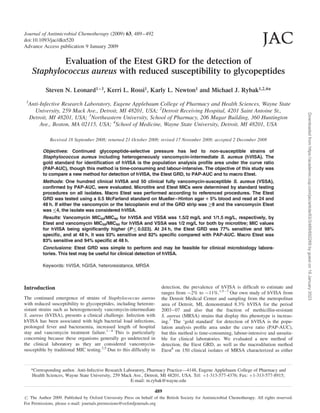
Examples of GRD strips at 48 h are shown in Figure 1. Macro
Etest was 83% sensitive and 94% specific at 48 h.
Discussion
Vancomycin remains the mainstay of therapy for infections
caused by MRSA. Unfortunately, strains of MRSA with reduced
susceptibility to vancomycin, including hVISA, are increasing in
prevalence, and infection with hVISA has been associated with
vancomycin treatment failures.1 –4
Therefore, early detection of
hVISA is of paramount importance. As demonstrated in this
investigation, MIC values for hVISA by both microtitre and
Etest methods are in the susceptible range, although there is a
tendency for a higher percentage of hVISA strains detected at an
MIC of 2 mg/L. This is similar to the findings reported pre-
viously.7
Currently, there is no standardized method for detec-
tion of hVISA though PAP-AUC is considered the gold
standard, a method too time-consuming and labour-intensive for
a clinical laboratory. Other methods of detection of hVISA are
available including BHIA plus 6 mg/L vancomycin (BHIA6V),
MHA plus 5 mg/L teicoplanin (MHA5T) and macro Etest. In an
investigation comparing these methods using PAP-AUC as the
gold standard, both macro Etest and MHA5T performed simi-
larly while BHIA6V displayed a poor sensitivity of 11%.8
We evaluated a new method for detection of hVISA that
offers the possibility of being read at 24 h. While the sensitivity
observed at 24 h (77%) was not as high as at 48 h, the high
degree of specificity at 24 h (98%) indicates that a positive
result at 24 h may have high significance. Positive and negative
Table 1. Etest vancomycin MIC distributions for hVISA and VSSA
isolates
Percentage of isolates with an MIC (mg/L) of
0.38 0.5 0.75 1 1.5 2
hVISA 0 0 2 8 41 49
VSSA 2 2 20 38 30 8
Leonard et al.
490
Downloaded
from
https://academic.oup.com/jac/article/63/3/489/692269
by
guest
on
19
January
2023
3. predictive values, however, were not calculated due to the artifi-
cially high prevalence of hVISA in our cohort (100 of 150
strains, 67%) compared with the prevalence reported in the
population (2–11%). We found the sensitivity of this test was
improved to 93% by incubation for 48 h, underscoring the
importance of reading the test not only at 24 h but also after a
full 48 h of incubation, though this occurred at the expense of a
lower specificity at this timepoint. This improvement in, and
level of, sensitivity is consistent with recent data reported on the
performance of the Etest GRD,10
though the specificity reported
at 48 h (95%) was higher than that reported by our investigation.
The reason for this difference is not immediately clear as we
used the same interpretive criteria, similar numbers of VSSA
(n ¼ 50 for our investigation and n ¼ 70 for their investigation)
and the same source for MHB plates (Becton, Dickinson and
Company).
In conclusion, we evaluated a new Etest method for the
detection of S. aureus with reduced susceptibility to glycopep-
tides. The test was simple to perform using standard media and
inocula utilized in clinical microbiology laboratories. At 24 h,
the test was sensitive at 77% with a very high specificity. The
sensitivity was improved to .90% with 48 h of incubation,
though the specificity declined at 48 h. Further research is
warranted to determine the value of this test, particularly an
evaluation of the positive and negative predictive values.
Acknowledgements
A portion of this work was presented at the Eighteenth European
Congress of Clinical Microbiology and Infectious Diseases,
Barcelona, Spain, 2008 (Abstract P1737).
Funding
No specific funding was received for this project. Vancomycin
and GRD Etests were provided by AB Biodisk.
Transparency declarations
None to declare.
References
1. Charles PG, Ward PB, Johnson PD et al. Clinical features asso-
ciated with bacteremia due to heterogeneous vancomycin-intermediate
Staphylococcus aureus. Clin Infect Dis 2004; 38: 448–51.
2. Howden BP, Johnson PD, Ward PB et al. Isolates with low-level
vancomycin resistance associated with persistent methicillin-resistant
(a)
(b)
(c)
Figure 1. Etest GRD for hVISA Mu3 (a), a clinical hVISA with a positive Etest GRD (b) and a clinical VSSA with a negative Etest GRD (c).
Evaluation of the Etest GRD for the detection of hGISA
491
Downloaded
from
https://academic.oup.com/jac/article/63/3/489/692269
by
guest
on
19
January
2023
4. Staphylococcus aureus bacteremia. Antimicrob Agents Chemother
2006; 50: 3039–47.
3. Howden BP, Ward PB, Charles PG et al. Treatment outcomes for
serious infections caused by methicillin-resistant Staphylococcus aureus
with reduced vancomycin susceptibility. Clin Infect Dis 2004; 38: 521–8.
4. Moore MR, Perdreau-Remington F, Chambers HF. Vancomycin
treatment failure associated with heterogeneous vancomycin-intermediate
Staphylococcus aureus in a patient with endocarditis and in the rabbit
model of endocarditis. Antimicrob Agents Chemother 2003; 47: 1262–6.
5. Liu C, Chambers HF. Staphylococcus aureus with hetero-
geneous resistance to vancomycin: epidemiology, clinical significance,
and critical assessment of diagnostic methods. Antimicrob Agents
Chemother 2003; 47: 3040–5.
6. Garnier F, Chainier D, Walsh T et al. A 1 year surveillance study
of glycopeptide-intermediate Staphylococcus aureus strains in a
French hospital. J Antimicrob Chemother 2006; 57: 146–9.
7. Rybak MJ, Leonard SN, Rossi KL et al. Characterization of
vancomycin-heteroresistant Staphylococcus aureus from the metro-
politan area of Detroit, Michigan, over a 22-year period (1986 to 2007).
J Clin Microbiol 2008; 46: 2950–4.
8. Wootton M, MacGowan AP, Walsh TR et al. A multicenter study
evaluating the current strategies for isolating Staphylococcus aureus
strains with reduced susceptibility to glycopeptides. J Clin Microbiol
2007; 45: 329–32.
9. Clinical and Laboratory Standards Institute. Methods for Dilution
Antimicrobial Susceptibility Tests for Bacteria That Grow Aerobically—
Seventh Edition: Approved Standard M7-A7. CLSI, Wayne, PA, USA,
2006.
10. Yusof A, Engelhardt A, Karlsson A et al. Evaluation of a new
Etest vancomycin–teicoplanin strip for detection of glycopeptide-
intermediate Staphylococcus aureus (GISA), in particular, hetero-
geneous GISA. J Clin Microbiol 2008; 46: 3042–7.
Leonard et al.
492
Downloaded
from
https://academic.oup.com/jac/article/63/3/489/692269
by
guest
on
19
January
2023
![hVISA or not hVISA [vancomycin-susceptible S. aureus (VSSA)]
by PAP-AUC.
Materials and methods
Bacterial strains
One hundred clinical hVISA, whose origins have been described
previously,7
and 50 clinical VSSA obtained from patients at the
Detroit Medical Center were evaluated.
Susceptibility testing
Microtitre dilution methods and Etest using standard reference
methods were performed on all isolates.9
PAP-AUC and macro Etest
All strains were characterized as either hVISA or not hVISA
(VSSA) by PAP-AUC as described previously.7
Briefly, 50 mL of a
bacterial suspension at an inoculum of 108 –9
was plated on brain
heart infusion agar (BHIA; Difco, Detroit, MI, USA) plates contain-
ing increasing concentrations of vancomycin (0, 0.5, 1, 2, 3, 4 and
8 mg/L) using an automated spiral plater (WASP, DW Scientific,
West Yorkshire, UK) and read using a laser colony counter
(ProtoCOL, Synoptics Limited, Frederick, MD, USA) after 48 h of
incubation at 358C. All PAP-AUC were performed in duplicate
using Mu3 as a positive control. Interpretation of PAP-AUC was as
follows: ratio of the AUC of the test isolate to Mu3 ,0.9 was con-
sidered VSSA, ratio of the AUC of the test isolate to Mu3 0.9 and
,1.3 was considered hVISA and ratio of the AUC of the test isolate
to Mu3 1.3 was considered VISA. Macrodilution method Etests
were done on all strains as described previously.8
Etest GRD
Evaluation with the Etest GRD was done according to the manufac-
turer’s instructions. A bacterial suspension corresponding to a 0.5
McFarland standard was lawned on a Mueller–Hinton agar þ 5%
blood (MHB; Becton, Dickinson and Company, Sparks, MD, USA)
plate and on a Mueller–Hinton agar (MHA; Difco) plate. A GRD
strip consisting of a double-sided gradient with vancomycin and
teicoplanin was then applied to the MHB plate and a standard vanco-
mycin Etest was applied to the MHA plate. The standard vanco-
mycin Etest was read and recorded after 18–24 h of incubation. The
zone of the Etest GRD strip was also read, at complete inhibition of
growth, at 24 and 48 h. The test isolate was considered positive for
hVISA if the Etest GRD strip was 8 mg/L for either vancomycin or
teicoplanin and the standard vancomycin Etest MIC was 4 mg/L.
Statistical analysis
Differences in vancomycin MIC between hVISA and VSSA were
evaluated by the Mann–Whitney U-test using SPSS statistical
software (Release 16.0, SPSS, Inc., Chicago, IL, USA). A P value
of 0.05 was considered significant. Sensitivity and specificity
analyses were performed to evaluate the performance of the GRD
test versus PAP-AUC. Sensitivity analysis describes the fraction of
correctly identified true positives (hVISA) by the Etest GRD strip,
while specificity analysis describes the fraction of correctly ident-
ified negatives (VSSA).
Results
The vancomycin MIC50, MIC90 and range were 1.5, 2 and
0.75–2 mg/L for hVISA and 1, 1.5 and 0.38–2 mg/L for VSSA,
respectively, by Etest; this difference in MIC distribution was
statistically significant (P,0.001). Most of the hVISA isolates
(90%) had a vancomycin MIC .1 mg/L. Vancomycin MIC dis-
tributions for hVISA and VSSA isolates are displayed in Table 1.
Overall, MIC by Etest tended to be slightly higher than microdi-
lution MIC with a vancomycin MIC50, MIC90 and range of 1, 2
and 0.5–2 mg/L for hVISA and 1, 2 and 0.5–2 mg/L for VSSA,
respectively. Still, the difference in MIC distribution between
hVISA and VSSA remained significant, with MIC values of
hVISA tending to be higher, though not to the same degree as by
Etest (P ¼ 0.023). At 24 h, the Etest GRD was 77% sensitive and
98% specific. At 48 h, the Etest GRD displayed a sensitivity of
93% and a specificity of 82%. Of those tests that were positive at
24 h, 100% were positive at the teicoplanin end of the strip only.
Examples of GRD strips at 48 h are shown in Figure 1. Macro
Etest was 83% sensitive and 94% specific at 48 h.
Discussion
Vancomycin remains the mainstay of therapy for infections
caused by MRSA. Unfortunately, strains of MRSA with reduced
susceptibility to vancomycin, including hVISA, are increasing in
prevalence, and infection with hVISA has been associated with
vancomycin treatment failures.1 –4
Therefore, early detection of
hVISA is of paramount importance. As demonstrated in this
investigation, MIC values for hVISA by both microtitre and
Etest methods are in the susceptible range, although there is a
tendency for a higher percentage of hVISA strains detected at an
MIC of 2 mg/L. This is similar to the findings reported pre-
viously.7
Currently, there is no standardized method for detec-
tion of hVISA though PAP-AUC is considered the gold
standard, a method too time-consuming and labour-intensive for
a clinical laboratory. Other methods of detection of hVISA are
available including BHIA plus 6 mg/L vancomycin (BHIA6V),
MHA plus 5 mg/L teicoplanin (MHA5T) and macro Etest. In an
investigation comparing these methods using PAP-AUC as the
gold standard, both macro Etest and MHA5T performed simi-
larly while BHIA6V displayed a poor sensitivity of 11%.8
We evaluated a new method for detection of hVISA that
offers the possibility of being read at 24 h. While the sensitivity
observed at 24 h (77%) was not as high as at 48 h, the high
degree of specificity at 24 h (98%) indicates that a positive
result at 24 h may have high significance. Positive and negative
Table 1. Etest vancomycin MIC distributions for hVISA and VSSA
isolates
Percentage of isolates with an MIC (mg/L) of
0.38 0.5 0.75 1 1.5 2
hVISA 0 0 2 8 41 49
VSSA 2 2 20 38 30 8
Leonard et al.
490
Downloaded
from
https://academic.oup.com/jac/article/63/3/489/692269
by
guest
on
19
January
2023](data:image/gif;base64,R0lGODlhAQABAIAAAAAAAP///yH5BAEAAAAALAAAAAABAAEAAAIBRAA7)