
Recomendados
Más contenido relacionado
La actualidad más candente
La actualidad más candente (20)
Similar a Genome organization in eukaryotes (molecular biology)
Similar a Genome organization in eukaryotes (molecular biology) (20)
Más de IndrajaDoradla
Más de IndrajaDoradla (20)
Último
Último (20)
Genome organization in eukaryotes (molecular biology)
- 1. genome organization in eukaryotes D.INDRAJA
- 2. Gene: It is a unit of heridity which is transferred from a parent to offspring and held to determine some charecteristic to offspring Genome: The entire set of genetic information in an organism It is encoded in DNA or RNA in case of many viruses It includes different types of genes they are Structural genes: DNA segments that code for some specific RNAs or proteins encode for mRNAs, tRNAs, SnRNAs Functional sequences: The sequences that are regulatory elements such as initiation site, promoter site, operator site Non functional sequences: It includes introns and repetetive sequences
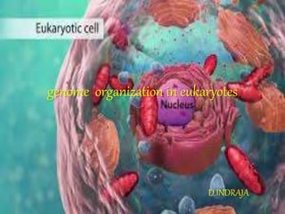
- 3. Nucleus: In eukaryotes nucleus is the heart of the cell which serves as distinguish feature of eukaryotic cell.the genomic material is present in the nucleus which separates cytoplasm with the nuclear membrane • Nucleus contain many thread like coiled structures which remain suspended in the nucleoplasm which are known as chromatin substance • Chromatin is the complex combination of DNA and proteins that makes up chromosomes • The major proteins involved in chromatin are histone proteins although many other chromosomal proteins have prominent roles too • The function of chromatin is to package DNA in to smaller volume to fit in the cell,to strengthen the DNA to allow mitosis and meiosis and to serve as mechanism to control gene expression and DNA replication
- 4. • The information stored in DNA is organized and replicated and read with the help of a variety of DNA binding proteins Structural proteins-histones (packing proteins): • Main structural proteins found in eukaryotic cell • Low molwt basic proteins with high proportion of positively charged aminoacids(lysine and arginine) • Bound to DNA along most of its length • The positively charged histones bind to the negatively charged DNA and play a crucial role in packaging of long DNA molecules • Types of histones H1,H2A,H2B,H3,H4 • Ratio H1:1, H2A:2, H2B:2, H3:2, H4:2 • The H1 histone is called linker histone and the remaining histones forms core particle
- 5. Non histone chromosomal proteins: • These serve as structural roles • These take part in genetic processes such as transcription and replication Eg: scaffold proteins • Scaffold means to provide or support with a raised frame network or platform • A protein whose main function is to bring other proteins together for them to interact • When chromatin is treated with a concentrated salt solution it removes histones and most of the other chromosomal proteins having a skeleton to which DNA was attached known as scaffold proteins • These scaffold proteins play a role in the folding and packaging of chromosome
- 6. GENOME ORGANIZATION MODELS They are different models explained for genome organization in eukaryotes 1. Multi stranded model 2. Folded fibre model 3. Nucleosome model most widely accepted Muti stranded model • This model was put forth by Ris in 1961 and Ris and Chandler in 1963. According to this model, the chromosome is multi-stranded, i.e., it contains several DNA double helices arranged parallel to each other. Each chromosome is divided into two chromatids, each chromatid is made of two “half chromatids” and each half chromatid is composed of two “quarter chromatids.” • Each “quarter chromatid” is, in turn, made of four chromatin fibres and each chromatin fibre contains 2 DNA double helices. The diameter of the DNA double helix is 2 nm, and two DNA molecules are associated with protein to make the chromatin fibre. • However, according to the recent studies, the chromosome is definitely not multi-stranded.
- 7. Folded fibre model DuPraw in 1965 proposed this model on the basis of electron microscopic studies of human chromosomes. the feautres of this model are • Each chromosome contains a single but long and coiled chromatin fibre • The chromatin fibre has DNA double helix with associated proteins .this DNA is packed spirally to form a fibre • The fibre is then coiled to form 10-100A°fibre called type A fibre and it is further coiled to form 200-250A° to form type B fibre. This is further folded to form chromatid • The fibre contain DNA and histones in super coiled condition ,histone proteins bound on outer side of DNA and form a shell aroud the DNA . Dupraw called this as histone shell • The chromatin fibre (chromatid) replicates during S phase of call cycle to produce two sister chromatids which are held together by the un-replicated regions
- 9. Nucleosome model Roger Kornberg proposed that DNA and histones were organized into repeated units called nucleosome. • Nucleosome model is the most accepted model of chromatin. • Nucleosomes are the fundamental repeating units of chromatin. • Nucleosome represents the ‘beads’ as proposed in the ‘beads on string’ organization of chromatin. Each nucleosome contains a nucleosome core particle. composed of a disc shaped structure of eight histone proteins. • The nucleosome core composed of two molecules of each of the four histones H2A, H2B, H3 and H4 and his structure is called the histone octamer. • The DNA helix is wrapped as super helical left handed turn around this histone octamer core. • Each histone core is encircled by 1.8 turns of DNA. • This 1.8 turn of DNA represents about 146 base pairs.
- 10. • Each nucleosome is about 10 nm in diameter. • The H1 histone stays outside the histone octamer. • Adjacent nucleosomes are connected by a short stretch of DNA called linker DNA. • Linker DNA is about 10 to 80 bp in length. H1 histones bind to the liner DNA. • H1 histone binds near the site where DNA enters and exits the nucleosome.
- 11. • The interaction of histones and DNA in nucleosome is stabilized by several types of non-covalent bonds. • Among these bonds, the ionic bonds formed between the negatively charged phosphate groups in the DNA with the positively charged amino groups of histones were very important • Nucleosome units organized into more compact structure of 30 nm in diameter called 30 nm fibers • The H1 histone plays very important role in the formation of the 30-nm fiber. • The formation of 30 nm fiber shortens genetic material (DNA) another seven-fold. • The linker DNA regions in 30-nm structure are variably bent and twisted to attain the folding pattern. • This 30 nm fibres are further folded to 300nm loop model with the help of scaffold and other proteins and these are further condensed to 700nm higher condensed loop model and finally to form 1400nm metaphase chromosome during cell division
- 12. Heirarchy of chromatin organization
- 14. The Importance of DNA supercoiling • DNA supercoiling is important for DNA packaging within all cells. Because the length of DNA can be thousands of times that of a cell, packaging this genetic material into the nucleus is a difficult . Supercoiling of DNA reduces the space and allows for much more DNA to be packaged. • DNA packaging is greatly increased during nuclear division events such as mitosis or meiosis, where DNA must be compacted and segregated to daughter cells. Condensins and cohesins are structural maintenance of chromosome (SMC) proteins that aid in the condensation of sister chromatids and the linkage of the centromere in sister chromatids. These SMC proteins induce positive supercoils.