2. munication between the MJ Research PTC-200 and the
Biomek was mediated by a serial connection between the
thermal cycler and the computer and by Jeff Cahlik’s
(Beckman Coulter) MJR Biomek 2000 Driver. While the
MJR thermal cycler could also have been directly inter-
faced with one of the Biomek’s connection ports, Cahlik’s
driver allowed suspension of the BioWorks program until
the completion of the thermal cycler’s run. This obviated
the problem of having to guess how long the thermal
cycler might take to complete its program. The program
for automated selection is available on request.
Selection Regime. At the beginning of the experi-
ment, Dynabeads Oligo(dT)25 (20 µL, corresponding to 6.6
× 106
beads) in a high-salt binding solution (80 µL; 20
mM Tris-HCl (pH 7.5), 1.0 M LiCl, and 2 mM EDTA)
were loaded into a well in a microplate on the magnetic
bead separator. Roughly 1 µg (∼1013 sequences) of a
random sequence RNA library (N30; 80 µL in transcrip-
tion buffer, see below) was added to the beads. The
binding reaction was thoroughly mixed by 10 cycles of
rapid aspiration and dispensing. The binding reaction
was incubated at room temperature for 2.5 min, mixed
again (10 cycles), and incubated an additional 2.5 min.
At the end of incubation, the MPC-auto96 raised magnets
between the rows of wells in the microplate and captured
the magnetic beads along the side of the well. The
supernatant was removed (less than 0.5 µL typically left
behind). The magnets were then lowered, and the beads
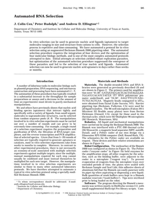
Figure 1. Selection robot. (a, top) The augmented Biomek 2000 used for automated selection. As landmarks, the PTC-200 thermal
cycler is on the upper left, while the pipet tools are on the upper right. Radioactive tips from the experiment were discarded onto a
sheet of plastic wrap covering the PTC-200 and dropped into a radioactive waste container at the lower left. (b, bottom) Schematic
of the x-y plane of the robot. The functions associated with the various positions are described in the text.
Figure 2. Random sequence pool. The N30 pool has previously been described (9). The random sequence core is flanked by constant
sequences required for enzymatic amplification. Residues that will be found in the transcribed RNA pool are capitalized; primer
sequences are in lower case. The primer containing the T7 RNA polymerase promoter is at the left, while the primer containing the
reverse transcription start site is at the right. Restriction sites that facilitate cloning are indicated.
846 Biotechnol. Prog., 1998, Vol. 14, No. 6
3. were resuspended and washed with 100 µL of low-salt
wash buffer (10 mM Tris-HCl (pH 7.5), 0.15 M LiCl, and
1 mM EDTA) to remove nonspecifically binding nucleic
acid species. As before, the beads and buffer were mixed
by 10 cycles of aspiration and dispensing. The wash step
was repeated once more before resuspending the beads
in 53 µL of water. The bead slurry was transferred to a
well in the microplate in the PTC-200 thermal cycler, the
lid of the machine was closed, and the slurry was heated
to 65 °C for 3 min to dissociate nucleic acid binding
species from the derivatized beads. During the elution,
the lid of the thermal cycler was kept at 80 °C. After
the elution step, the magnetic bead mixture was trans-
ferred back to the magnetic particle separator to ensure
the complete removal of magnetic beads from the eluate.
To amplify the selected RNA molecules, the eluate (51
µL, purposefully leaving behind a small amount of liquid)
was transferred to a new well of the microplate on the
thermal cycler.
Reverse Transcription and PCR. Conversion of the
selected RNAs into double-stranded DNAs was carried
out in a single step. RT-PCR reaction buffer (45 µL) was
added to the 51 µL of selected RNA eluate. Upon mixing
and dilution the amplification reaction contained 10 mM
Tris-HCl (pH 8.4), 50 mM KCl, 1.5 mM MgCl2, 0.2 mM
dNTPs, 5% acetamide, 0.05% Nonidet P40, and 0.5 µM
each of 5′ and 3′ primers. To follow the progress of the
selection, 5 µCi of R-32P-labeled dATP was also included
in the amplification reaction. The reaction mixture was
incubated at 65 °C for 10 min (lid ) 80 °C) to facilitate
denaturation of secondary structures present in the
selected RNAs that could inhibit reverse transcription.
Finally, the temperature of the reaction was reduced to
50 °C, and 4 µL of RT-PCR enzyme solution was added
to the prewarmed mixture. The RT-PCR enzyme solution
contained 0.2 units of Display Taq (Promega, Madison,
WI) and 5 units of AMV reverse transcriptase (RT)
(Amersham Pharmacia Biotech, Arlington Heights, IL).
To increase the volume of the enzyme mixture to 4 µL,
these enzymes were diluted into a solution containing
50% glycerol, 10 mM Tris-HCl (pH 8.4), 50 mM KCl, and
1.5 mM MgCl2. Enzymes were not diluted until the start
of the experiment and were kept in the enzyme cooler
(-20 °C). The addition of the enzyme mixture brought
the final volume of the RT-PCR reaction to 100 µL.
Reverse transcription proceeded at 50 °C for 10 min, and
the amplification reaction was then thermally cycled (45
s at 94 °C, 60 s at 50 °C, and 90 s at 72 °C) seven times.
At the end of the RT-PCR reaction, 10 µL (one-tenth) of
the products was transferred to a new well of the
microplate on the thermal cycler.
In Vitro Transcription. Transcription reaction buffer
(87 µL) was added to the newly amplified DNA templates.
Upon mixing and dilution, the transcription reaction
contained 40 mM Tris-HCl (pH 7.9), 26 mM MgCl2, 0.01%
Triton X-100, 2.5 mM spermidine, 5 mM dithiothreitol,
and 2.5 mM of each NTP. To follow the progress of the
selection, 5 µCi of R-32P-labeled UTP was included in the
transcription reaction. Transcription was initiated by the
addition of 100 units of T7 RNA polymerase (Stratagene,
La Jolla, CA) and 40 units of RNasin (Promega) in a total
volume of 3 µL. The transcription reaction was allowed
to proceed at 37 °C for 75 min.
Additional Rounds of Selection. A majority of the
transcription reaction (80 µL) was transferred to a well
on the magnetic particle separator that contained mag-
netic beads (6.6 × 10⁶ beads) in the high-salt buffer (80
µL). The binding reaction was mixed as described above,
and separation of bound and unbound RNAs proceeded
as before. The entire selection and amplification proce-
dure was repeated without change for four additional
rounds.
Sequencing. At the end of the five cycles of selection
and amplification, 1 µL of the final transcription reaction
was used to seed 100 µL of a RT-PCR reaction similar to
those described above. Double-stranded products were
cloned into pCR2.1 with Invitrogen’s (Carlsbad, CA) TA
cloning kit according to the manufacturer’s instructions.
Individual colonies were screened for the presence of
insert using a colony PCR protocol (10). Purified plas-
mids containing inserts were then sequenced using
Epicentre’s (Madison, WI) Sequitherm Excel II DNA
sequencing kit according to the manufacturer’s directions.
Because long runs of a single base were expected, the
isothermal (rather than thermal cycle) sequencing reac-
tion directions supplied with the kit were employed.
Results and Conclusion
Design of the Automated Selection Station. The
Beckman Biomek 2000 is an automated pipetting robot
that can manipulate liquids in a limited x-y-z volume.
To satisfy all of the requirements for a typical in vitro
selection experiment, we augmented the Biomek 2000
with a magnetic bead separator, the Dynal MPC-auto96
(to sieve target:nucleic acid complexes from free nucleic
acids), and an MJ Research PTC-200 PCR machine (for
nucleic acid amplification). In addition, we built a small
Peltier cooler to hold enzyme solutions necessary for
amplification. The augmented robot is shown in Figure
1a, and the layout of the x-y plane for selection experi-
ments is shown in Figure 1b. It should be noted that
the x-y grid has been laid out to avoid cross-contamina-
tion problems that might arise between parallel or serial
selection experiments: “clean” tips that have not seen
amplicons always move from right to left on the surface,
eventually being discarded at the far left. Other precau-
tions against cross-contamination include the use of
barrier tips and puncturable seals on microplates.
Selection Experiments. A brief description of the
automated selection procedure follows; a more complete
description can be found in Materials and Methods. A
random sequence RNA pool (N30; Figure 2) that had
previously yielded aptamers (9) was used as a starting
point for selection. The pool was loaded into a well in
the “reagent” microplate. The Biomek transferred a
portion of the randomized RNA pool (1 µg, ∼1013 different
sequences) into a solution containing magnetic beads
derivatized with a target molecule, in this case (dT)25.
Oligo(dT) was chosen as a target to evaluate the efficiency
of the robot and to facilitate interpretation of the results
of the automated selection. The binding reaction was
pipetted up and down several times to ensure adequate
mixing of pool and target, and then incubated for 5 min
at room temperature to allow nucleic acid:target com-
plexes to form (total time ) 7 min). The magnetic beads
and associated RNA molecules were captured using a
magnetic particle separator, and unbound RNAs were
removed. To ensure that only high-affinity species were
retained, the beads were then washed several times with
a low salt solution and the washes removed (10 min). Any
remaining RNA molecules were eluted from the target
by the addition of a no-salt (water) solution. To ensure
efficient elution, the beads were transferred to the
thermal cycler, and the elution step was carried out at
65 °C (10 min). All of the eluted species were used to
seed a RT-PCR reaction; the selected population, buffers
and substrates, and an enzyme mixture were separately
added and mixed by the Biomek (110 min). One-tenth
Biotechnol. Prog., 1998, Vol. 14, No. 6 847
4. of the amplified template was then used for in vitro
transcription (75 min). Finally, the amplified RNA, still
in its transcription buffer, was introduced into a new
round of selection. The total time required for a round
of selection was 212 min.
Five rounds of automated selection were carried out
using this procedure. To follow the production of double-
stranded DNA templates and single-stranded RNA tran-
scripts, small amounts of radiolabeled nucleotides (R-32
P
dATP for DNA and R-32P UTP for RNA) were included
in the amplification reactions. A portion of each RT-PCR
and transcription reaction was analyzed by gel electro-
phoresis (Figure 3). As can be seen, the robot initially
faithfully reproduced the sizes of the DNA and RNA
products, despite the fact that a constant number of
thermal cycles had been programmed into each round of
automated amplification. We had anticipated this result,
since the N30 pool was designed to undergo many rounds
of amplification without the production of artifacts (11)
and had, in fact, previously yielded no replication para-
sites during selection experiments. However, by the
third round of selection, a higher molecular weight
species appeared and became even more prominent in
both RNA and DNA populations in rounds 4 and 5. We
initially assumed that the size heterogeneity was the
result of imprecise automated amplification that had
previously not been observed during manual amplifica-
tion; however, these bands are, in fact, the result of a
selected expansion of the random sequence region (see
below).
Sequences of Selected RNA Molecules. Amplified
RNA molecules from the last round of selection were
further amplified using a RT-PCR protocol similar to that
employed during robotic selection. The amplified, double-
stranded DNA molecules were cloned, individual clones
were screened for the presence of inserts using a standard
colony PCR protocol (10), and clones containing inserts
were sequenced. The sequences of several individual
clones are shown in Figure 4. As expected, the success
of the selection experiment was immediately apparent:
each clone contained long, uninterrupted runs of aden-
osine. It is anticipated that (dT)25 can readily be em-
ployed as a positive control in future automated selection
experiments.
The sequences of the selected clones revealed the
provenance of the larger molecular weight bands ob-
served in rounds 3-5. The random sequence tracts had
expanded to include 44-66 residues. The expansion
appeared to be due primarily to an expansion of the
target binding site, since runs of 21-39 contiguous
adenosine residues were present. The runs were not
preferentially localized within the transcripts and could
occur at either end of the original random sequence tract.
No shorter clones were obtained, likely because the
majority of clones had become elongated by the conclusion
of the selection (Figure 3).
The expansion of the random sequence region to
include additional adenosine residues had previously
been observed in manual selection experiments and was
likely due to “stuttering” in the register of T7 RNA
polymerase and/or reverse transcriptase as homopolymer
tracts were copied (12, 13). However, the fact that such
long runs or adenosines are produced so soon during the
course of the selection experiment indicates that the
selection procedure employed was quite stringent yet
consistently yielded enough selected molecules for ad-
ditional amplification. While molecules such as proteins
will undoubtedly prove to be more difficult selection
targets, these results bode well for the general application
of the automated selection procedures.
Parameters That Influence the Success of Auto-
mated Selection Experiments. During the develop-
Figure 3. Progress of automated selection. RNA molecules corresponding in size to the N30 pool are shown at the left and right;
the sizes of the RNA pool and DNA pool are indicated with arrows. A fraction (5 µL; 5%) of each RT-PCR (“RT-PCR”) or in vitro
transcription (“TXN”) reaction was pipetted into separate wells in a microplate during the selection. The wells contained an equal
volume of sequencing stop dye. At the conclusion of the selection, the samples were electrophoresed on a 8% denaturing polyacrylamide
gel, the gel was exposed to a Phosphor Screen, and the screen was read using a Phosphorimager SI (Molecular Dynamics, Sunnyvale,
CA).
Figure 4. Sequences of selected clones. Individual clones were isolated and sequenced as described in Materials and Methods. The
sequences of the random sequence regions from these clones are shown. The adenosine run length and total length of the random
sequence tract are shown at the right.
848 Biotechnol. Prog., 1998, Vol. 14, No. 6
5. ment of the automated selection method described above
several variations were introduced. The amount or
proportion of selected RNA that was introduced into
amplification reactions for subsequent cycles of selection
did not appear to influence the course of the selection,
nor did the amount or proportion of newly transcribed
RNA that was used for selection. The amount of enzymes
used for amplification could be varied by factors of 2-3
without any observable changes in the dynamics of the
selection experiment. The apparent robustness of the
automated protocol to these modifications was especially
encouraging, given that several different aspects of
selection procedures (e.g., the amount of nucleic acid
population bound in each cycle, the number of cycles of
amplification necessary to obtain a PCR product, the
amount of RNA obtained from transcription) are typically
closely monitored by experimentalists during manual
selection protocols. Overall, it appears as though these
and other variables can be subsumed within common
mechanized manipulations without significantly altering
or belaying the progress of the selection.
However, one parameter that had a decided effect on
the success of the selection experiments was the ampli-
fication method that was employed. We originally at-
tempted to perform automated selection using an iso-
thermal amplification procedure (3SR) that combined
reverse transcription and transcription in one tube (14).
The successful implementation of this procedure would
have allowed at least one step to be excised from the
protocol and would have sped up the turnaround time
for each round of selection. However, attempts to amplify
random sequence RNA molecules using the isothermal
amplification protocol consistently led to the generation
of amplification parasites that overran the selected RNAs
(see, for example, Figure 5). The amplification parasites
that were observed were themselves interesting, given
that they were larger than the expected product, rather
than smaller as are expected of amplification parasites,
and have been further characterized. In contrast to the
larger bands observed during a successful selection
experiment (Figure 3), the parasites were quasispecies
centered on a single molecule, did not contain runs of
adenosines, and arose irrespective of whether oligo(dT)25
was included in the automated selection protocol or
whether the selections were carried out manually or by
the robot. These results are especially significant given
that previous selection experiments carried out with
partially (as opposed to completely) randomized popula-
tions successfully employed 3SR as an amplification
procedure (15). Unfortunately, while partially random-
ized nucleic acid populations centered on a particular
molecular species generally reproduce faithfully, com-
pletely randomized populations offer many more poten-
tial routes to replication parasites. The likely success of
“discontinuous” amplification procedures (RT-PCR fol-
lowed by transcription) for molecular engineering relative
to “continuous” amplification procedures (3SR) had previ-
ously been predicted by evolutionary biologists on theo-
retical grounds alone (16).
Prospects for Automated Selection. Just as in
vitro selection techniques have been applied to a wide
variety of targets from small molecules to proteins to
supramolecular structures such as viruses and tissues,
it should be possible to apply automated selection pro-
cedures to a similar range of targets. For example, Eigen
and co-workers have previously built a PCR machine
coupled with a liquid delivery system that allowed the
parallel amplification and serial transfer of multiple
nucleic acid samples (17). The automated protocol we
have developed was designed to be carried out with off-
the-shelf robotics and to be readily adaptable to a variety
of selection modalities and conditions. A number of
different derivatization chemistries can be used to con-
jugate target molecules to magnetic beads, and the
amplification protocols are amenable to double-stranded
DNA libraries, single-stranded DNA libraries, and modi-
fied nucleic acid libraries, as well as conventional RNA
libraries of any length.
Using the duration of the current experiments as a
guide, it should be possible to carry out at least 10
rounds/day of automated selection against a single target.
By using multipipettor tools (e.g., the Beckman MP200),
it should be possible to simultaneously select against
8-96 or more different targets or to assess 8-96 different
selection conditions.
A final, often unappreciated, advantage of automated
procedures relative to their manual counterparts is the
consistency of repetitive tasks. In vitro selection experi-
ments have previously been used to address evolutionary
questions, but the relevance and uniformity of the data
sets that have been derived have been open to question
because there is large variation between how individual
researchers or laboratories carry out the plethora of
manual procedures that are required to extract “winning”
sequences from random sequence libraries. Automated
selection procedures should provide a reproducible bench-
mark for comparison, and may allow results from many
different labs to ultimately be combined and interpreted.
Acknowledgment
This work was supported by NIH Grant No. AI 36083-
03.
References and Notes
(1) Boland, E. J.; Pillai, A.; Odom, M. W.; Jagadeeswaran, P.
Automation of the Maxam-Gilbert chemical sequencing
reactions. Biotechniques 1994, 16, 1088-1092; 1094-1095.
(2) Civitello, A. B.; Richards, S.; Gibbs, R. A. A simple protocol
for the automation of DNA cycle sequencing reactions and
polymerase chain reactions. DNA Sequence 1992, 3, 17-23.
Figure 5. Isothermal amplification yields replication parasites.
Experiments similar to those described in Materials and
Methods were carried out, except that the 3SR procedure of
Guatelli et al. (14) was used for amplification rather than RT-
PCR followed by in vitro transcription. Fractions (5 µL; 5%) of
the selected RNAs (“selected RNA”) and isothermal amplifica-
tion reactions (“selected RNA amplified”) were run on an 8%
denaturing gel. In the gel that is shown, only RNA molecules
were labeled. No bands are seen in the “selected RNA” lanes,
because early in the selection few RNA molecules are extracted
from the population by the Dynabeads. The relative sizes of the
RNA pool and the replication parasite are indicated by arrows.
Biotechnol. Prog., 1998, Vol. 14, No. 6 849
6. (3) Wang, D. G.; Fan, J. B.; Siao, C. J.; Berno, A.; Young, P.; et
al. Large-scale identification, mapping, and genotyping of
single- nucleotide polymorphisms in the human genome.
Science 1998, 280, 1077-1082.
(4) Meier-Ewert, S.; Maier, E.; Ahmadi, A.; Curtis, J.; Lehrach,
H. An automated approach to generating expressed sequence
catalogues. Nature 1993, 361, 375-376.
(5) Saborio, J. L.; Galvin, N. M.; Lee, M.; Cahlik, J. A.; Reddy,
M. P. A fully automated method for rapid purification of
plasmid DNA using the Biomek 2000 and Qiagen’s “QIAwell
96 Ultra” kit; Beckman Coulter, Inc., A-1849A; Beckman:
Fullerton, CA, 1998.
(6) Gonzalez, D.; Sasaki, G.; Threadgill, G. High-throughput
purification of plasmid DNA using the 96-Plasmid Purifica-
tion Kit on the Biomek 2000 Laboratory Automated Worksta-
tion, Beckman Coulter, Inc., A-1826A; Beckman: Fullerton,
CA, 1996.
(7) Macas, J.; Nouzova, M.; Galbraith, D. W. Adapting the
Biomek 2000 laboratory automation workstation for printing
DNA microarrays. Biotechniques 1998, 25, 106-110.
(8) Gold, L.; Polisky, B.; Uhlenbeck, O.; Yarus, M. Diversity of
oligonucleotide functions. Annu. Rev. Biochem. 1995, 64, 763-
797.
(9) Bell, S. D.; Denu, J. M.; Dixon, J. E.; Ellington, A. D. RNA
molecules that bind to and inhibit the active site of a tyrosine
phosphatase. J. Biol. Chem. 1998, 273, 14309-14314.
(10) Clackson, T.; D. Gu¨ssow, D.; Jones, P. T. In PCR 1;
Rickwood, D., Hames, B. D., Eds.; Oxford University Press:
Oxford, 1991; pp 210-212.
(11) Crameri, A.; Stemmer, W. P. 10(20)-fold aptamer library
amplification without gel purification. Nucleic Acids Res.
1993, 21, 4410.
(12) Cunningham, P. R.; Weitzmann, C. J.; Ofengand, J. SP6
RNA polymerase stutters when initiating from an AAA...
sequence. Nucleic Acids Res. 1991, 19, 4669-4673.
(13) Macdonald, L. E.; Zhou, Y.; McAllister, W. T. Termination
and slippage by bacteriophage T7 RNA polymerase. J. Mol.
Biol. 1993, 232, 1030-1047.
(14) Guatelli, J. C.; Whitfield, K. M.; Kwoh, D. Y.; Barringer,
K. J.; Richman, D. D. et al. Isothermal, in vitro amplification
of nucleic acids by a multienzyme reaction modeled after
retroviral replication. Proc. Natl. Acad. Sci. U.S.A. 1990, 87,
1874-1878.
(15) Wright, M. C.; Joyce, G. F. Continuous in vitro evolution
of catalytic function. Science 1997, 276, 614-617.
(16) Bull, J. J.; Pease, C. M. Why is the polymerase chain
reaction resistant to in vitro evolution? J. Mol. Evol. 1995,
41, 1160-1164.
(17) Schober, A.; Walter, N. G.; Tangen, U.; Strunk, G.; Ederhof,
T.; et al. Multichannel PCR and serial transfer machine as a
future tool in evolutionary biotechnology. Biotechniques 1995,
18, 652-661.
Accepted October 7, 1998.
BP980097H
850 Biotechnol. Prog., 1998, Vol. 14, No. 6