Recomendados

Recomendados
Más contenido relacionado
La actualidad más candente
La actualidad más candente (20)
Similar a ACS Poster TV4 TM2 JE
Similar a ACS Poster TV4 TM2 JE (20)
ACS Poster TV4 TM2 JE
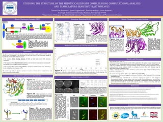
- 1. STUDYING THE STRUCTURE OF THE MITOTIC CHECKPOINT COMPLEX USING COMPUTATIONAL ANALYSIS AND TEMPERATURE-SENSITIVE YEAST MUTANTS Trevor Van Eeuwen*^, James Luginsland^, Patricia Melloy*, Gloria Anderle^ Fairleigh Dickinson University, Madison, New Jersey 07940 * Department of Biological and Allied Health Sciences, ^ Department of Chemistry and Pharmaceutical Science Cellular Studies of cdc20-1 References Acknowledgements Hypothesis The cdc20-1 mutation, the result of a glycine to arginine substitution at amino acid 544 (G544R), causes cells to arrest in metaphase.2 The mechanism by which this occurs is not understood, though we hypothesize that one of the following occurs: The mutation affects binding domains of Mad2 or Mad3 and restricts APC substrate recognition. The mutation affects thermodynamic stability at non-permissive temperatures, disrupting the hydrophobic core causing the protein to denature. The mutation affects protein folding kinetics, causing the unfolded protein to be disposed of by the unfolded protein response (UPR). Research was funded in part by Pfizer PURE Grant, the Novo Nordisk Summer Research Fellowship, the Novartis Research Scholarship and an NSF Equipment grant number 0721251 for which we are immeasurably thankful. Funding also provided Becton College Grant–in-Aid. 25oC 27oC 30oC 32oC 37oC Wildtype +++ +++ +++ +++ +++ cdc20-1 +++ +++ ++ +/- -25oC 27oC 30oC 32oC 37oC wt cdc20-1 wt cdc20-1 wt cdc20-1 wt cdc20-1 We wanted to use molecular dynamic simulations of protein models to observe protein behavior and compare this with cellular studies. Figure 5A. Analyzing the temperature-sensitive phenotype of cdc20-1. Shown are serial dilutions of the wildtype control (top row) and cdc20-1 (second row) budding yeast strains grown at permissive temperature (25oC), non-permissive temperature (37oC) and three intermediate temperatures for 7 days on YPD media. The temperature-sensitive phenotype is first visible at 32oC (red arrow) and confirmed with no growth seen at 37oC (yellow arrow). Figure 5B. Cdc20-1-GFP is visible in the nucleus and then the vacuole depending on growth temperature. Shown are images of the candidate cdc20-1-GFP strain at 25oC and 37oC. At 25oC, the GFP signal (orange arrow) localizes to bud neck of mitotic cells and nucleus and is distinct from the central vacuole (blue arrow)5. At 37oC, the GFP signal (yellow arrow) colocalizes with the central vacuole (purple arrow).6 Images were taken using the Leica DM5500 microscopy system and an ORCA ER camera(Hamamatsu). Expos ure times were 5ms (DIC) , 2s (GFP) and 2s (RHO) 2x2 binning. 1. Hartwell LH, Mortimer RK, Culotti J, Culotti M (1973). Genetic control of the cell division cycle in yeast. V. Genetic analysis of cdc mutants. Genetics, 74:267-286. 2. Schott EJ, Hoyt MA (1998). Dominant alleles of Sacchromyces cerevisiae CDC20 reveal its role in promoting anaphase. Genetics 148(2):599-610. 3. Chao WCH, Kulkarni K, Zhang Z, Kong EH, Barford D (2012). Structure of the mitotic checkpoint complex. Nature, 484: 208-214 4. Chen et al. (2010). MolProbity: all-atom structure validation for macromolecular crystallography. Acta Crystallographica D66:12-21. 5. Melloy PM, Holloway S (2004). Changes in the Localization of the Saccharomyces cerevisiae Anaphase-Promoting Complex Upon Microtubule Depolymerization and Spindle Checkpoint Activation. Genetics, 167:1079-1094. 6. Bertolotti, A., et al., Dynamic interaction of BiP and ER stress transducers in the unfolded-protein response. Nat Cell Biol, 2000. 2(6): p. 326-32. 7. Chaudhury S, Lyskov S, Gray JJ (2010). PyRosetta: a script-based interface for implementing molecular modeling algorithms using Rosetta. Bioinformatics., 26:689–691 8. Tan KP, Khares S, Varadarajan R, Madhusudhan MS (2014). Tspred: a web server for the rational design of temperature sensitive mutants. Nucelic Acids Research. doi: 10.1093/nar.gku319. Results and Discussion Mitotic Checkpoint Complex (MCC) Figure 1A. The MCC helps to regulate the cell division cycle by working with the anaphase-promoting complex (an E3 ubiquitin ligase). In the event of unattached kinetochores or other spindle damage, the MCC forms and inhibits the APC to block cell cycle progression until damage is repaired. Mad1 and Mad3 are recruited to unattached kinetochores, facilitating MCC formation. Mad2, Mad3 and Bub3 bind to Cdc20 to form MCC which in turn binds APC thereby blocking substrate recognition. The inability of APC to ubiquitinate securins and cyclins maintains cells in metaphase until kinetochores are properly attached. Mad1 Bub3 MCCCdc20 binding event Cdc20 APC Unattached Kinetochore Inactivated APC Mad3 Bub3 Mad2 Figure 1B. The MCC (left) is responsible for maintaining chromosome and spindle integrity during the cell cycle. Mutations in these genes can cause errors in response to spindle damage. The Cdc20-1 mutant (obtained from the ATCC) causes cells to arrest in metaphase.1 Major Results of our study are: Fluorescence Microscopy indicates cdc20-1-GFP is localized to the nucleus at room temperature and the vacuole at 37 oC. Comparative studies of Cdc20-GFP localization at 25 oC and 37 oC are in progress. We have created Saccharomyces cerevisiae homology models of Cdc20p, Mad2p, and Mad3p using an existing S. pombe crystal structure (4AEZ) as a template. Structures have been validated using the MolProbity structural validation server. Using a molecular dynamics in-silico simulation, we have shown that a folded cdc20-1 protein is predicted to be stable at 37 oC. Our current data suggests that cdc20-1 causes defects in protein folding. Localization to central vacuole suggests involvement of Unfolded Protein Response (UPR) in removing misfolded cdc20-1. Current simulations at permissive and non-permissive temperatures indicate no major changes in thermodynamic stability. Recent studies found that centrally located hydrophobic amino acids in protein structure were the most successful at producing a temperature sensitive phenotype by protein destabilization8. Based on our model and the sequence, residues surrounding G544 were identified as potential disrupters of the hydrophobic core. Bub3 Mad3 Cdc20 Mad2 Cdc20 Mad3 Bub3 Mad2 Cdc20 Mad2 Mad3 Cell-division cycle protein 20 (Cdc20) Figure 4. RMS plot of molecular dynamic simulations in AMBER indicate wild type and cdc20-1 have relatively similar thermodynamic stability after running simulations at permissive and non-permissive temperatures for 1000 picoseconds. This suggests all models reside in a potential well and are not experiencing short-timeframe structural changes. Molecular Dynamics of Cdc20p and cdc20-1p Figure 2A. Cdc20 is an Anaphase Promoting Complex (APC) activator characterized by the beta sheets arranged in seven beta propellers, known as a WD40 repeat motif.2 Saccharomyces cerevisiae Cdc20p is 640 amino acids long.3 We generated the 313 residue long Cdc20 model pictured, that focuses on the functional regions near the N-terminus. Residues in green indicate the location of the D box receptor (a motif used to bind APC substrates), red residues indicates the KEN box receptor residues (another substrate binding motif) and blue indicates location of cdc20-1 (GR).3 Cdc20 wild type (Glycine) cdc20-1 (Arginine) Figure 2C. The cdc20-1 mutation is characterized by a glycine to arginine point mutation at amino acid 544.1,2 This single change renders the cdc20-1 protein temperature-sensitive, meaning that the protein is stable at permissive temperature, but non-functional at higher temperatures. We are attempting to understand the nature of the temperature-sensitivity on the protein level with this study. Pictured here is a computational model of the wild type protein with the small, achiral glycine (blue, left) at the top of a beta sheet. The mutant cdc20-1 has a larger, sterically hindered and charged arginine (blue, right) in the same position. Mitotic Checkpoint Complex Docking Figure 6A. Areas of Mad3p and Cdc20p interface are of great interest as they include the KEN Box of Mad3 (K30 E31 N32) and the KEN Box Receptor (D260 D261 F262 Y263) on Cdc20.3 The KEN Box Receptor has been identified, along with the D Box, as sites involved in ubiquitination of cyclin and securin. Amino acids typically act by hydrogen bonding though side chains and with the protein backbone. Homology Modeling Cdc20 Figure 3B. The S. cerevisiae homology model of Cdc20p (purple) exhibits an extremely high degree of structural similarity with the template structure (textured). The S. pombe (fission yeast) structure (4AEZ chains A, D and G) and the S. cerevisiae homology model are both characterized by the WD40 repeats and beta sheet secondary structure.3 DIC GFP VACUOLE MERGE Cdc20-1at25oCCdc20-1at37oC Figure 6B. Docking the homology models of Mad3 (orange), Mad2 (green) and Cdc20 (purple) with PyRosetta will help us with further computational analysis of the MCC and associated proteins.7 Future research will focus on modeling the Cdc20-Mad2 interaction points, an area out of the scope of our current model. Figure 3A. Structural analysis via Ramachandran plots and MolProbity structural validation server indicate a stable and realistic model. Analysis shows all rotamers in favored position, five Ramachandran outliers, no bad backbone angles and ranks in the 83rd percentile of structures as ranked by MolProbity score.4 Figure 2B. A linear representation of S. cerevisiae (budding yeast) Cdc20 protein. The functional features relative to sequence including Beta-propellers (blue), D box receptor (green star) and KEN box receptor (red star).