tracheostomy.pptx
•Descargar como PPTX, PDF•
8 recomendaciones•73 vistas

A tracheostomy is an opening (made by an incision) through the neck into the trachea (windpipe). A tracheostomy opens the airway and aids breathing. A tracheostomy may be done in an emergency, at the patient’s bedside or in an operating room. Anesthesia pain relief medication may be used before the procedure. Depending on the person’s condition, the tracheostomy may be temporary or permanent

Recomendados
Más contenido relacionado
La actualidad más candente
La actualidad más candente (20)
Similar a tracheostomy.pptx
Similar a tracheostomy.pptx (20)
Más de Krishna Krish Krish
Más de Krishna Krish Krish (20)
Último
Último (20)
tracheostomy.pptx
- 2. Overviews of the topic: • Anatomy and Physiology: • Introduction: • Anatomic landmarks for tracheostomy: • Indications: • Contraindication: • Technique: (equipment) • Complication:
- 3. Anatomy and Physiology: • The trachea is a structure composed of incomplete cartilaginous rings (except for the first ring, which is complete) beginning at the subglottic larynx and terminating at the carina and mainstem bronchi. • The posterior wall of the trachea is shared with the anterior wall of the esophagus. • The first ring which connects the trachea to the larynx is called the cricoid cartilage, which is a complete ring and also contains the cricothyroid joint of the larynx. • The trachea lies deep to the sternohyoid and sternothyroid muscles, with the thyroid gland typically overlying the second to fourth tracheal rings in the neck. Immediately lateral to the cervical trachea lie
- 4. Conti… • the recurrent laryngeal nerves and some peritracheal lympho-fatty tissue. These structures are surrounded by the middle (or pretracheal) layer of the deep cervical fascia. • Lateral to these structures lies the common carotid arteries, which are encased in the carotid sheath, a component of the deep layer of the deep cervical fascia. • The thymus and anterior mediastinal contents overlie the thoracic trachea as it courses posterior to the heart. • The innominate artery crosses over the trachea as it arises from the aorta.
- 5. Introduction: • Tracheostomy is an operative procedure that creates a surgical airway in the cervical trachea. • It is most often performed in patients who have had difficulty weaning off a ventilator, followed by those who have suffered trauma or a catastrophic neurologic insult. • Infectious and neoplastic processes are less common in diseases that require a surgical airway. • Tracheostomy is a utilitarian surgical procedure of access; therefore, it should be discussed in light of the problem it addresses: access to the tracheobronchial tree. • The trachea is a conduit between the upper airway and the lungs that delivers moist warm air and expels carbon dioxide and sputum.
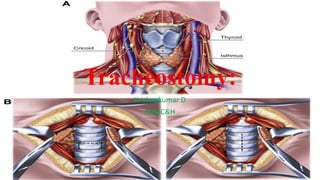
- 6. Anatomic landmarks for tracheostomy: • Thyroid notch - a palpable landmark to identify the superior aspect of the larynx in the midline. • Cricothyroid membrane - a palpable depression between cricoid and thyroid cartilages. This is the location for an emergent cricothyrotomy. • Cricoid cartilage - a palpable landmark to identify the junction of the larynx and trachea. The skin incision is typically placed 1-2cm inferior to the cricoid. • Sternal notch - a palpable landmark to identify the thoracic inlet. It is important to palpate here to the possibility of a high-riding innominate artery that may be encountered during tracheostomy.
- 8. Indications: • Acute upper airway obstruction with failed endotracheal intubation (foreign body, angioedema, infection, anaphylaxis, etc.) • Post-cricothyrotomy (if a cricothyrotomy has been placed it should be immediately formalized into a tracheostomy once an airway has been secured) • Penetrating laryngeal trauma • LeFort III fracture • Obstruction of the mouth or throat • Breathing difficulty caused by edema (swelling), injury or pulmonary (lung) conditions • Airway reconstruction following tracheal or laryngeal surgery • Airway protection from secretions or food because of swallowing problems • Airway protection after head and neck surgery • Long-term need for ventilator (breathing machine) support
- 9. Indications for elective tracheostomy include: • Prolonged ventilator dependence • Prophylactic tracheostomy prior to head and neck cancer treatment • Obstructive sleep apnoea refractory to other treatments • Chronic aspiration • Neuromuscular disease • Subglottic stenosis
- 12. Technique: • Open Tracheostomy • Anatomic landmarks such as the thyroid notch, cricoid cartilage, and sternal notch are palpated and marked. • The surgeon should pay close attention to palpation in the sternal notch to detect a high-riding innominate artery. • A skin incision is then marked in the midline anterior neck 1 to 2 cm inferior to the carotid cartilage. A horizontal or vertical incision may be utilized. • The incision is extended through the platysma muscle to expose the strap muscles (sternohyoid and sternothyroid), identifying the median raphe. • The strap muscles are then retracted laterally, exposing the cricoid cartilage and thyroid gland. • The thyroid isthmus is identified and ligated, if necessary, depending on its location along the trachea.
- 14. • Hemostats can be utilized to cross-clamp the isthmus, subsequently oversewing each stump with a silk suture to ensure hemostasis of thyroid tissue. • A cricoid hook is then placed under the cricoid cartilage to elevate the larynx and trachea into the operative field. • The second and third tracheal rings are identified. • Stay ligatures may be placed laterally to facilitate traction on the trachea for tube placement, as well as tube security in the postoperative period. • An incision is made between the second and third rings, and tracheostomy tube placed. • Various modifications have been proposed including removal of an anterior window of cartilage (often removing a segment of 1 to 2 rings), the use of a vertical anterior incision across 1-2 rings (used in pediatric tracheostomy), or the creation of a Bjork flap,
- 15. Technique: • in which an inferiorly-based cartilage flap is created and secured to the subcutaneous tissues. • The first ring is avoided to decrease the risk of subsequent stenosis. • With the tube in place, it is connected to the anesthesia circuit, and end-tidal CO2 confirmed. • Only then is the cricoid hook released. • The tracheostomy tube is secured with a soft trans-cervical tie as well as sutured to the anterior neck skin until the first tracheostomy tube change on postoperative day five.
- 17. Percutaneous Tracheostomy: • This method uses a dilatational process via a modified Seldinger technique under bronchoscopy guidance. • There have been numerous studies comparing the outcomes of the two techniques, suggesting several potential advantages of each technique over the other. • The percutaneous technique is more amenable to bedside performance, avoiding the transport of potentially critically ill patients to the operating room. • The percutaneous technique has also been associated with less blood loss and lower infection rates than the open technique. • The percutaneous technique has been associated with several significant devastating complications such as tracheal laceration, aortic injury, and esophageal perforation, which are extremely unusual after the open procedure.
- 19. Thank you