1. We are two students from Taft, and both of us personally know of
people that have fallen victim to HIV and AIDS. Pushed by a
initially broad desire to contribute to a better world, we decided to
conduct research on how we could help.
In 2004, Merck, one of the largest pharmaceutical companies in
the world, began testing a newly developed HIV vaccine on
humans after the vaccine had past preliminary FDA approval.
Confident that the vaccine would lead to a breakthrough in the
fight against HIV, the lab was shocked by the results that they had
in fact placed their 3,000 volunteers at a much higher risk for
contracting HIV. Having realized this trend, the testing was
abruptly stopped in 2008, and the 1,836 men from the Merck trial
were followed until the end of 2009 . The results were
disheartening as 172 men, nearly 9.4% percent, from this group
contracted HIV by the end of 2009. This numbers are colossal
compared to the .13% of American males who currently live with
HIV . So the big question that is on everyone’s mind is, why was
this vaccine permitted to be tested on human subjects? And the
answer to this lies in the current and conventional methods of
analyzing test cells.
Table 1. Merck Vaccine Trial Results (data table from thebodypro.com). The results of
this experimental trial was criticized to be a huge setback and failure in the history of
human experimentation. Results showed a huge yield of patients acquiring HIV/AIDS
after vaccination.
Merck used the ELISPOT method for vaccine efficacy testing,
which analyzes bulk secretions of a single cytokine. CD4+ T-
helper cells relay messages to killer T-cells and other important
supporters of the active immune response through their multi-
functional cytokine secretion. These effector molecules are
paramount to a high quality immune response. In Merck’s vaccine
testing, the ELISpot test was positive, in bulk, for a single
cytokine parameter IFN-ϒ. However, when moved into STEP
trials the vaccine did not elicit a protective immune response and,
in fact, put subjects at risk. The efficacy testing should have
instead looked into multiple cytokine secretions per each single
cell to reveal whether or not multifunctional CD4+ T cell clusters
were active with multiple protective cytokine secretions instead of
averaging responses for a single cytokine (which is also terminally
secreted). A better efficacy test is necessary to prevent trial
failures and give more accurate data on predicted immune
response.
Our project presents a new method of single-cell multiplexing
developed by IsoPlexis. The work of the lab presents a method
that can analyze up to 45 different proteins per tested cell, a
number unbeatable by any other system, even flow cytometry,
ELISA and other methods. The IsoPlexis model shows promise
for revolutionizing the analysis of single immune cells and the
proteins they secrete for furthered understanding of protective
immunity and immunogenicity.
The Single-Cell
Immunoplex Device
Development and Application
of the Single-Cell Immunoplex Device
James Lee and Leon Vortmeyer
Rong Fan Lab of the Yale Department of Biomedical Engineering &
Isoplexis Inc.
Device Assembly
Bibliography
The Immuno Single Cell Analysis Device is a secretomic analysis
platform that overcomes the aforementioned disadvantages of
ELISA and ELISPOT assays (clustering data loss due to
averaging), and even those of other single-cell devices such as
CYTOF and ICS flow cytometry (i.e. cell fixing, expensive).
This platform incorporates a microchamber array along with a
dense antibody barcode that can simultaneously detect up to 45
different cytokines in each trial. The device is a handheld
immunological laboratory assay capable of analyzing single
immune cells in parallel for up to 10,000 cellular measurements
for 45 protein parameters which include the aforementioned
cytokines, chemokines, growth factors, and other secreted
proteins.
The device proved effective in areas such as cell capture, rapid
quantification and computer-automated analysis in real time,
proving itself to be a utility that can help immensely in areas
such as cellular diagnosis.
Faced with a need to create a distinct clamp that will epitomize
many different parameters at once, 3D design and rapid prototyping
was necessary.
Introduction Research Results
Figure 1. The conventional ELISA
(Enzyme-Linked Immunosorbent Assay).
Utilizing rows and columns to analyze
secretions, it is commonly used in labs
today.
Original drawing from ebioscience.com
As mentioned above, the device is composed of two distinct parts:
a subnanoliter microchamber array and an antibody barcode
for secretion analysis.
The microchamber is a chip fabricated from PDMS
(polydimethylsiloxane), which is a transparent silicone-based
fabrication that serves in many microfluidics laboratories. It
contains 10,000 microchambers for single cell analysis. The
second component, the barcode slide, consists of 30 repeats of
each detection barcode, which can each contain 20 or more stripes
to detect proteins in each barcode (spatially and specretally
encoded). The stripes are 20um in width and is coated on a poly-
L-lysine surface. Manufactured separately, the two components
are ultimately assembled at the time of the array.
After the process of cell suspension, the cells are loaded directly
on to the surface of the chip, where gravity takes over and pulls
the cells into the microchambers. The barcode slide is then placed
face-down on the microchambers.
Figure 2. Immunoplex device used for protein secretion
detection with the use of a microchamber and antibody barcodes.
Original illustration from the Yale Department of Biomedical
Engineering
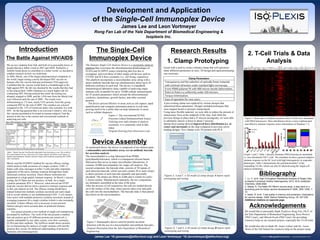
1. Clamp Prototyping
2. T-Cell Trials & Data
Analysis
Acknowledgements
We would like to personally thank Professor Rong Fan, Ph.D. of
the Yale Department of Biomedical Engineering, Kara Brower
(PhD Cand.), and Minsuk Kwak (PhD Cand.) for providing
extensive help on our research project--especially the T-Cell trials.
We would also like to thank Mr. James Lehner and Ms. Laura
Monti of the Taft School for extensive help on the project setup.
The Battle Against HIV/AIDS
A pre-existing clamp was replaced by various designs that
epitomized these parameters. Designs included prototypes that
were magnet-based or pressure clamp-based.
Using more flexible material, we were able to reduce the amount of
unnecessary force at the midpoint of the chip. And while the
previous design in place had a 35 micron incongruity, we were able
to ultimately narrow it down to about 7 microns.
Spring force testing showed improved force distribution (i.e. at
center and surround) on the chip compared to previous screw-
loading designs. New clamps were 3D printed with PLA.
Figure 5. Heat map of cytokine secretion when T-Cells were stimulated
with PMA/lomomycin. More distribution shows a more sophisticated
and effective immune response (more information on Handout 1.1)
Graph 1 and 2. Table 1 directly illustrates PMA/lomomycin stimulated
vs. non stimulated CD4 T cells. The stimulant invokes a general adaptive
immune response on the SC level with high heterogeneity in responder
phenotypes. Table 2 demonstrates the polyfunctionality* of the
responding T-Cells, which can only be detected using the single-cell
analysis technology.
Figures 3, 4 and 5. A 3D model of clamp design A before rapid
prototyping and testing.
Figures 6, 7 and 8. A 3D model of clamp design B before rapid
prototyping and testing.
1. Lu, Y., et Al. High-Throughput Secretomic Analysis of Single Cells
to Assess Functional Cellular Heterogeneity, 2013. ACS, Analytical
Chemistry. 2548-2556.
2. Sekaly, R. The failed HIV Merck vaccine study: a step back or a
launching point for future vaccine development? 2008. JEM, V205, 7-
12.
3. Seder, R. et Al. T-cell quality in memory and protection: implications
for vaccine design, 2008. Nature Publishing Group, V8, 247-259.
Additional citations on separate page.
James Lee ‘16 (jameslee@taftschool.org) and Leon Vortmeyer ‘16 (lavortmeyer@taftschool.org)