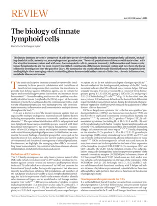
The biology of innate lymphoid cells

El sistema inmune innato se compone de una serie diversa de tipos de células hematopoyéticas evolutivamente antiguas, incluyendo células dendríticas, monocitos, macrófagos y granulocitos. Estas poblaciones de células colaboran entre sí, con el sistema inmune adaptativo y con las células no hematopoyéticas para promover la inmunidad, inflamación y reparación de tejidos. células linfoides innatas son los constituyentes más recientemente identificados del sistema inmune innato y han sido objeto de intensa investigación en los últimos cinco años. Resumimos los estudios que identifican formalmente células linfoides innatas y ponemos de relieve sus roles emergentes para el control de la homeostasis del tejido en el contexto de la infección, la inflamación crónica, enfermedades metabólicas y cáncer.

Recomendados
Más contenido relacionado
La actualidad más candente
La actualidad más candente (20)
Similar a The biology of innate lymphoid cells
Similar a The biology of innate lymphoid cells (20)
Último
Último (20)
The biology of innate lymphoid cells
- 1. 1 Weill Cornell Medical College, Cornell University, New York, New York 10021, USA.2 Academic Medical Center at the University of Amsterdam, 1105 AZ Amsterdam, the Netherlands. T he innate and adaptive immune systems have evolved to simul- taneously facilitate peaceful cohabitation with the trillions of beneficial microorganisms that constitute the microbiota, to provide host defence against infectious agents, and to initiate the repair and remodelling processes that restore and maintain tissue homeostasis1,2 . Groundbreaking studies over the past five years for- mally identified innate lymphoid cells (ILCs) as part of the innate immune system; these cells can directly communicate with a wide variety of haematopoietic and non-haematopoietic cells to orches- trate immunity, inflammation and homeostasis in multiple tissues throughout the body3–5 . ILCs are a distinct arm of the innate immune system that are regulated by multiple endogenous mammalian cell-derived factors including neuropeptides, hormones, eicosanoids, cytokines and other alarmins3–5 . The specialized distribution of ILCs in lymphoid and non-lymphoid tissues across multiple species, coupled with their functional heterogeneity3 , has provoked a fundamental reassess- ment of how ILCs integrate innate and adaptive immune responses and control diverse physiological processes. In this Review, we sum- marize the recent findings of multiple groups that converged on the identification of ILCs and discuss our current understanding of the developmental and functional heterogeneity of this cell population. Furthermore, we highlight the emerging roles of ILCs in control- ling tissue homeostasis in the context of infectious diseases, chronic inflammation, metabolic homeostasis and cancer. Definition of ILC subsets The ILC family encompasses not only classic cytotoxic natural killer (NK) cells (which were discovered6 in 1975 and are involved in pro- tection against certain viruses and tumours) and lymphoid tissue inducer (LTi) cells (which were discovered7 in 1997 and promote the formation of lymph nodes during embryogenesis), but also more recently described non-cytotoxic ILC populations. All members of the ILC family are characterized by a classic lymphoid cell morphol- ogy, but lack the expression of cell-surface molecules that identify other immune cell types, and so are defined as cell lineage marker- negative (Lin− ) cells3,4,7–11 . ILCs express subunits of cytokine receptors including interleukin (IL)-2 receptor-α (also called CD25) and IL-7 receptor-α (also known as CD127), but unlike adaptive T and B lym- phocytes, ILCs lack expression of somatically rearranged antigen receptors and so do not exhibit any degree of antigen specificity3–5 . Recent analysis of the developmental pathways of the ILC family members indicates that NK cells and non-cytotoxic helper ILCs are separate lineages. The non-cytotoxic ILCs consist of three distinct groups: group 1 ILCs (ILC1s), group 2 ILCs (ILC2s) and group 3 ILCs (ILC3s) including LTi cells3,12–14 (Fig. 1). As described later, the non-cytotoxic ILC subsets are defined on the basis of their differential requirements for transcription factors during development, their pat- terns of expression of effector cytokines and the acquisition of other distinct effector functions3,4 . ILC1s are largely non-cytotoxic Lin− cells that are capable of pro- ducing interferon-γ (IFNγ) and tumour necrosis factor (TNF) and that have been implicated in immunity to intracellular bacteria and parasites13,15–17 . By contrast, ILC2s produce T helper 2 (TH2)-cell- associated cytokines (including IL-4, IL-5, IL-9 and IL-13) and/ or the epidermal growth factor receptor ligand amphiregulin and promote type 2 inflammation required for anti-helminth immunity, allergic inflammation and tissue repair8–10,18–20 . Finally, depending on the stimulus, ILC3s produce IL-17A, IL-17F, IL-22, granulocyte macrophage (GM) colony-stimulating factor (CSF) and TNF and can promote antibacterial immunity, chronic inflammation or tissue repair21–25 . ILC3s are heterogeneous in both mice and humans. In mice, two subsets can be distinguished on the basis of their expression of the chemokine receptor CCR6. CCR6+ ILC3s encompass CD4+ and CD4− LTi cells. The CCR6− ILC3 population consists of two subpopu- lations that can be distinguished based on expression patterns of the natural cytotoxicity receptor (NCR) NKp46. In humans, almost all ILC3s express CCR6 and CD117 (also known as c-kit), and at least two subsets can be distinguished on the basis of the expression of the NCR NKp44 (refs 21–23). Strikingly, the ILC1, ILC2 and ILC3 sub- sets exhibit remarkable functional similarity with T-helper-cell sub- sets in terms of cytokine expression and potential effector function, although these cells perform their diverse functions in the absence of antigen specificity. Developmental requirements for ILCs It is well established that all lymphocytes arise from a common lym- phoid progenitor (CLP) that differentiates into precursors that are committed to particular cell lineages13,26,27 . Whereas precursor cell pop- ulations committed to the T- and B-cell lineages have been extensively The innate immune system is composed of a diverse array of evolutionarily ancient haematopoietic cell types, includ- ing dendritic cells, monocytes, macrophages and granulocytes. These cell populations collaborate with each other, with the adaptive immune system and with non-haematopoietic cells to promote immunity, inflammation and tissue repair. Innate lymphoid cells are the most recently identified constituents of the innate immune system and have been the focus of intense investigation over the past five years. We summarize the studies that formally identified innate lymphoid cells and highlight their emerging roles in controlling tissue homeostasis in the context of infection, chronic inflammation, metabolic disease and cancer. The biology of innate lymphoid cells David Artis1 & Hergen Spits2 1 5 J A N U A R Y 2 0 1 5 | V O L 5 1 7 | N A T U R E | 2 9 3 REVIEW doi:10.1038/nature14189 © 2015 Macmillan Publishers Limited. All rights reserved
- 2. characterized, CLP-derived committed ILC precursors have only recently been identified. Precursors that can develop into all ILC subsets and NK cells (but not into T cells and B cells) are contained within a population that seems to be very similar to that of CLPs but that express the integrin α4β7, referred to as α-lymphoid precursor (αLP) cells. αLP cells expressing the chemokine receptor CXCR6 are heterogeneous and can develop into conventional NK (cNK) cells and ILC3s, but not T cells or B cells and may include the common ILC and cNK cell precursors13,28,29 . Downstream of these cells are at least two precursors that express the transcriptional repressor Id2, which can develop into ILCs and NK cells (see ‘Transcriptional regulation of development and function’)13,14,28 . One of these Id2+ ILC precursor populations expressed CD127 and the integrin α4β7, and could be distinguished from CLPs by the absence of FLT3 and CD93 (ref. 13). The Id2+ ILC precursors could develop into ILC1s, ILC2s and ILC3s, including LTi cells, but were unable to develop into cNK cells13 . Another ILC precursor was identified on the basis of expression of the transcription factor promyeloid leukaemia zinc finger (PLZF; encoded by Zbtb16), which is important for the maturation of NK T cells30,31 . PLZF+ CD3ε− cells were found in fetal liver and adult bone marrow, expressed CD127, α4β7, Thy1 and CD117, and could give rise to CD127+ ILC1s, ILC2s and ILC3s in vitro and in vivo but were unable to develop into LTi cells or cNK cells14 . These data suggest that Id2+ PLZF− LTi cell or ILC precursors are upstream of an Id2+ PLZF+ ILC precursor population. Thus, our present knowledge indicates that there are at least three populations of ILC precursors with pro- gressively limited precursor potential (Fig. 2). A bone-marrow- resident precursor that is committed to the ILC2 lineage has also been identified32,33 , and there is also evidence for the existence of precursors of committed ILC1 and ILC3 subsets in mice13,28,34 . Further research will be required to refine the precursors and developmental checkpoints in the formation of the three ILC subsets, and additional analyses of people with primary immunodeficiencies may provide more insight into the developmental requirements of human ILCs. Transcriptional regulation of development and function The transcription factors and related molecules that control the devel- opment of ILCs are being identified at a rapid pace, building on our knowledge of transcriptional control of T-helper-cell subsets. Two tran- scription factors that affect early differentiation of ILCs and NK cells have been identified, TOX and NFIL3 (refs 14, 16, 35–38). These tran- scription factors do not affect early development of T cells and B cells, although TOX is important at later stages of T-cell development for the differentiation of CD4+ T cells39 . Id2 is required for the develop- ment of ILCs at an early stage, as Id2-deficient mice lack ILCs and NK cells8,19,28,33,40 . One pathway by which Id2 may allow ILC development to proceed is through sequestration of the basic helix–loop–helix transcription factor E47, which was shown to block development of LTi and NK cells41 , although it has yet to be determined whether this pathway also functions in the development of ILC1s and ILC2s. It should be noted that although Id2 is required for optimal develop- ment of NK cells, this factor seems to act at a later developmental stage in the transition of pre-NK to immature NK cells13,40,41 . TCF-1 and GATA3 also seem to be key transcription factors that drive the development of all CD127+ ILCs12,42–44 . Genetic ablation of GATA3 in haematopoietic stem cells (HSCs) resulted in the inhibi- tion of all ILC subsets and T cells but not of NK cells or B cells12 . Deletion of GATA3 in mature ILCs affected ILC2, but not ILC3 populations12,33,45,46 , suggesting that GATA3 is crucial for the post- development maintenance and survival of ILC2s. Other recent data indicate that multiple transcription factors regulate the development and function of ILC subsets. For instance, the transcription factor GFI1 might contribute to the function of GATA3 in ILC2s; this factor targets the Gata3 gene, and levels of GATA3 expression were reduced in ILC2s deficient in GFI1 (ref. 47), providing a potential mechanism through which GATA3 expression is maintained in mature ILC2s. These findings indicate that the influence of GATA3 and associated transcriptional regulatory networks are dependent on the develop- mental stage of the ILC precursors, similar to their role in the devel- opment of T cells48 . The dependence of NK cells, ILC1s and ILC3s on shared transcrip- tion factors for their development and function further emphasizes the remarkable functional similarity of ILCs with corresponding T-helper-cell subsets. For example, eomesodermin and T-bet have both redundant and non-redundant roles in the development, func- tion and migration of NK and CD8+ T cells49 , whereas the master transcription factor of TH1 cells, T-bet50 , is important for the devel- opment and IFNγ-producing function of non-cytotoxic ILC1s. It should also be noted that T-bet is required for upregulation of NKp46 expression and IFNγ production by CCR6− ILC3s13,51,52 . RORγt is Cytotoxic ILCs Non-cytotoxic ILCs NK IFNγPerforin and granzyme IL-12 IL-15 IL-18 NK cells T-bet+ ILC1s GATA3+ ILC2s RORγt+ ILC3s ILC1 CD127– CD127+ LTi cells CCR6+ T-bet– ILC1 IL-12 IL-15 IL-18 IL-12 IL-18 ILC2 CD4+ CD4– IL-25 IL-33 TSLP TL1A IL-1β IL-23 AHR ligands CCR6– T-bet+ NCR+ NCR– IL-1β IL-23 AHR ligands Immunity to viruses and cancer Chronic inflammation Immunity to intracellular bacteria and protozoa Chronic inflammation Immunity to helminths Asthma and allergic diseases Metabolic homeostasis Lymphoid tissue development Intestinal homeostasis Immunity to extracellular bacteria Chronic inflammation IFNγIFNγTNF TNF TNF IL-22IL-17A GM-CSFIL-5IL-4 IL-9 IL-13 Areg IL-22IFNγ GM-CSF Figure 1 | The innate lymphoid cell family. Group 1, group 2 and group 3 innate lymphoid cells (ILCs) are defined by differential expression of cell- surface markers, transcription factors and patterns of expression of effector cytokines. ILCs can be activated by a diverse range of stimuli including neuropeptides, hormones, eicosanoids, cytokines and other alarmins, and can contribute to immunity, inflammation and maintenance of tissue homeostasis. Dysregulated ILC responses can also contribute to chronic inflammatory diseases, metabolic disorders and cancer. AHR, aryl hydrocarbon receptor; Areg, amphiregulin; GM-CSF, granulocyte macrophage colony-stimulating factor; IFNγ, interferon-γ; IL, interleukin; LTi, lymphoid tissue inducer; NCR, natural cytotoxicity receptor; NK, natural killer; TNF, tumour necrosis factor; TSLP, thymic stromal lymphopoietin. 2 9 4 | N A T U R E | V O L 5 1 7 | 1 5 J A N U A R Y 2 0 1 5 REVIEWINSIGHT © 2015 Macmillan Publishers Limited. All rights reserved
- 3. indispensable for the development of all ILC3 subsets11,23,53 , as it is for TH17 cells54 . In addition, the aryl hydrocarbon receptor (AHR), a ligand-activated transcription factor that acts as a sensor for multiple exogenous and endogenous compounds including toxins, tryptophan metabolites, dietary products and bacterial pigments, controls the survival and function of ILC3 subsets55,56 and TH17 cells57 . Finally, Notch signalling, which is required for T-cell development, has also been implicated in the development of ILC3s28,29,56 and ILC2s42,58 . It is important to note that there are also transcription factors that act on ILC subsets but not on the corresponding T-helper cell subset. For example, RORα is important for ILC2s but does not seem to play a T-cell intrinsic part in TH2 cell development and function32,58 . As with T-helper-cell subsets, there is some evidence that ILCs can show functional plasticity in response to environmental cues. In mouse models it was shown that the function of ILC3s can be affected by a gradient of expression of the transcription factors RORγt and T-bet51 . Following activation by cytokines such as IL-12 and IL-18, ILC3s exhibit increased expression of T-bet and decreased expres- sion of RORγt, resulting in increased IFNγ production and loss of their capacity to produce IL-17 and IL-22 (refs 17, 51). These murine cells, termed ex-RORγt+ ILC3s, demonstrate ILC1-like function. In humans, an apparently similar switch in ILC3 to ILC1 effector func- tion has been observed15,59 . Whether human or mouse ILC2s exhibit plasticity in their effector functions and what the functional signifi- cance of this might be remains unclear at present. Heterogeneous functions of ILCs Emerging studies have identified diverse functions for ILCs, including promoting host defence against infection and regulating interactions with the microbiota. In addition, ILCs can orchestrate wound healing and tissue repair, whereas in other circumstances they can promote inflammation and tumour progression. ILCs promote immunity to infection ILCs are enriched at barrier surfaces that are common sites of colo- nization or invasion by pathogens. It is now clear that all ILC subsets can have an important role in innate immune responses to viruses, bacteria, fungi and both intracellular and extracellular parasites at these barrier surfaces. ILC recruitment to barrier tissues takes place during embryonic development and further migration of ILCs is likely to occur in the context of ongoing inflammation. Constitu- tively present within adult tissues, ILCs are poised for rapid activa- tion by host-derived cytokines and growth factors3−5 . Epithelial cells and myeloid cell lineages act cooperatively to sense infection and/ or tissue damage and produce cytokines and alarmins that mobi- lize the rapid recruitment of distinct ILC populations3 . For example, IL-12, IL-15 and IL-18 activate NK cells and ILC1s13,15,16 , whereas IL-2 (refs 60, 61), IL-4 (refs 62, 63), IL-25, IL-33 (refs 8−10,19, 64−67), thymic stromal lymphopoietin (TSLP)46,68 , IL-9 (refs 20, 69) and TL1A70,71 activate ILC2s. By contrast, IL-1β and IL-23 stimulate ILC3 responses59,72 (Fig. 1). The role of NK cells in killing virus-infected cells through gran- zyme and/or perforin-mediated cytolysis is well established and has been reviewed elsewhere73 . In the context of immunity to intracellular bacteria and parasites, there are important roles for ILC1s and ILC3s in host defence. For example, IFNγ-producing ILC1s contribute to resistance to Salmonella enterica subsp. enterica serovar Typhimu- rium and Toxoplasma gondii infection in the intestine13,51 (Fig. 3). In addition, before the development of adaptive immune responses, innate immunity to the extracellular Gram-negative bacterium Cit- robacter rodentium is critically dependent on ILC3-derived IL-22 (refs 23, 74), which has an important role in promoting STAT3- dependent expression of antimicrobial peptides and maintenance of intestinal epithelial barrier function (Fig. 3)74,75 . Mice deficient in IL-22 exhibited exaggerated intestinal inflammation and ero- sion of the epithelial barrier and rapidly succumbed to infection23,74 . ILC3-derived IL-22 also works cooperatively with lymphotoxin to induce fucosylation of intestinal epithelial cells, contributing to resistance to S. Typhimurium infection76 . In addition, ILC3s play an important part in regulating host–commensal-bacteria relationships (see ‘Interactions of ILCs with the microbiota’). The host protective effects of ILC3s are not limited to bacterial infection in the intestine. ILC3s have also been implicated as an important source of IL-22 in the lungs following infection with the fungus Candida albicans77 or the bacterium Streptococcus pneumoniae78 , and an innate source of IFNγ and IL-17 that resembles ILC3s in the lungs of mice subjected to a bacillus Calmette-Guérin (BCG) vaccination protocol pro- vided enhanced protection against challenge with Mycobacterium tuberculosis79 . Whereas ILC1s and ILC3s promote innate immunity to viruses, intracellular bacteria and parasites, and fungi (Fig. 3), ILC2s are essential to the promotion of type 2 inflammation required for immunity to some extracellular helminth parasites1,3,4 . The type 2 inflammatory response is characterized by production of cytokines, including IL-4, IL-5, IL-9 and IL-13 that regulate the alternative acti- vation of macrophages, granulocyte responses, goblet cell hyperplasia and smooth muscle contractility that promote parasite expulsion and associated tissue-repair processes1 . In studies of the mouse-adapted intestinal nematode parasite Nippostrongylus brasiliensis, ILC2s were Pre NK NK cells LTi cellsEx-RORγt+ ILC3 CLP NFIL3 TOX Eomes T-bet Id2 ILC1 T-bet T-bet NKp46+ ILC3 ILC precursor Id2+ PLZF+ LTi/ILC precursor Id2+ ILC3 ILC/NK precursor NFIL3+ TOX+ α4β7+ T-bet ILC2 GATA3 TCF1 RORα GFI1 Id2, GATA3 RORγt RORγt AHR Figure2|Modelofdevelopmentalpathwaysofinnatelymphoidcellsand conventionalnaturalkillercells. Like all lymphocytes, innate lymphoid cells (ILCs) are derived from a common lymphoid progenitor (CLP). The common ILC or natural killer (NK) cell precursor is enclosed in a population of cells that have the same phenotype as the CLP but that also express the α4β7 integrin. Downstream of the common ILC/NK cell precursor (preNK) is an Id2-expressing precursor that can give rise to all ILCs, including lymphoid tissue inducer (LTi) cells and an Id2+ PLZF+ precursor that is restricted to ILCs but is unable to develop into LTi cells. In mice, RORγt+ ILC3s require T-bet to develop into interferon-γ (IFNγ)-producing cells. Cytokines that upregulate T-bet such as interleukin (IL)-12 and IL-18 lead to downregulation of RORγt, abrogation of IL-22 and increase of IFNγ-producing capacity. These IFNγ-producingILC1- likecellsarealsocalledex-RORγt+ ILC3s.AHR,arylhydrocarbonreceptor. 1 5 J A N U A R Y 2 0 1 5 | V O L 5 1 7 | N A T U R E | 2 9 5 REVIEW INSIGHT © 2015 Macmillan Publishers Limited. All rights reserved
- 4. identified as the dominant non-T-cell source of IL-13, which is cru- cial for the expulsion of N. brasiliensis8–10 . ILC2-deficient mice failed to efficiently expel their parasites, but adoptive transfer of wild-type ILC2s into ILC- or IL-13-deficient mice was sufficient to restore effi- cient worm expulsion, suggesting that these cells are crucial for the development of protective immune responses to N. brasiliensis in the intestine8−10 . During N. brasiliensis infection, IL-25 and IL-33 (refs 8−10), the signalling adaptor molecule Act1 (ref. 80) and the transcription fac- tors GATA3 (ref. 81), TCF-1 (ref. 42) and GFI1 (ref. 47) have been shown to be essential for the population expansion and IL-13 expres- sion by ILC2s that contribute to worm expulsion. These data sug- gest that a complex network of regulatory factors promote optimal ILC2 responses that directly contribute to protective immunity to helminths. In addition, recent work has shown that ILC2s interact with adaptive immune cells to indirectly promote protective type 2 immune responses. For example, ILC2s express major histocompat- ibility complex (MHC) class II and can activate T cells (albeit less efficiently than dendritic cells) to induce IL-2, which in turn elicits ILC2 proliferation and production of TH2-associated cytokines that promote worm expulsion61 . Thus, ILC2s participate in both innate and adaptive immune responses to directly and indirectly facilitate expulsion of N. brasiliensis. There is evidence that ILC2 popula- tions also expand and contribute to protective immunity following infection with other helminth species, although the role of ILC2s in expulsion of these helminths remains less well defined. ILC2s can express amphiregulin19 , which is required for immunity to the gastrointestinal nematode parasite Trichuris muris82 , and IL-33 pro- motes the expansion of ILC2-like cells in the lungs of mice infected with the helminth Strongyloides venezuelensis83 . Collectively, these studies provide the foundation for further investigation required to comprehensively define the mechanistic role of ILC2s in immune responses that coordinate protective immunity to diverse species of helminth parasites. Interactions of ILCs with the microbiota In contrast to their role in promoting antimicrobial responses to patho- gens, ILCs also regulate interactions between the host and the diverse array of commensal bacterial species that constitute the microbiota. ILCs do so by regulating non-haematopoietic and haematopoietic cell functions to limit inappropriate immune responses to commensal bacteria3–5 . Although the accumulation of most ILC populations in the murine intestinal tissues and gut-associated lymphoid tissues can occur independently of colonization by the microbiota19,56,84–86 , ILC3s seem to have a crucial role in the anatomical containment of lymphoid tissue- resident commensal bacteria86 . For example, in the intestine, loss of ILC3s was associated with reduced expression of IL-22 and lower levels of antimicrobial peptides expressed by intestinal epithelial cells. These effects were coincident with the dissemination of bacteria that belong to the genus Alcaligenes and the development of low-grade systemic inflammation86 . Intestinal ILC3s also interact with various immune cells to prevent dissemination of commensal bacteria or limit inappro- priate immune responses to them. For example, ILC3s can activate B cells through membrane lymphotoxin α1β2 and induce the production of immunoglobulin A (IgA) that subsequently regulates dendritic cell activity and promotes immunological exclusion of commensal bacte- ria87 . These data suggest that ILC3-dependent maintenance of intesti- nal epithelial and immune-cell responses limits the dissemination of select commensal bacterial species and maintains appropriate separa- tion between inflammatory microbial-derived products and the host immune system. ILC3s can also promote an immunologically tolerogenic state in the intestine that limits the magnitude of potentially damaging T-cell responses against commensal bacteria. ILC3s express MHC class II and are able to process and present antigens to T cells, although they lack expression of the co-stimulatory molecules required to activate T-cell responses88 . In this context, depletion of ILC3s or selective deletion of MHC class II on ILC3s was associated with exaggerated commensal- bacteria-specific TH17 cell responses and, in some circumstances, the developmentofintestinalinflammation,indicatingthatintestinalILC3s dampen commensal bacteria-specific T-cell responses in an MHC- class-II-dependent manner76,88,89 . The antigen-presenting capacity of ILC3s may depend on the tissue and activation state of the cell because, in contrast to intestinal ILC3s, spleen-derived ILC3s that are cultured on feeder cells ex vivo can express co-stimulatory molecules and prime CD4+ T cells in vitro90 . In addition, T cells can regulate the population size and function of ILC3s in the intestine91 , suggesting that dynamic cross-regulation exists between T cells and ILC3s in this tissue site. Pro- ductionofGM-CSFisanothermechanismbywhichILC3scanpromote a tolerogenic state. A recent study demonstrated that cues from the intestinal microbiota elicit GM-CSF from ILC3s to promote intestinal homeostasis through enhancing dendritic cell and regulatory T-cell function72 . Human and murine ILC3s can regulate the proliferation of B cells, particularly of marginal zone B cells92 , that could also contribute toimmunologicalexclusionandanatomicalcontainmentofcommensal bacteria. Collectively, the effects of ILC3s on the epithelium, dendritic cells, T cells and B cells supports the capacity of the mammalian host to tolerate colonization by the microbiota. NK ILC1 ILC2 ILC3 Macrophage Immunity to viruses, intracellular bacteria and parasites Immunity to helminths Immunity to extracellular bacteria Antimicrobial peptides Eosinophil Smooth muscle Dying cell Alternatively activated macrophage Virus Bacterium IL-4, IL-5, IL-9 and IL-13 IFNγ TNF LT, TNF, IL-17A and IL-22 Goblet cell Mucus Figure 3 | Host-protective effector functions of innate lymphoid cells at barrier surfaces. In addition to their functions in lymphoid tissue development and metabolic homeostasis, innate lymphoid cells (ILCs) can orchestrate multiple antimicrobial effector functions at barrier surfaces in the context of exposure to viruses, bacteria, protozoa and helminths. Both natural killer (NK) cells and ILC1s produce interferon-γ (IFNγ) and contribute to protective immunity against viruses, intracellular bacteria and protozoan parasites. Production of type-2 cytokines, including interleukin (IL)-4, IL-5, IL-9 and IL-13 promote alternative activation of macrophages, eosinophilia, goblet-cell hyperplasia and smooth-muscle contractility that contribute to expulsion of helminth parasites. ILC3s produce IL-17A, IL-22, lymphotoxin (LT) and tumour necrosis factor (TNF) and contribute to control of extracellular bacterial infection. 2 9 6 | N A T U R E | V O L 5 1 7 | 1 5 J A N U A R Y 2 0 1 5 REVIEWINSIGHT © 2015 Macmillan Publishers Limited. All rights reserved
- 5. ILC-mediated tissue remodelling, healing and repair ILCs also contribute to the maintenance of tissue homeostasis by con- tributing to tissue remodelling, wound healing and repair processes at multiple tissues sites3,4 . LTi cells, a subset of ILC3s, have long been recognized for their ability to promote tissue modelling in the fetus and after birth5,7,11 . During embryonic development, LTi cells promote the formation of secondary lymphoid organs such as Peyer’s patches in the gut11 . LTi cells produce LTα1β2 that binds to LTβR on stromal cells, resulting in stromal cell secretion of chemokines (CXCL13, CCL21 and CCL19) and upregulation of adhesion molecules (VCAM1, MadCam1 and ICAM1) that attract and bind leukocytes to ultimately form lym- phoid structures93 . After birth, LTi cells use the LTα1β2–LTβR pathway to aid in the formation of isolated lymphoid follicles, structures that are important for immune reactions in the gut87 . Other tissue remodelling functions of ILCs include wound healing and repair of damaged tissues. ILC3s have been implicated in repair of lymphoid tissues damaged following acute viral infection and an ensuing cytotoxic T-cell response against the virus-infected lymph node stromal cells94 . IL-22-producing ILC3s also promote tissue repair and regeneration in the inflamed intestine95 and in radiation- damaged thymic tissue96 . Furthermore, they can limit allergic airway hyper-responsiveness in the lung97 , indicating that ILC3s participate in the maintenance of homeostasis in multiple tissues following inflammation or damage. In addition, ILC2s express amphiregulin, a member of the epidermal growth factor family, and can restore bronchial epithelium that has been damaged by infection with influ- enza virus19 (Fig. 4). Together, these studies suggest that ILC2s and ILC3s contribute to the diverse processes of tissue remodelling that promote tissue repair and homeostasis. ILCs and chronic inflammatory diseases In addition to their ability to promote tissue homeostasis, ILCs can also promote inflammation at mucosal and barrier surfaces. In this context, chronic ILC activation can contribute to pathology in a wide range of inflammatory disorders3,4 . In mice, intra-epithelial ILC1s and IFNγ-producing ILC3s can induce inflammation, and block- ing IFNγ could ameliorate this disease in some models of colitis16,25 . IFNγ-producing ILCs may also be involved in human inflamma- tory bowel disease because the population of ILC1s expands and IL- 22-producing ILC3s decrease in inflamed intestinal tissues of patients with Crohn’s disease15,16 . IL-17-producing ILC3s have been shown to play a part in inflammatory bowel disease in T-cell-independent mouse models25,98 . In one of these models, loss of T-bet resulted in a substantial population expansion of IL-17-producing ILCs, driven by TNF and IL-23 produced by dendritic cells98 . In this context, CD127 blockade reduced intestinal ILC3 numbers and attenuated disease98 . Together, these findings indicate that ILC1s and ILC3s can contribute to the development of inflammation in the intestine (Fig. 4). ILC2s have been shown to be detrimental in a large variety of inflammatory disorders in experimental animals3,4 . In mice with allergic lung inflammation, ILC2 populations expand in response to cytokines and alarmins, including IL-25, TSLP, IL-33, eicosanoids and TL1A produced by type 2 pneumocytes, alveolar macrophages and granulocytes; ILC2s also contribute to type 2 inflammation in the lung through production of IL-4, IL-5, IL-9 and IL-13 (refs 47, 70, 71, 99–101). Recent work has also suggested that ILC2s coordinate with dendritic cells and CD4+ T cells at mucosal sites to shape T-cell responses that contribute to allergic airway inflammation102,103 . It is unclear to what extent ILC2s are involved in human lung inflam- mation, but it is striking that ILC2s are increased in the peripheral blood of people with asthma relative to those without the condi- tion104 , and several genes associated with ILC2s are enriched in people with asthma, including the gene that encodes IL1RL1 (a component of the IL-33 receptor), RORα and IL-13, suggesting a role for ILC2s in disease pathogenesis105 . Human ILC2s are robustly expanded in another type 2 inflammatory disease, chronic rhinosinusitis, which is characterized by pronounced eosinophilia and high IgE levels in serum18,46 . Epithelial cells of resected nasal polyps are capable of ILC1 Macrophage Alternatively- activated macrophage Gut: Crohn’s disease Eosinophil Mast cell Virus IFNγ ILC3 IL-17A Intestinal epithelial cell Gut: Colorectal cancer Lung: Tissue repair after virus infection ILC3 ILC2 IL-22 IL-22BP Dendritic cell Areg IL-33 TSLP IL-33 IL-25 IL-5, IL-13 IL-4, IL-9 Allergen Lung: Airway hypersensitivity ILC2 TSLP IL-33 IL-25 Eosinophil Basophil Mast cell IL-5, IL-13 ILC2 Areg IL-9 Mucus Skin: Psoriasis and skin thickening NCR+ ILC3 IL-22 IL-4 ILC3 IL-17A Skin: Atopic dermatitis Inflamed cell Tumour cell ILC3 Stratum corneum Figure 4 | Pro-inflammatory and tissue reparative functions of innate lymphoid cells. Innate lymphoid cells (ILCs) are amplified in a variety of inflammatory diseases that affect barrier functions, suggesting that they contribute to pathology. In the skin disorder atopic dermatitis, ILC2s increase in numbers. NKp44+ ILC3s (NCR+ ) are amplified in skin lesions of people with psoriasis and produce interleukin (IL)-22, possibly contributing to acanthosis (skin thickening), which is characteristic for this disease. IL-17-producing ILCs may increase skin inflammation. Interferon-γ (IFNγ)- and IL-17- producing ILCs may contribute to inflammatory bowel disease in mice, and IFNγ-producing ILC1 are strongly amplified in inflamed intestinal tissues of patients with Crohn’s disease. ILC3-derived IL-22 promotes proliferation of tumour cells in a mouse model of infection-induced colorectal cancer. IL-22 binding protein (BP) secreted by dendritic cells can counteract the effect of IL-22. ILC2s cause airway hypersensitivity in a variety of mouse models of allergic asthma. Airway epithelial cells triggered by allergens produce thymic stromal lymphopoietin (TSLP), IL-25 and IL-33, which activate ILC2s to produce IL-5, IL-4, IL-9 and IL-13 that lead to airway hyper-reactivity. In one mouse model, ILC3s have been shown to dampen ILC2 hyper-reactivity. Lung ILC2s can mediate repair of tissue damaged by a virus through production of amphiregulin (Areg). 1 5 J A N U A R Y 2 0 1 5 | V O L 5 1 7 | N A T U R E | 2 9 7 REVIEW INSIGHT © 2015 Macmillan Publishers Limited. All rights reserved
- 6. producing large amounts of TSLP and IL-33 that may lead to the enhanced IL-5 and IL-13 production by ILCs observed in patients with this disease18,46 . Another prominent type 2 disease is atopic dermatitis, an inflam- matory skin condition characterized by the presence of TH2 cells and high levels of IL-5 and IL-13 in the skin. Recent work has sug- gested that ILC2s might be involved in the pathogenesis of atopic dermatitis, because the ILC2 population is expanded in the lesions of patients with atopic dermatitis67,68 . These cells are stimulated by TSLP68 and are also likely to be stimulated by IL-25 and IL-33, because increased transcripts of these cytokines have been detected in atopic dermatitis skin lesions65,67,68 . Notably, the lectin inhibitory receptor KLRG1 may have a regulatory role in the control of ILC2 activation during atopic dermatitis, because the KLRG1 ligand epi- thelial cadherin (E-cadherin) diminishes ILC2 cytokine production, and E-cadherin is downregulated in atopic dermatitis lesions67 . Stud- ies in mouse models of atopic dermatitis support the idea that ILC2s can play a T-cell-independent part in establishing skin lesions68 . In addition, ILC2s can interact with other innate immune cell types such as mast cells and basophils to promote type 2 inflammation in the skin62,63,106 (Fig. 4). Similar to the role for ILC2s in regulating type 2 inflammation in the skin, IL-17- and IL-22-producing ILC3s have been associated with the inflammatory skin disease psoriasis vulgaris. ILC2s and NKp44− ILC3s but not NKp44+ ILC3s are present in the peripheral blood and skin of healthy people, whereas the blood and inflamed skin of those with psoriasis contained NKp44+ ILC3s, indicating that dynamic changes in ILC population structure are associated with psoriatic inflammation107 . In support of this idea, one patient who showed a reduction of psoriatic plaques in response to treatment with anti-TNF monoclonal antibodies also showed a decrease in cir- culating NKp44+ ILC3s107 . Studies in mouse models of psoriasis also suggest that ILC3s contribute to this disease, because mice treated with cream containing imiquimod (a Toll-like receptor 7 agonist that elicits a psoriasis-like skin inflammation) had IL-17A-, IL-17F- and IL-22-producing ILC3s and γδ T cells in the skin108 . The involvement of ILCs in various inflammatory and autoim- mune disorders, as already discussed, has raised considerable inter- est in developing strategies to modify ILC functions to treat these diseases. Antibodies against ILC2-activating cytokines or monoclo- nal antibodies targeting the cytokines produced by ILC2s, including IL-5 and IL-13, are in clinical trials for the treatment of allergic dis- eases such as asthma and chronic rhinosinusitus109 . ILC3 functions have also recently been targeted in the context of multiple sclerosis. Patients with multiple sclerosis had increased frequencies of ILC3s, and treatment with an anti-IL-2R antibody resulted in alleviation of inflammation and was associated with a reduction of ILC3s in peripheral blood110 . Although this study suggests that ILC3s have a role in promoting multiple sclerosis, ILC3s seem to be dispensable in a murine model of experimental autoimmune encephalomyeli- tis111 . In addition, small molecular compounds (SMCs) that target ILCs could also potentially be used to modify ILC functions to treat disease. In particular, arachidonic acid metabolites that regulate ILC2 function have attracted interest. Signalling by leukotriene D4 to ILC2s that express the cysteinyl leukotriene receptor 1 (CysLT1R) results in secretion of type 2 cytokines, which can be inhibited by montelukast, a leukotriene receptor antagonist101 . Prostaglandin D2 (PGD2; which is produced by mast cells) binds to one of its receptors, CRTH2, on human ILC2s18 to promote migration and cytokine pro- duction, which can be blocked by CRTH2 antagonists100,112 . Notably, mast cells, which are major producers of PGD2, stably interact with ILC2s, and the resulting dialogue might induce IL-13 production106 . Finally, lipoxin A4, an anti-inflammatory metabolite of arachidonic acid, binds to ILC2s and inhibits IL-13 production100 . SMCs that may target ILC3 activities include antagonists of RORγt, which were shown to affect biological activities of RORγt-dependent TH17 cells113 . Although the action of these various monoclonal antibodies or SMCs might go beyond ILCs, these data suggest that targeting ILC functions might be clinically efficacious in the context of multiple disease states. ILC2s and metabolic homeostasis Emerging data highlighting an association between altered ILC responses and obesity, malnutrition and metabolic homeostasis sug- gest that ILCs may be crucial responders to nutrient and metabolic stress. For example, a population of IL-5- and IL-13-producing ILC2s in murine white adipose tissue114,115 maintains eosinophil and alterna- tively activated macrophage responses that limit high-fat-diet-induced obesity and insulin resistance114 . Consistent with this, IL-25-elicited ILC2 responses were associated with lower weight gain following exposure to a high fat diet115 , suggesting that ILC2 responses may play an important part in regulating adipocyte development and/or function in the context of obesity. Before the discovery of ILCs another report demonstrated that delivery of IL-33 to genetically obese mice could reduce adiposity and improve insulin tolerance, whereas dele- tion of the IL-33 receptor was associated with exaggerated high-fat- diet-induced obesity116 , supporting a potential role for IL-33 and ILC2s in metabolic homeostasis. In addition to regulating metabolic homeostasis in white adipose tis- sue, ILC2s seem to be poised to rapidly respond to changes in nutrient status. Effector cytokine production by ILC2s in the small intestine was suppressed following fasting, and is regulated by the circadian clock through vasoactive intestinal peptide117 . Furthermore, in the setting of malnutrition elicited by vitamin A deficiency in mice, an imbalance in ILC2 and ILC3 responses was evident, resulting in defective ILC3- dependent antibacterial immunity but enhanced ILC2-dependent anti- helminth immunity118 . Together, these data provoke a model whereby alterations in nutritional status directly influence ILC-dependent maintenance of tissue homeostasis and host defence against infection. However, the mechanisms through which ILCs recognize metabolic or dietary stress and how they control metabolic homeostasis remain poorly defined. ILCs can promote tumour development There is ample evidence that NK cells are involved in protection against cancer in humans and experimental animals. NK cells are particularly efficacious in killing tumour cells that have lost class I MHC antigens, a process that could be exploited for cancer therapy119 . However, studies that investigate the involvement of CD127+ ILCs in tumour immunity are limited. One study has shown that CCR7+ CD4+ ILC3s promote tumour outgrowth of melanoma cells that were engi- neered to express the chemokine CCL21, which was associated with the development of a suppressive tumour microenvironment120 . In this context, whether ILC3s can directly tolerize T cells, as has been observed in the gut88 , remains to be established. Other evidence of a role for ILCs in tumorigenesis comes from stud- ies investigating the pro-carcinogenic roles of cytokines involved in the activation and effector functions of ILCs, including IL-23 and IL-22. IL-23 receptor expression is increased in human colorectal cancer (CRC), and transgenic expression of IL-23 in mice induced adenomatous tumours that originated in the duodenum121 . In this model, a crucial role for ILC3s in tumorigenesis in lymphocyte- deficient mice was identified, although the contribution of adaptive cells remains unclear122 . In other models, a possible role for IL-22 in tumorigenesis in the gut has been described. IL-22 is important for colonic epithelial cell repair, but this activity must be tightly controlled by the soluble IL-22 receptor (IL-22 binding protein, IL-22BP) to pro- tect against tumorigenesis, because genetic ablation of IL-22BP led to uncontrolled IL-22 production that facilitated tumour development123 . In addition, in a mouse model for inflammation induced by CRC, IL-22- and IL-17-producing RORγt-dependent ILC3s in the colon caused inflammation and in the presence of a carcinogen promoted 2 9 8 | N A T U R E | V O L 5 1 7 | 1 5 J A N U A R Y 2 0 1 5 REVIEWINSIGHT © 2015 Macmillan Publishers Limited. All rights reserved
- 7. development and growth of CRC124 . IL-22 might also be involved in human CRC because IL-22-producing tumour-infiltrating lympho- cytes, including both T cells and non-T cells, were frequently observed in CRC, and IL-22 production in the tumour was significantly higher than in non-tumour tissue sections from the same patients124 . How- ever, the exact role of IL-22 in tumour growth and the relative impor- tance of IL-22-producing ILC3s and TH22 cells in CRC has yet to be determined. Together, these studies suggest that ILCs can promote tumour growth through production of tumour-promoting cytokines and by creating a suppressive tumour microenvironment. However, one study has reported an ILC3-mediated regression of a skin-residing B16 melanoma cell line that was engineered to express IL-12 (ref. 125). These ILC3s produced IFNγ and IL-17, but the mechanism of tumour rejection induced by ILC3s in this model remains unclear125 . Although it is possible that tumour-derived IL-12 induced differentiation of IL-22-producing ILC3s into protective IFNγ-producing ex-RORγt+ ILC3s, as has been observed in the gut15,17 , further studies are required to understand the plasticity and function of ILCs within the tumour microenvironment. Outlook and future directions The identification of ILCs and the subsequent recognition of their diverse functions in integrating non-haematopoietic and haemat- opoietic cell responses have provided new insights into how innate immune responses and tissue homeostasis are regulated in health and disease and how these innate responses affect the adaptive immune system. Although present in tissues in relatively low numbers, the selective distribution of ILCs within lymphoid and non-lymphoid tissues seems to confer a remarkable ability to regulate multiple physiological processes throughout the body. A limitation of many ILC-related studies has been the inability to genetically target select ILC populations in the presence of the adaptive immune system. The development of new genetic tools will address this challenge and could also lead to the identification of previously unrecognized innate immune cell populations that will allow greater understanding of the complexity of the immune cell network. Finally, understand- ing how ILC responses are dysregulated in the context of infectious, metabolic and chronic autoimmune and inflammatory conditions in humans might offer therapeutic potential in the treatment of a wide range of debilitating diseases. Note added in proof: Two papers recently appeared online while the current Review was in press reporting a crucial role for ILC2s in promoting the beiging of white fat and the regulation of meta- bolic homeostasis (J. R. Brestoff et al. Group 2 innate lymphoid cells promote beiging of white adipose tissue and limit obesity. Nature http://dx.doi.org/10.1038/nature14115 (2014); M. -W. Lee et al. Acti- vated type 2 innate lymphoid cells regulate beige fat biogenesis. Cell http://dx.doi.org/10.1016/j.cell.2014.12.011 (2014)). ■ Received 26 September; accepted 4 November 2014. 1. Pulendran, B. & Artis, D. New paradigms in type 2 immunity. Science 337, 431–435 (2012). 2. Iwasaki, A. & Medzhitov, R. Regulation of adaptive immunity by the innate immune system. Science 327, 291–295 (2010). 3. Spits, H. et al. Innate lymphoid cells – a proposal for uniform nomenclature. Nature Rev. Immunol. 13, 145–149 (2013). This is a key review describing the consensus nomenclature for ILC subsets developed by experts in the field. 4. Spits, H. & Di Santo, J. P. The expanding family of innate lymphoid cells: regulators and effectors of immunity and tissue remodeling. Nature Immunol. 12, 21–27 (2011). 5. Eberl, G. Development and evolution of RORγt+ cells in a microbe’s world. Immunol. Rev. 245, 177–188 (2012). 6. Kiessling, R., Klein, E. & Wigzell, H. “Natural” killer cells in the mouse. I. Cytotoxic cells with specificity for mouse Moloney leukemia cells. Specificity and distribution according to genotype. Eur. J. Immunol. 5, 112–117 (1975). 7. Mebius, R. E., Rennert, P. & Weissman, I. L. Developing lymph nodes collect CD4+ CD3− LTβ+ cells that can differentiate to APC, NK cells, and follicular cells but not T or B cells. Immunity 7, 493–504 (1997). This is the first description of a role for ILCs that express lymphotoxin in the establishment of lymphoid structures. 8. Moro, K. et al. Innate production of TH2 cytokines by adipose tissue-associated c-Kit+ Sca-1+ lymphoid cells. Nature 463, 540–544 (2010). 9. Neill, D. R. et al. Nuocytes represent a new innate effector leukocyte that mediates type-2 immunity. Nature 464, 1367–1370 (2010). 10. Price, A. E. et al. Systemically dispersed innate IL-13-expressing cells in type 2 immunity. Proc. Natl Acad. Sci. USA 107, 11489–11494 (2010). This article and refs 8 and 9 provide three seminal reports identifying murine ILC2s that produce type 2 cytokines and contribute to anti-helminth immunity and type 2 inflammation. 11. Eberl, G. et al. An essential function for the nuclear receptor RORγt in the gen- eration of fetal lymphoid tissue inducer cells. Nature Immunol. 5, 64–73 (2004). This report describes for the first time the critical dependence of LTi cells, which help to direct the formation of secondary lymphoid structures, on the transcription factor RORγt. 12. Yagi, R. et al. The transcription factor GATA3 is critical for the development of all IL-7Rα-expressing innate lymphoid cells. Immunity 40, 378–388 (2014). This cutting edge report describes all ILCs, not just ILC2s, as being dependent on the transcription factor GATA3 for development. 13. Klose, C. S. et al. Differentiation of type 1 ILCs from a common progenitor to all helper-like innate lymphoid cell lineages. Cell 157, 340–356 (2014). 14. Constantinides, M. G., McDonald, B. D., Verhoef, P. A. & Bendelac, A. A committed precursor to innate lymphoid cells. Nature 508, 397–401 (2014). Both this article and ref. 13 are ground-breaking studies describing committed progenitor cells of ILCs. 15. Bernink, J. H. et al. Human type 1 innate lymphoid cells accumulate in inflamed mucosal tissues. Nature Immunol. 14, 221–229 (2013). 16. Fuchs, A. et al. Intraepithelial type 1 innate lymphoid cells are a unique subset of IL-12- and IL-15-responsive IFN-gamma-producing cells. Immunity 38, 769–781 (2013). This article, along with ref. 15, provides evidence of non-NK cell ILC1s in humans and mice. 17. Vonarbourg, C. et al. Regulated expression of nuclear receptor RORγt confers distinct functional fates to NK cell receptor-expressing RORγt+ innate lymphocytes. Immunity 33, 736–751 (2010). 18. Mjösberg, J. M. et al. Human IL-25- and IL-33-responsive type 2 innate lymphoid cells are defined by expression of CRTH2 and CD161. Nature Immunol. 12, 1055–1062 (2011). This is one of the first reports of ILC2s in humans, implicating their pathological role in allergic inflammation. 19. Monticelli, L. A. et al. Innate lymphoid cells promote lung-tissue homeostasis after infection with influenza virus. Nature Immunol. 12, 1045–1054 (2011). This report provides the first description of a tissue-protective role for ILC2s, describing how ILC2s produce amphiregulin, a ligand of EGFR, and contribute to lung-tissue repair following influenza A virus infection in mice. 20. Wilhelm, C. et al. An IL-9 fate reporter demonstrates the induction of an innate IL-9 response in lung inflammation. Nature Immunol. 12, 1071–1077 (2011). 21. Cella, M. et al. A human natural killer cell subset provides an innate source of IL-22 for mucosal immunity. Nature 457, 722–725 (2009). This is one of the first descriptions of IL-22-producing ILC3s in humans and mice, implicating their role in anti-pathogen immunity. 22. Cupedo, T. et al. Human fetal lymphoid tissue-inducer cells are interleukin 17-producing precursors to RORC+ CD127+ natural killer-like cells. Nature Immunol. 10, 66–74 (2009). 23. Satoh-Takayama, N. et al. Microbial flora drives interleukin 22 production in intestinal NKp46+ cells that provide innate mucosal immune defense. Immunity 29, 958–970 (2008). 24. Sonnenberg, G. F., Monticelli, L. A., Elloso, M. M., Fouser, L. A. & Artis, D. CD4+ lymphoid tissue-inducer cells promote innate immunity in the gut. Immunity 34, 122–134 (2011). 25. Buonocore, S. et al. Innate lymphoid cells drive interleukin-23-dependent innate intestinal pathology. Nature 464, 1371–1375 (2010). This paper was the first report of a role for ILC3-like cells in promoting intestinal inflammation. 26. Ichii, M. et al. Functional diversity of stem and progenitor cells with B-lymphopoietic potential. Immunol. Rev. 237, 10–21 (2010). 27. Yang, Q., Jeremiah Bell, J. & Bhandoola, A. T-cell lineage determination. Immunol. Rev. 238, 12–22 (2010). 28. Cherrier, M., Sawa, S. & Eberl, G. Notch, Id2, and RORγt sequentially orchestrate the fetal development of lymphoid tissue inducer cells. J. Exp. Med. 209, 729–740 (2012). 29. Possot, C. et al. Notch signaling is necessary for adult, but not fetal, development of RORγt+ innate lymphoid cells. Nature Immunol. 12, 949–958 (2011). 30. Kovalovsky, D. et al. The BTB-zinc finger transcriptional regulator PLZF controls the development of invariant natural killer T cell effector functions. Nature Immunol. 9, 1055–1064 (2008). 31. Savage, A. K. et al. The transcription factor PLZF directs the effector program of the NKT cell lineage. Immunity 29, 391–403 (2008). 32. Halim, T. Y. et al. Retinoic-acid-receptor-related orphan nuclear receptor alpha is required for natural helper cell development and allergic inflammation. Immunity 37, 463–474 (2012). 33. Hoyler, T. et al. The transcription factor GATA-3 controls cell fate and maintenance of type 2 innate lymphoid cells. Immunity 37, 634–648 (2012). 34. van de Pavert, S. A. et al. Maternal retinoids control type 3 innate lymphoid cells and set the offspring immunity. Nature 508, 123–127 (2014). 1 5 J A N U A R Y 2 0 1 5 | V O L 5 1 7 | N A T U R E | 2 9 9 REVIEW INSIGHT © 2015 Macmillan Publishers Limited. All rights reserved
- 8. 35. Aliahmad, P., de la Torre, B. & Kaye, J. Shared dependence on the DNA-binding factor TOX for the development of lymphoid tissue-inducer cell and NK cell lineages. Nature Immunol. 11, 945–952 (2010). 36. Geiger, T. L. et al. Nfil3 is crucial for development of innate lymphoid cells and host protection against intestinal pathogens. J. Exp. Med. 211, 1723–1731 (2014). 37. Seillet, C. et al. Nfil3 is required for the development of all innate lymphoid cell subsets. J. Exp. Med. 211, 1733–1740 (2014). 38. Yu, X. et al. The basic leucine zipper transcription factor NFIL3 directs the development of a common innate lymphoid cell precursor. eLife 3, e04406 (2014). 39. Aliahmad, P. & Kaye, J. Development of all CD4 T lineages requires nuclear factor TOX. J. Exp. Med. 205, 245–256 (2008). 40. Yokota, Y. et al. Development of peripheral lymphoid organs and natural killer cells depends on the helix-loop-helix inhibitor Id2. Nature 397, 702–706 (1999). 41. Boos, M. D., Yokota, Y., Eberl, G. & Kee, B. L. Mature natural killer cell and lymphoid tissue-inducing cell development requires Id2-mediated suppression of E protein activity. J. Exp. Med. 204, 1119–1130 (2007). 42. Yang, Q. et al. T cell factor 1 is required for group 2 innate lymphoid cell generation. Immunity 38, 694–704 (2013). 43. Serafini, N. et al. Gata3 drives development of RORγt+ group 3 innate lymphoid cells. J. Exp. Med. 211, 199–208 (2014). 44. Malhotra, N. et al. A network of high-mobility group box transcription factors programs innate interleukin-17 production. Immunity 38, 681–693 (2013). 45. Klein Wolterink, R. G. et al. Essential, dose-dependent role for the transcription factor Gata3 in the development of IL-5+ and IL-13+ type 2 innate lymphoid cells. Proc. Natl Acad. Sci. USA 110, 10240–10245 (2013). 46. Mjösberg, J. et al. The transcription factor GATA3 is essential for the function of human type 2 innate lymphoid cells. Immunity 37, 649–659 (2012). 47. Spooner, C. J. et al. Specification of type 2 innate lymphocytes by the transcriptional determinant Gfi1. Nature Immunol. 14, 1229–1236 (2013). 48. Ting, C. N., Olson, M. C., Barton, K. P. & Leiden, J. M. Transcription factor GATA-3 is required for development of the T-cell lineage. Nature 384, 474–478 (1996). 49. Gordon, S. M. et al. The transcription factors T-bet and Eomes control key checkpoints of natural killer cell maturation. Immunity 36, 55–67 (2012). 50. Szabo, S. J. et al. A novel transcription factor, T-bet, directs Th1 lineage commitment. Cell 100, 655–669 (2000). 51. Klose, C. S. et al. A T-bet gradient controls the fate and function of CCR6-RORγt+ innate lymphoid cells. Nature 494, 261–265 (2013). 52. Rankin, L. C. et al. The transcription factor T-bet is essential for the development of NKp46+ innate lymphocytes via the Notch pathway. Nature Immunol. 14, 389–395 (2013). 53. Sun, Z. et al. Requirement for RORγ in thymocyte survival and lymphoid organ development. Science 288, 2369–2373 (2000). 54. Ivanov, I. I. et al. The orphan nuclear receptor RORγt directs the differentiation program of proinflammatory IL-17+ T helper cells. Cell 126, 1121–1133 (2006). 55. Kiss, E. A. et al. Natural aryl hydrocarbon receptor ligands control organogenesis of intestinal lymphoid follicles. Science 334, 1561–1565 (2011). 56. Lee, J. S. et al. AHR drives the development of gut ILC22 cells and postnatal lymphoid tissues via pathways dependent on and independent of Notch. Nature Immunol. 13, 144–151 (2012). 57. Veldhoen, M. et al. The aryl hydrocarbon receptor links TH17-cell-mediated autoimmunity to environmental toxins. Nature 453, 106–109 (2008). 58. Wong, S. H. et al. Transcription factor RORα is critical for nuocyte development. Nature Immunol. 13, 229–236 (2012). 59. Cella, M., Otero, K. & Colonna, M. Expansion of human NK-22 cells with IL-7, IL-2, and IL-1β reveals intrinsic functional plasticity. Proc. Natl Acad. Sci. USA 107, 10961–10966 (2010). 60. Mirchandani, A. S. et al. Type 2 innate lymphoid cells drive CD4+ Th2 cell responses. J. Immunol. 192, 2442–2448 (2014). 61. Oliphant, C. J. et al. MHCII-mediated dialog between group 2 innate lymphoid cells and CD4+ T cells potentiates type 2 immunity and promotes parasitic helminth expulsion. Immunity 41, 283–295 (2014). This paper is a cutting-edge description of how ILC2s and T cells interact to promote type 2 inflammation that drives helminth expulsion from the gut. 62. Kim, B. S. et al. Basophils promote innate lymphoid cell responses in inflamed skin. J. Immunol. 193, 3717–3725 (2014). 63. Motomura, Y. et al. Basophil-derived interleukin-4 controls the function of natural helper cells, a member of ILC2s, in lung inflammation. Immunity 40, 758–771 (2014). 64. McHedlidze, T. et al. Interleukin-33-dependent innate lymphoid cells mediate hepatic fibrosis. Immunity 39, 357–371 (2013). 65. Imai, Y. et al. Skin-specific expression of IL-33 activates group 2 innate lymphoid cells and elicits atopic dermatitis-like inflammation in mice. Proc. Natl Acad. Sci. USA 110, 13921–13926 (2013). 66. Saenz, S. A. et al. IL-25 simultaneously elicits distinct populations of innate lymphoid cells and multipotent progenitor type 2 (MPPtype2) cells. J. Exp. Med. 210, 1823–1837 (2013). 67. Salimi, M. et al. A role for IL-25 and IL-33-driven type-2 innate lymphoid cells in atopic dermatitis. J. Exp. Med. 210, 2939–2950 (2013). 68. Kim, B. S. et al. TSLP elicits IL-33-independent innate lymphoid cell responses to promote skin inflammation. Sci. Transl. Med. 5, 170ra116 (2013). 69. Turner, J. E. et al. IL-9-mediated survival of type 2 innate lymphoid cells promotes damage control in helminth-induced lung inflammation. J. Exp. Med. 210, 2951–2965 (2013). 70. Meylan, F. et al. The TNF-family cytokine TL1A promotes allergic immunopathology through group 2 innate lymphoid cells. Mucosal Immunol. 7, 958–968 (2014). 71. Yu, X. et al. TNF superfamily member TL1A elicits type 2 innate lymphoid cells at mucosal barriers. Mucosal Immunol. 7, 730–740 (2014). 72. Mortha, A. et al. Microbiota-dependent crosstalk between macrophages and ILC3 promotes intestinal homeostasis. Science 343, 1249288 (2014). 73. Biron, C. A., Nguyen, K. B., Pien, G. C., Cousens, L. P. & Salazar-Mather, T. P. Natural killer cells in antiviral defense: function and regulation by innate cytokines. Annu. Rev. Immunol. 17, 189–220 (1999). 74. Sonnenberg, G. F., Fouser, L. A. & Artis, D. Border patrol: regulation of immunity, inflammation and tissue homeostasis at barrier surfaces by IL-22. Nature Immunol. 12, 383–390 (2011). 75. Zheng, Y. et al. Interleukin-22 mediates early host defense against attaching and effacing bacterial pathogens. Nature Med. 14, 282–289 (2008). 76. Goto, Y. et al. Innate lymphoid cells regulate intestinal epithelial cell glycosylation. Science 345, 1254009 (2014). 77. Gladiator, A., Wangler, N., Trautwein-Weidner, K. & LeibundGut-Landmann, S. Cutting edge: IL-17-secreting innate lymphoid cells are essential for host defense against fungal infection. J. Immunol. 190, 521–525 (2013). 78. Van Maele, L. et al. Activation of type 3 innate lymphoid cells and interleukin 22 secretion in the lungs during Streptococcus pneumoniae infection. J. Infect. Dis. 210, 493–503 (2014). 79. Pitt, J. M. et al. Blockade of IL-10 signaling during bacillus Calmette-Guerin vaccination enhances and sustains Th1, Th17, and innate lymphoid IFN- gamma and IL-17 responses and increases protection to Mycobacterium tuberculosis infection. J. Immunol. 189, 4079–4087 (2012). 80. Kang, Z. et al. Epithelial cell-specific Act1 adaptor mediates interleukin-25- dependent helminth expulsion through expansion of Lin− c-Kit+ innate cell population. Immunity 36, 821–833 (2012). 81. Liang, H. E. et al. Divergent expression patterns of IL-4 and IL-13 define unique functions in allergic immunity. Nature Immunol. 13, 58–66 (2012). 82. Zaiss, D. M. et al. Amphiregulin, a TH2 cytokine enhancing resistance to nematodes. Science 314, 1746 (2006). 83. Yasuda, K. et al. Contribution of IL-33-activated type II innate lymphoid cells to pulmonary eosinophilia in intestinal nematode-infected mice. Proc. Natl Acad. Sci. USA 109, 3451–3456 (2012). 84. Ganal, S. C. et al. Priming of natural killer cells by nonmucosal mononuclear phagocytes requires instructive signals from commensal microbiota. Immunity 37, 171–186 (2012). 85. Sawa, S. et al. Lineage relationship analysis of RORγt+ innate lymphoid cells. Science 330, 665–669 (2010). 86. Sonnenberg, G. F. et al. Innate lymphoid cells promote anatomical containment of lymphoid-resident commensal bacteria. Science 336, 1321–1325 (2012). This report identifies a crucial role for ILC3s in regulating host–microbiota interactions. 87. Kruglov, A. A. et al. Nonredundant function of soluble LTα3 produced by innate lymphoid cells in intestinal homeostasis. Science 342, 1243–1246 (2013). 88. Hepworth, M. R. et al. Innate lymphoid cells regulate CD4+ T-cell responses to intestinal commensal bacteria. Nature 498, 113–117 (2013). This paper provides the first evidence that ILC3s directly regulate adaptive immune responses. 89. Qiu, J. et al. The aryl hydrocarbon receptor regulates gut immunity through modulation of innate lymphoid cells. Immunity 36, 92–104 (2012). 90. von Burg, N. et al. Activated group 3 innate lymphoid cells promote T-cell- mediated immune responses. Proc. Natl Acad. Sci. USA 111, 12835–12840 (2014). 91. Korn, L. L. et al. Conventional CD4+ T cells regulate IL-22-producing intestinal innate lymphoid cells. Mucosal Immunol. 7, 1045–1057 (2014). 92. Magri, G. et al. Innate lymphoid cells integrate stromal and immunological signals to enhance antibody production by splenic marginal zone B cells. Nature Immunol. 15, 354–364 (2014). 93. van de Pavert, S. A. et al. Chemokine CXCL13 is essential for lymph node initiation and is induced by retinoic acid and neuronal stimulation. Nature Immunol. 10, 1193–1199 (2009). 94. Scandella, E. et al. Restoration of lymphoid organ integrity through the interaction of lymphoid tissue-inducer cells with stroma of the T cell zone. Nature Immunol. 9, 667–675 (2008). 95. Sawa, S. et al. RORγt+ innate lymphoid cells regulate intestinal homeostasis by integrating negative signals from the symbiotic microbiota. Nature Immunol. 12, 320–326 (2011). 96. Dudakov, J. A. et al. Interleukin-22 drives endogenous thymic regeneration in mice. Science 336, 91–95 (2012). 97. Taube, C. et al. IL-22 is produced by innate lymphoid cells and limits inflammation in allergic airway disease. PLoS ONE 6, e21799 (2011). 98. Powell, N. et al. The transcription factor T-bet regulates intestinal inflammation mediated by interleukin-7 receptor+ innate lymphoid cells. Immunity 37, 674–684 (2012). 99. Chang, Y. J. et al. Innate lymphoid cells mediate influenza-induced airway hyper-reactivity independently of adaptive immunity. Nature Immunol. 12, 631–638 (2011). 100. Barnig, C. et al. Lipoxin A4 regulates natural killer cell and type 2 innate lymphoid cell activation in asthma. Sci. Transl. Med. 5, 174ra126 (2013). This is one of the only reports to describe a factor or pathway that limits ILC2 function. 3 0 0 | N A T U R E | V O L 5 1 7 | 1 5 J A N U A R Y 2 0 1 5 REVIEWINSIGHT © 2015 Macmillan Publishers Limited. All rights reserved
- 9. 101. Doherty, T. A. et al. Lung type 2 innate lymphoid cells express cysteinyl leukotriene receptor 1, which regulates TH2 cytokine production. J. Allergy Clin. Immunol. 132, 205–213 (2013). 102. Drake, L. Y., Iijima, K. & Kita, H. Group 2 innate lymphoid cells and CD4+ T cells cooperate to mediate type 2 immune response in mice. Allergy 69, 1300–1307 (2014). 103. Halim, T. Y. et al. Group 2 innate lymphoid cells are critical for the initiation of adaptive T helper 2 cell-mediated allergic lung inflammation. Immunity 40, 425–435 (2014). 104. Bartemes, K. R., Kephart, G. M., Fox, S. J. & Kita, H. Enhanced innate type 2 immune response in peripheral blood from patients with asthma. J. Allergy Clin. Immunol. 134, 671–678 (2014). 105. Moffatt, M. F. et al. A large-scale, consortium-based genomewide association study of asthma. N. Engl. J. Med. 363, 1211–1221 (2010). 106. Roediger, B. et al. Cutaneous immunosurveillance and regulation of inflammation by group 2 innate lymphoid cells. Nature Immunol. 14, 564–573 (2013). 107. Villanova, F. et al. Characterization of innate lymphoid cells in human skin and blood demonstrates increase of NKp44+ ILC3 in psoriasis. J. Invest. Dermatol. 134, 984–991 (2014). 108. Pantelyushin, S. et al. Rorγt+ innate lymphocytes and γδ T cells initiate psoriasiform plaque formation in mice. J. Clin. Invest. 122, 2252–2256 (2012). 109. Hambly, N. & Nair, P. Monoclonal antibodies for the treatment of refractory asthma. Curr. Opin. Pulm. Med. 20, 87–94 (2014). 110. Perry, J. S. et al. Inhibition of LTi cell development by CD25 blockade is associated with decreased intrathecal inflammation in multiple sclerosis. Sci. Transl. Med. 4, 145ra106 (2012). This paper identifies that targeting pathways that regulate ILCs, such as IL-2 signalling, can be efficacious in treating diseases associated with aberrant ILC responses. 111. Mair, F. & Becher, B. Thy1+ Sca1+ innate lymphoid cells infiltrate the CNS during autoimmune inflammation, but do not contribute to disease development. Eur. J. Immunol. 44, 37–45 (2014). 112. Xue, L. et al. Prostaglandin D2 activates group 2 innate lymphoid cells through chemoattractant receptor-homologous molecule expressed on TH2 cells. J. Allergy Clin. Immunol. 133, 1184–1194 (2014). 113. Solt, L. A. et al. Suppression of TH17 differentiation and autoimmunity by a synthetic ROR ligand. Nature 472, 491–494 (2011). 114. Molofsky, A. B. et al. Innate lymphoid type 2 cells sustain visceral adipose tissue eosinophils and alternatively activated macrophages. J. Exp. Med. 210, 535–549 (2013). 115. Hams, E., Locksley, R. M., McKenzie, A. N. & Fallon, P. G. Cutting edge: IL-25 elicits innate lymphoid type 2 and type II NKT cells that regulate obesity in mice. J. Immunol. 191, 5349–5353 (2013). 116. Miller, A. M. et al. Interleukin-33 induces protective effects in adipose tissue inflammation during obesity in mice. Circ. Res. 107, 650–658 (2010). 117. Nussbaum, J. C. et al. Type 2 innate lymphoid cells control eosinophil homeostasis. Nature 502, 245–248 (2013). This report establishes a connection between eosinophil homeostasis and ILC2 activity in the context of the regulation of food intake and metabolic homeostasis that is controlled by circadian rhythms in the intestine. 118. Spencer, S. P. et al. Adaptation of innate lymphoid cells to a micronutrient deficiency promotes type 2 barrier immunity. Science 343, 432–437 (2014). 119. Vivier, E., Ugolini, S., Blaise, D., Chabannon, C. & Brossay, L. Targeting natural killer cells and natural killer T cells in cancer. Nature Rev. Immunol. 12, 239–252 (2012). 120. Shields, J. D., Kourtis, I. C., Tomei, A. A., Roberts, J. M. & Swartz, M. A. Induction of lymphoid-like stroma and immune escape by tumors that express the chemokine CCL21. Science 328, 749–752 (2010). 121. Langowski, J. L. et al. IL-23 promotes tumour incidence and growth. Nature 442, 461–465 (2006). 122. Chan, I. H. et al. Interleukin-23 is sufficient to induce rapid de novo gut tumorigenesis, independent of carcinogens, through activation of innate lymphoid cells. Mucosal Immunol. 7, 842–856 (2014). 123. Huber, S. et al. IL-22BP is regulated by the inflammasome and modulates tumorigenesis in the intestine. Nature 491, 259–263 (2012). 124. Kirchberger, S. et al. Innate lymphoid cells sustain colon cancer through production of interleukin-22 in a mouse model. J. Exp. Med. 210, 917–931 (2013). 125. Eisenring, M., vom Berg, J., Kristiansen, G., Saller, E. & Becher, B. IL-12 initiates tumor rejection via lymphoid tissue-inducer cells bearing the natural cytotoxicity receptor NKp46. Nature Immunol. 11, 1030–1038 (2010). Acknowledgements We thank L. Osborne, K. Germar, M. R. Hepworth, E. Tait Wojno and G. F. Sonnenberg for discussions and critical reading of the manuscript. We apologize to colleagues whose work could not be directly quoted due to space constraints. Research in the Artis laboratory is supported by the US National Institutes of Health (AI061570, AI095608, AI074878, AI095466, AI106697, AI102942, AI097333), the Crohns and Colitis Foundation of America and the Burroughs Wellcome Fund. Research in the Spits lab is supported by an advanced grant (341038) of the European Research Council. Author Information Reprints and permissions information is available at www.nature.com/reprints. The authors declare competing financial interests: see go.nature.com/9rjgkc for details. Readers are welcome to comment on the online version of this paper at go.nature.com/9rjgkc. Correspondence should be addressed to D.A. (dartis@med.cornell.edu) or H.S. (hergen.spits@amc.uva.nl). 1 5 J A N U A R Y 2 0 1 5 | V O L 5 1 7 | N A T U R E | 3 0 1 REVIEW INSIGHT © 2015 Macmillan Publishers Limited. All rights reserved