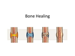
Bone healing occurs in overlapping stages of soft callus formation, bony callus formation, and remodeling. It is a highly regulated process involving the expression of many genes and signaling molecules. During soft callus formation, hematoma, fibroblasts, and new blood vessels form a framework for bone regeneration. Bony callus formation involves the deposition of woven bone through endochondral or intramembranous ossification. Finally, remodeling occurs where the callus is reduced and remodeled into lamellar bone through weight bearing and muscle action. Impediments to bone healing include movement, infection, malnutrition, genetic disorders, and bone density issues.
10. Stage of Fracture Repair Biological Processes Expression of Signaling Molecules and their Proposed Functions
Inflammation Hematoma IL-1, IL-6, and TNF-α play a role in initiating the repair cascade.
Inflammation TGF-β, PDGF, and BMP-2 expression increases to initiate callus formation.
Recruitment of mesenchymal
stem cells
GDF-8 is restricted to day 1, suggesting its role in controlling cellular proliferation.
Cartilage Formation and
Periosteal Response
Chondrogenesis and
endochondral ossification begins
TGF-β2, -β3, and GDF-5 peak due to their involvement in chondrogenesis and
endochondral bone formation.
Cell proliferation in
intramembranous ossification
BMP-5 and -6 rise.
Vascular in-growth Angiopoietins and VEGFs are induced to stimulate vascular in growth from vessels
in the periosteum.
Neo-angiogenesis
Cartilage Resorption and
Primary Bone Formation
Phase of most active
osteogenesis
TNF-α rises in association with mineralized cartilage resorption. This promotes the
recruitment of mesenchymal stem cells and induces apoptosis of hypertrophic
chondrocytes.
Bone cell recruitment and woven
bone formation
RANKL and MCSF rise in association with mineralized cartilage resorption.
Chondrocyte apoptosis and
matrix proteolysis
Osteoclast recruitment and
cartilage resorption
BMP-3, -4, -7, and -8 rise in association with the resorption of calcified cartilage.
They promote recruitment of cells in the osteoblastic lineage.
Neo-angiogenesis BMP-5 and -6 remain high during this stage, suggesting a regulatory effect on both
intramembranous and endochondral ossification.
VEGFs are up-regulated to stimulate neo-angiogenesis.
Secondary Bone Formation
and Remodeling
Bone remodeling coupled with
osteoblast activity
IL-1 and IL-6 rise again in association with bone remodeling, whereas RANKL and
MCSF display diminished levels.
Establishment of marrow Diminished expression of members of the TGF-β superfamily.