![M.A.A.Al-Maqrashi
T9-L3 Costal Margin ▪ Costal slips of diaphragm
T10
▪ Esophagus passes through diaphragm behind 7th
-8th
costal
cartilage just to the left [1-2cm] of median plane and Terminates
at the cardiac orifice of the stomach
T12
▪ Aorta, azygos v. & thoracic duct pass through diaphragm
▪ Upper border of the kidneys
Lumbar
L1
Trans-Pyloric plane
[found at midpoint b/w
jugular notch & pubic
symphysis]
[tip of 9th costal cartilage]
▪ Pyloris of stomach immediately above and to the right of the
midline.
▪ Beginning of duodenum
▪ Duodenojejunal flexure to the left of midline and immediately
below it
▪ Course of splenic vessels along Pancreas
▪ Root of transverse mesocolon
▪ Position of renal pelvis
▪ Hilum of kidneys: left is above, and right is below.
▪ R. renal a. originate just below this line & L. renal a. is just above
▪ Celiac a. originates just above
▪ Origin of Superior Mesenteric artery
▪ Termination of spinal cord
L2
▪ Thoracic duct begins
▪ Azygos & hemiazygos v. begin
L3
Sub-Costal Plane
[Through 10th costal
cartilage]
▪ Lowest point of costal margin
▪ Lower border of the kidneys
L4
Supra-Cristal Plane
[Through the highest point
of Iliac Crest]
▪ Bifurcation of aorta to R & L common iliac a.
▪ Inf. Vena cava begins.
▪ Important to locate 4th
, 3rd
or 2nd
IV space for lumbar puncture
▪ Spinous process of L3 lies just above → identifying vertebral
levels
L5
Trans-Tubercular Plane
[passes through the iliac
tubercles, small elevations
found in the iliac crest of the
iliac bone.]
▪ Continuation of cecum as ascending colon
Sacral
S2
Posterior Superior iliac spine
[dimple]
▪ End of sural sac
▪ Middle of sacroiliac joint
S3 Posterior inferior iliac spine
▪ End of sigmoid colon → rectum starts
[imp. For recto-sigmoid carcinoma surgery]](data:image/gif;base64,R0lGODlhAQABAIAAAAAAAP///yH5BAEAAAAALAAAAAABAAEAAAIBRAA7)
Recomendados
Más contenido relacionado
La actualidad más candente
La actualidad más candente (20)
Similar a Anatomy-Important vertebral level
Similar a Anatomy-Important vertebral level (20)
Más de Maryam Al-Maqrashi
Más de Maryam Al-Maqrashi (7)
Último
Último (20)
Anatomy-Important vertebral level
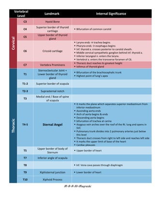
- 1. M.A.A.Al-Maqrashi Vertebral Level Landmark Internal SignificanceCervical C3 Hyoid Bone C4 Superior border of thyroid cartilage ▪ Bifurcation of common carotid C5 Upper border of thyroid gland C6 Cricoid cartilage ▪ Larynx ends → trachea begins ▪ Pharynx ends → esophagus begins ▪ Inf. thyroid a. crosses posterior to carotid sheath. ▪ Middle cervical sympathetic ganglion behind inf. thyroid a. ▪ Inferior laryngeal n. enters the larynx. ▪ Vertebral a. enters the transverse foramen of C6. C7 Vertebra Prominens ▪ Thoracic duct reaches its greatest height ▪ Isthmus of thyroid gland Thoracic T1 Sternoclavicular Joint + Lower border of thyroid gland ▪ Bifurcation of the brachiocephalic trunk ▪ Highest point of lung’s apex T1-2 Superior border of scapula T2-3 Suprasternal notch T3 Medial end / Base of spine of scapula T4-5 Sternal Angel ▪ It marks the plane which separates superior mediastinum from inferior mediastinum ▪ Ascending aorta ends ▪ Arch of aorta begins & ends ▪ Descending aorta begins ▪ bifurcation of trachea at carina ▪ Azygous vein arches over the roof of the Rt. lung and opens in SVC ▪ Pulmonary trunk divides into 2 pulmonary arteries just below this level ▪ Thoracic duct crosses from right to left side and reaches left side ▪ It marks the upper limit of base of the heart ▪ Cardiac plexuses T5 Upper border of body of Sternum ▪ Upper border of heart T7 Inferior angle of scapula T8 ▪ Inf. Vena cava passes through diaphragm T9 Xiphisternal junction ▪ Lower border of heart T10 Xiphoid Process
- 2. M.A.A.Al-Maqrashi T9-L3 Costal Margin ▪ Costal slips of diaphragm T10 ▪ Esophagus passes through diaphragm behind 7th -8th costal cartilage just to the left [1-2cm] of median plane and Terminates at the cardiac orifice of the stomach T12 ▪ Aorta, azygos v. & thoracic duct pass through diaphragm ▪ Upper border of the kidneys Lumbar L1 Trans-Pyloric plane [found at midpoint b/w jugular notch & pubic symphysis] [tip of 9th costal cartilage] ▪ Pyloris of stomach immediately above and to the right of the midline. ▪ Beginning of duodenum ▪ Duodenojejunal flexure to the left of midline and immediately below it ▪ Course of splenic vessels along Pancreas ▪ Root of transverse mesocolon ▪ Position of renal pelvis ▪ Hilum of kidneys: left is above, and right is below. ▪ R. renal a. originate just below this line & L. renal a. is just above ▪ Celiac a. originates just above ▪ Origin of Superior Mesenteric artery ▪ Termination of spinal cord L2 ▪ Thoracic duct begins ▪ Azygos & hemiazygos v. begin L3 Sub-Costal Plane [Through 10th costal cartilage] ▪ Lowest point of costal margin ▪ Lower border of the kidneys L4 Supra-Cristal Plane [Through the highest point of Iliac Crest] ▪ Bifurcation of aorta to R & L common iliac a. ▪ Inf. Vena cava begins. ▪ Important to locate 4th , 3rd or 2nd IV space for lumbar puncture ▪ Spinous process of L3 lies just above → identifying vertebral levels L5 Trans-Tubercular Plane [passes through the iliac tubercles, small elevations found in the iliac crest of the iliac bone.] ▪ Continuation of cecum as ascending colon Sacral S2 Posterior Superior iliac spine [dimple] ▪ End of sural sac ▪ Middle of sacroiliac joint S3 Posterior inferior iliac spine ▪ End of sigmoid colon → rectum starts [imp. For recto-sigmoid carcinoma surgery]