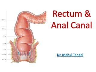
Rectum & anal canal
•
340 recomendaciones•97,011 vistas

Detailed anatomy of rectum and anal canal along with applied aspects. very useful for students.

Recomendados
Más contenido relacionado
La actualidad más candente
La actualidad más candente (20)
Similar a Rectum & anal canal
Similar a Rectum & anal canal (20)
Más de Mehul Tandel
Último
Último (20)
Rectum & anal canal
- 2. Objectives • Rectum – Introduction – Course – Measurements – Curvatures – Relations – Structure – Blood supply – Nerve supply – Applied
- 3. Rectum • Rectum = ? • Straight in quadrupeds • Not straight in man • But curved in A-P & Side to side. • Misnomer Straight
- 4. Rectum • Lower dilated part of large gut lies in pelvis • B/w Sigmoid colon and Anal canal • Devoid of taenia coli, sacculations, appendices epiploicae and mesentery
- 5. Ano-rectal Junction Rectum – Extent & Course Begins as continuation of sigmoid colon Opposite S3 Ends at a point: 2-3cm in front & little below tip of coccyx Lies at level of apex of prostate
- 6. Rectum - Measurements • Length = 12cm • Breadth = 4cm • Lower dilated part – Ampulla • When empty – Transverse slit
- 7. Rectum - Curvatures • 2 types :- Antero-posterior & Lateral Antero-posterior Curves Convex backward Convex Forward
- 8. Rectum - Curvatures Lateral Curves Upper Convex on right Middle Convex on Left S3 – S4 Sacro-coccygeal Junction Lower Convex on right Tip of Coccyx
- 9. Relations Peritoneal Relations Non- peritoneal Posterior Anterior Upper 2/3rd - peritoneal
- 10. Reflection Differ in Male & Female
- 11. Recto-vesical pouch In Males 7.5 cm above anus
- 13. In Females Recto-uterine pouch of Douglas Utero-vesical pouch 5.5 cm above anus
- 14. Relations Peritoneal Relations On each Side Para-rectal Fossa
- 15. Relations General Relations - Male Recto-vesical fascia of Denonviller In Front
- 16. Relations General Relations - Female Recto-vaginal fascia In Front
- 17. Relations General Relations - Posterior
- 18. Relations General Relations On each Side Para-rectal Fossa Pelvi-rectal Space & Levator ani Ischio-rectal Fossa
- 19. Interior of Rectum • Mucosal folds • 2 types • Temporary • Permanent • Temporary: • Longitudinal • In Lower part • Disappear on distension
- 20. Permanent Mucous folds • Horizontal • Semilunar • Along Concavity of lateral curves Houston’s Valves Reduplication of mucous membrane containing submucosa & thickening of circular muscle
- 21. Permanent Mucous folds • 1 & 3rd are constant • 2nd & 4th are inconstant Total 4 Valves
- 22. 1st Valve Recto-Sigmoid Jn S3 Right / Left wall 12 – 14 cm above Anus
- 23. 3rd Valve Most important & constant Anterior & Right wall Along concavity of middle lateral curve Nelaton’s Sphincter S5, 5cm above Anus Divide rectum in to upper & lower part
- 24. Upper Chamber Pre-allantoic hind gut May contain faeces without reflex act of defaecation Lower Chamber Endodermal Cloaca Entry of faeces – Start defaecation
- 25. 2nd Valve 2.5cm above 3rd Left wall Along concavity of upper lateral curve
- 26. 4th Valve 2.5cm below 3rd valve Left wall Along concavity of lower lateral curve
- 27. • Support weight of faeces • Prevent passage of instrument • Rectal washing should be done in left lateral position • To prevent injury to 3rd valve Importance of Valves
- 28. Arterial Supply Five rectal arteries
- 29. Arterial Supply Superior rectal artery Posterior View Continuation of IMA Principal artery of rectum Entire mucosa and upper musculature Straight vessels arise from plexus – anastomose with inferior rectal artery
- 30. Arterial Supply Middle rectal artery Posterior View Branch of Anterior division of Internal Iliac Artery Supply lower part
- 31. Arterial Supply Inferior rectal artery Posterior View Branch of Internal Pudendal Artery Supply peri-anal skin & Sphincter ani
- 32. Arterial Supply Median Sacral artery Posterior View Branch of Aorta Supply posterior wall of Ano-rectal Junction
- 33. Venous Drainage Arranged in plexus Annulus Haemorrhoidalis 2 sets Internal rectal venous plexus External rectal venous plexus
- 34. Venous Drainage Superior Rectal Vein Internal rectal venous plexus External rectal venous plexus Inferior Mesenteric Vein Middle Rectal Vein Internal iliac Vein Inferior Rectal Vein Internal Pudendal Vein Six Radical Veins Porto-Caval Anastomosis
- 35. Lymphatic Drainage From Upper PartFrom Middle Part From Lower Part
- 36. Nerve Supply Sympathetic: •Sup Hypogastric plexus (L1 & L2) •Vasomotor •Stimulate Sphincter & Inhibit musculature Parasympathetic: •Pelvic splanchnic nerves (S2, S3 & S4) •Secreto-motor •Stimulate peristalsis & Inhibit sphincter
- 37. • Pelvic floor by Levator ani muscles • Fascia of Waldeyer • Lateral ligaments of the Rectum • Rectovesical fascia of Denonvillers • Pelvic peritoneum • Perineal body with its muscles • Pelvi-rectal & Ischio-rectal fat Support of Rectum
- 38. Anal Canal
- 39. Downwards & Backwards Anal Canal – Extent & Course Begins at ano-rectal jn At a point 2-3cm in front & little below tip of coccyx Opens at anal orifice – 4cm below & in front of tip of coccyx Terminal part of Large Intestine
- 40. Anal Canal – Pecularities • Ant wall shorter • Surrounded by Sphincter Ani • Tone of it close the anal canal • Opens only during defaecation
- 41. Anal Canal- Measurements • Length = 3.8cm • Breadth • When empty – laterall wall approximated Antero-posterior slit
- 42. Relations • Anteriorly: Perineal body Males: membranous urethra bulb of penis Females: lower end of vagina • Posteriorly: Anococcygeal ligament Tip of coccyx • Laterally: Ischiorectal fossae • All round: Sphincteric muscles, tone of which keeps the anal closed...
- 46. Interior of Anal Canal Divided in to 3 parts Pectinate Line Hilton’s White Line Upper area Intermediate area / Pecten Lower area / Anal Verge
- 47. Upper Muscular part • 15 mm • Endodermal origin • Lined by semitransparent mucous membrane – Simple columnar / Stratified columnar or squamous • Plum red due to?
- 48. Upper Muscular part - Features Anal Columns / Columns of Morgagni Permanent longitudinal mucosal folds Contains Radicals of Sup rectal veins Reduplication of mucous membrane
- 49. Upper Muscular part - Features Anal Valves / Valves of Ball Crescentic mucosal folds Connect lower ends of anal columns Pectinate Line / Dentate Line Upper surface Lower surface Injury – Anal Fissure
- 50. Importance of Pectinate Line Divide canal in to Upper & Lower Areas Development Arterial Supply Pectinate Line / Dentate Line Venous Drainage Lymphatic Drainage Nerve Supply 85% diseases starts here
- 51. Upper Muscular part - Features Anal Papillae Epithelial processes Remnant of anal membrane
- 52. Upper Muscular part - Features Anal Sinuses Recesses above valves between anal columns Foreign body
- 53. Upper Muscular part - Features Anal Glands Open in floor of sinuses Infection - Fistula
- 54. Intermediate area / Area of Pectane • 15 mm • Ectodermal origin • Anal column: Absent • Bluish pink appearance of mucosa • St sq epithelium • No sweat / Sebaceous gland
- 55. Lower area / Anal Verge • 8 mm • Ectodermal origin • True skin with pigmentation • Corrugation due to corrugator cutis ani • Course hairs
- 56. Intermediate area / Area of Pectane • Color contrast between Bluish pink mucosa above & black skin below • Lies at lower end of internal anal sphincter White line of Hilton
- 58. Musculature of Rectum & Anal Canal
- 59. Musculature of Rectum Outer longitudinal Anterior Thick Band Posterior Thick Band
- 60. Musculature of Rectum Longitudinal Muscle Pubo-rectalis / Levator Ani Conjoint Fibro-elastic sheath Internal Anal Sphincter External Anal Sphincter
- 61. Musculature of Rectum Conjoint Fibro-elastic sheath Splits into number of septa Corrugator Cutis Ani Anal Intermuscular Septum
- 62. Musculature of Rectum Conjoint Fibro-elastic sheath Laterally Medially Recto-urethral muscle of Roux Recto-coccygeal muscle
- 63. Musculature Anal Canal • Anal Sphincters: • Internal: – Thickened lower circular muscle of rectum – Involuntary • External: – Striated muscles – Voluntary, – 3 parts: 1) Subcutaneous 2) Superficial 3) Deep
- 65. External Anal Sphincter Subcutaneous Superficial Deep Inf Rectal nerve Perineal Br of S4
- 66. Clinical Anatomy • Digital Per Rectal Examination • Proctoscopy/Sigmoidoscopy • Prolapse of Rectum • Neurological disturbances of Rectum • Rectal incontinence • Carcinoma of Rectum • Piles/Haemorrhoids • Anal Fissure/Fissure in ano • Fistula in ano
- 67. DPRE
- 69. Internal hemorrhoids: • Tributary of sup rectal • Above white line • Generally painless External hemorrhoids: • Tributary of inf rectal • Below white line • Generally painful Hemorrhoids / Piles
- 73. 11 o’clock 7 o’clock 3 o’clock
- 75. Anal Fissure Rupture of anal valves Painful
- 76. Anal Fissure
- 78. Fistula in Ano A fistula-in-ano is a hollow tract lined with granulation tissue connecting a primary opening inside the anal canal to a secondary opening in the perianal skin. Secondary tracts may be multiple and from the same primary opening.