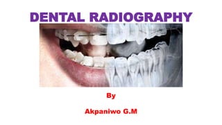
2. DENTAL RADIOGRAPHY
• Dental Radiography, is a Radiographic procedure that is used or
employed to take images of the teeth, bones, and soft tissues around
them, in order to identify, diagnose, plan treatments and monitor
both treatments and lesion development.
• The Radiographers practicing dental radiography are called Dental
Radiographers and provide dental images/radiographs for the dentist.
• The first dental clinic was established by Dr.C.E.Kells in July 1896,
using Xray machine.
3. DENTAL RADIOGRAPHY
REASONS FOR REQUESTING DENTAL RADIOGRAPHS.
1. To detect pathology associated with teeth and their supporting
structures, such as caries, periodontal disease and periapical
pathology.
2. To detect anomalies/injuries associated with the teeth, their
supporting structures, the maxilla and the mandible.
3. To determine the presence/absence of teeth and to localize Un-
erupted teeth.
4. To measure the length of the roots of teeth before endodontic
therapy
4. DENTAL RADIOGRAPHY
REASONS FOR REQUESTING DENTAL RADIOGRAPHS.
5. To detect the presence/absence of radio-opaque salivary calculi
and foreign bodies.
6. To detect anomalies/injuries/pathology of adjacent facial
structures.
7. To evaluate skeletal and/or soft tissues before orthodontic
treatment.
8. To monitor the progression of orthodontic treatment and dental
disease.
9. To enable a preoperative assessment of skeletal and soft tissue
patterns before orthognathic surgery.
10. To assess bony healing and effectiveness of surgical treatment of
the patient postoperatively.
5. DENTAL RADIOGRAPHY
RISK INVOLVE IN DENTAL XRAY PROCEDUE
• Dental Xray are considered to be safe for both adult and children.
• This is due to the low exposed levels used in carrying out this Xray.
RADIATION PROTECTION IN DENTAL XRAY PROCEDUE
• When carrying out dental xray, the patient should be protected by
wearing the patient a lead aprons and thyroid collars. This is to
protect the reproductive organ and Thyroid glad of the patient.
• Ask for pregnancy information, and do not proceed if the patient is
pregnant, consult the dental officer.
6. DENTAL RADIOGRAPHY
RADIATION PROTECTION IN DENTAL XRAY PROCEDUE
• When you are taking radiographs on a patient, observe the following
precautions to avoid unnecessary exposure to radiation:
1. NEVER stand in the path of the central X-ray beam during exposure.
2. NEVER hold the X-ray film packet in the patient's mouth during
exposure.
3. NEVER hold the tube head or the tube head cylinder of the X-ray
machine during exposure.
4. ALWAYS stand behind a lead-lined screen during an exposure.
7. DENTAL RADIOGRAPHY
CARE OF YOUR PATIENT FOR DENTAL RADIOGRAPHY
• Dental X-rays require no special preparation.
• The only thing you may ask your patient to brush he/her teeth before
coming for the appointment. That will create a more hygienic
environment for you to work inside mouth.
8. DENTAL RADIOGRAPHY
TERMINOLOGY USED IN DENTAL RADIOGRAPHY
• Buccal/Labial:- The (Outer) aspect of the teeth that lies between
the teeth and the cheeks or lips.
• Lingual/palatal:- The (Inner) aspect of the teeth that lies between the
teeth and the tongue.
• Distal:- The direction of the dental arch towards the molars,
posteriorly and outwards away from the MSP. It is
used to describe beam shift, tube shift or
angulation.
• Mesial:- The direction of the dental arch towards the
incisors, anteriorly and inwards towards the MSP. It
is used to describe beam shift, tube shift or angulation,
and it is in the opposite direction to distal movement.
9. DENTAL RADIOGRAPHY
TERMINOLOGY USED IN DENTAL RADIOGRAPHY
• Alatragal line:- An imaginary line from the tragus of the ear to the
middle of the ala of the nose(the area of soft tissue
around the nostril).
• Occlusal plane (upper):- This is the line of the biting surface of
the upper teeth. When the mouth is
closed this is deemed to be the occlusal
plane rather than the upper occlusal
plane. The line lies parallel to the
anthropological baseline and alatragal
line. It lies 4cm below the alatragal line.
10. DENTAL RADIOGRAPHY
TERMINOLOGY USED IN DENTAL RADIOGRAPHY
• Occlusal plane (lower):- With the mouth open, this line lies parallel
to and approximately 2cm below the line which lies between the
tragus of the ear and the outer canthus of the mouth. Because all
radiography of the teeth should be undertaken with the mouth closed
around an ionizing radiation holder or occlusal film.
• Medial Sagital plane (MSP):- This plane runs vertically down the
middle of the face, separating the left and right sides.
12. DENTAL RADIOGRAPHY
Types of Dental Radiography Procedures
There are four types of dental radiography procedures and this are
1. Intra-Oral Radiography, and its projections are (bitewing, periapical
and occlusal)
2. Panoramic radiography
3. Oblique lateral radiography
4. Cephalometric radiography
5. Cone-beam CT (CBCT)
Extra- Oral Radiography
13. DENTAL RADIOGRAPHY
Types of Dental Radiography Procedures
There are four types of dental radiography procedures and this are
1. Intraoral Radiography, and the following projections are the most
frequently requested examination. (bitewing, periapical and occlusal)
Bitewing Radiography
16. DENTAL RADIOGRAPHY
Types of Dental Radiography Procedures
(a) Small volume cone-beam CT (b) Large volume cone-beam CT
17. DENTAL RADIOGRAPHY
Types of Dental Radiography Procedures
• Bitewing radiography is a lateral view of the posterior regions of the
jaws. The view demonstrates the crowns of the teeth and the alveolar
crestal bone of the premolar and molar regions of both the maxilla
and mandible.
• Periapical radiography is a lateral projection displaying both the
crown and the root of the tooth and the surrounding bone.
• Occlusal radiography comprises a number of views in which the film
is positioned in the occlusal plane.
18. DENTAL RADIOGRAPHY
Types of Dental Radiography Procedures
Extra-oral radiography :- The most frequently requested extra-oral
projections are dental panoramic radiography, oblique lateral
radiography and cephalometry.
Dental panoramic radiography is a projection that produces an
image of both jaws and their respective dentitions on a single extra-
oral film.
Oblique lateral radiography demonstrates large areas of the maxilla
and mandible, with the region imaged depending on the technique
chosen.
Cephalometry employs techniques to produce standardized and
reproducible films of the facial bones for use in orthodontic,
orthognathic and implant treatment.
19. DENTAL RADIOGRAPHY
DENTITIONS OF THE TEETH
• The primary or deciduous dentition of humans comprises of 20 teeth,
with five in each quadrant of the jaws.
• These are replaced from six years onwards by a permanent dentition
of 32 teeth. With eruption of all 32 permanent teeth, there will be
eight permanent teeth in each quadrant.
• Some teeth may fail to develop or erupt, a complication most
commonly affecting the third permanent molars (the wisdom teeth).
21. DENTAL RADIOGRAPHY
DENTAL FORMULAE
• There are several internationally
recognized methods of identifying
the teeth that require
radiography.
• The two most commonly used
methods of notation are:
1. Palmer notation and
2. Fédération Dentaire International
(FDI) notation.
22. DENTAL RADIOGRAPHY
DENTAL FORMULAE : Palmer notation
• In the Palmer notation, Each dental quadrant extends from the
midline of the oral cavity posteriorly and, individually, corresponds to
the upper left and right quadrants in the maxilla and the lower left
and right quadrants in the mandible.
• The Palmer notation is depicted schematically, with a vertical line
between the central maxillary and mandibular incisors and a
horizontal line between the maxilla and mandible, dividing the oral
cavity into quadrants.
• The clinician requesting intraoral radiography uses these vertical and
horizontal lines to denote the quadrant to which the tooth/teeth to
be radiographed belong.
23. DENTAL RADIOGRAPHY
DENTAL FORMULAE : Palmer notation
• In using palmer notation, it is important to avoid confusion between
the permanent and deciduous dentition.
• The following conventions is used.
For the deciduous dentition: five teeth in each quadrant are assigned
the letters A–E, from the central deciduous incisor to the second
deciduous molar, respectively.
24. DENTAL RADIOGRAPHY
DENTAL FORMULAE : Palmer notation
For the permanent dentition: eight teeth in each quadrant are
assigned the numbers 1–8, from the central incisor to the third
permanent molar, respectively.
25. DENTAL RADIOGRAPHY
DENTAL FORMULAE : Palmer notation
A request card for dental radiography using the palmer notation must
contain the following.
• The number of tooth to be radiographed
• The letter of the tooth to be radiographed
This is added to complete the notation, and will give the Radiographer
more information in carrying out the examination.
For Example
26. DENTAL RADIOGRAPHY
DENTAL FORMULAE : Fédération Dentaire International (FDI) notation
• In the FDI , The dentition is again divided into four quadrants. These
are assigned the numbers 1–4 for the permanent teeth and the
numbers 5–8 for the deciduous dentition.
• In both dentitions, the quadrants follow on numerically, starting from
the upper right, to the upper left, to the lower left and, finally, to the
lower right.
• The number of the quadrant precedes the number of the tooth to be
radiographed.
27. DENTAL RADIOGRAPHY
DENTAL FORMULAE : Fédération Dentaire International (FDI) notation
• Examples of requests
for dental examinations
using this formula are:
11 – upper left canine.
31, 32 – lower Right second and
third molars.
28. DENTAL RADIOGRAPHY
Features of X-ray equipment for dental
radiography
• Dental equipment for intra-oral radiography is designed in order to
comply with radiation protection legislation and to ensure that the
patient dose is minimized. This equipment will have the following
features:
• X-ray tube potential:
– nominal tube potential not lower than 50 kVp, with recommended
operating range of 60–70 kVp.
• X-ray tube filtration:
– 1.5 mm aluminium equivalent for dental units up to 70 kVp;
– 2.5 mm aluminium equivalent (of which 1.5 mm should be permanent)
for dental units over 70 kVp.
29. DENTAL RADIOGRAPHY
Features of X-ray equipment for dental
radiography
• X-ray beam dimensions:
– Beam diameter at the patient’s skin not greater than 60mm.
– Rectangular collimation to be provided on new equipment
and retro-fitted to existing equipment.
• Minimum focus-to-skin distance:
– 200 mm for dental units of 60 kVp or greater;
– 100 mm for dental units less than 60 kVp
32. DENTAL RADIOGRAPHY
Image Receptor in dental radiography
The image receptors used in dental radiography:
Intra-oral radiography:
– direct or non-screen film;
– digital receptors.
Extra-oral radiography:
– film-screen (usually rare-earth);
– digital receptors: storage phosphor and solid-
state.
33. DENTAL RADIOGRAPHY
Image Receptor in dental radiography
Direct or non-screen film:- This films has the
advantage of producing a high resolution image
that provides the fine detail needed to assess
pathological changes.
•The film is made up of the following:
1. Outer plastic wrapper, which is to prevent
moisture contamination. The reverse side of the
outer wrapper has a two-toned appearance to
differentiate it as the non-imaging side of the
film packet.
34. DENTAL RADIOGRAPHY
Image Receptor in dental radiography
2. Black paper, that is wrapped around the film to
protect it from light ingress and damage during
handling.
3. Lead foil with an embossed pattern is
positioned at the back of the film to reduce
film fogging from scattered radiation.
4. A single sheet of film comprising a plastic
base with emulsion adherent to both surfaces.
36. DENTAL RADIOGRAPHY
FILM SIZES FOR INTRA-ORAL Dental Radiography
• There are several film sizes for Intra-oral dental radiography:
1. Size 0 - 22 x 35 mm: used for small children and anterior
periapicals using the paralleling technique.
2. Size 1 - 24 x 40 mm: used for bitewings in small children
and also for anterior projections in adults.
3. Size 2 - 31 x 41 mm: used for bitewings in adults and older
(generally six years plus) children and periapical
projections. It can be used for occlusal views in young
children.
4. Size 3 - 57 x 76 mm: used for occlusal projections of the
maxilla and mandible.
37. DENTAL RADIOGRAPHY
FILM SIZES FOR INTRA-ORAL Dental Radiography
•NOTE:- Intra-oral film are also available in some
sizes as double film packets.
•The reason for this is
to enables the Radiographer
to release one of the film
to the patient and to retain
the other for archiving or for
research research purpose.
38. DENTAL RADIOGRAPHY
Film identification of intra-oral films
•There is an embossed dot at one corner of the
front of the film packet, this allows for correct film
orientation by denoting the front of the film.
•The radiographer should adopt a working practice
of positioning the dental film with this dot
towards the crown of the tooth, so that it will not
obscure pathology within areas of interest.
39. DENTAL RADIOGRAPHY
Principles for optimal image geometry
In order to minimize distortion effects and to achieve optimal
image geometry, the following principles should be applied for
intra-oral radiography:
1. The focal spot should be as small as possible.
2. The focal spot-to-object distance should be as great as
possible.
3. The object-to-film distance (OFD) should be as small as
possible.
4. The film should be parallel to the plane of the object.
5. The central ray should be perpendicular to both the object
and the film.
40. DENTAL RADIOGRAPHY
Principles for optimal image geometry
Note:-
• In Bitewing radiography, the beam is required to be in the horizontal
plane, and should meets the teeth and the film at right-angles and
passes through all the contact areas.
• However, for Periapical radiography, two techniques is employed for
optimal image geometry. These are
1. Bisecting angle technique.
2. Paralleling technique
41. DENTAL RADIOGRAPHY
Principles for optimal image geometry
Bisecting angle technique: this is based upon the geometric theorem
of isometry. It requires the central ray of the X-ray beam to pass
through the root of the tooth at right-angles to a plane that is the
bisector of the angle formed by the long axis of the tooth and the
plane of the film.
42. DENTAL RADIOGRAPHY
Principles for optimal image geometry
Paralleling technique: this requires that the X-ray film is positioned
parallel with the long axes of the teeth or tooth to be imaged. This
enables the central ray of the X-ray beam to pass at right-angles, i.e.
perpendicular, to the beam to the long axes of the teeth and the
plane of the film.
43. DENTAL RADIOGRAPHY
BITEWING RADIOGRAPHY
• Bitewing radiography is used for:
1. The detection of dental caries in the upper and lower premolar and
molar teeth;
2. The crowns, interproximal surfaces and gingival margins of the
premolars and molars.
3. Monitoring the progression of dental caries.
4. Assessment of existing restorations.
5. Assessment of the periodontal condition.
44. DENTAL RADIOGRAPHY
BITEWING RADIOGRAPHY
There are three methods used to position the film intra-orally:
1. Bitewing tab: a heavy-duty paper tab
attached to an intra-oral film.
The attachment can be either by
an adhesive backing to the tab or
by a bitewing loop with an attached
tab for the patient to bite on.
46. DENTAL RADIOGRAPHY
BITEWING RADIOGRAPHY
3. Film-holding beam-alignment instrument: a device with a bite
block, rigid backing and an extra-oral arm to correctly position the tube
relative to the film.
47. DENTAL RADIOGRAPHY
BITEWING RADIOGRAPHY
PATIENT POSITION
• The patient sit with their neck
leaning on a support,
• A bitewing film or bitewing holder is
placed with its tube side in contact with
the lingual surface of the teeth under
examination and the flap between the
occlusal surfaces of the teeth.
• The patient close their teeth over the flap
• The MSP is vertical and the upper
• occlusal plane horizontal
49. DENTAL RADIOGRAPHY
BITEWING RADIOGRAPHY
BEAM DIRECTION:-
• The beam direction is horizontal, with a 5 – 8 degree caudal
angulation.
CENTRING POINT:-
• Centre to the middle of the image receptor, over the occlusal pane
INCLUDE:-
Crowns of the teeth under examination and alveolar crests.
50. DENTAL RADIOGRAPHY
BITEWING RADIOGRAPHY
Criteria for assessing image quality:-
1. Crowns of the teeth and alveolar crests are demonstrated
2. No evidence of elongation or foreshortening of the teeth
3. No over lap of adjacent teeth
4. Slight separation of occlusal surfaces of the teeth
5. Sharp images demonstrating the enamel in contrast with pulp cavity
and the alveolar crests
52. DENTAL RADIOGRAPHY
PERIAPICAL RADIOGRAPHY
Periapical radiography provides an image of the teeth, the surrounding periodontal
tissues and the alveolar bone.
Indication for Periapical Radiography
1. Assessment of the periodontium encompassing the periapical and the
periodontal status.
2. Assessment of apical pathology and other lesions situated within alveolar
bone.
3. Pre- and postoperative assessment of alveolar surgery.
4. Following trauma to teeth and alveolar bone.
5. Localization of teeth and presence/absence of teeth.
6. Before extraction to assess root morphology and the relationship of roots to
vital structures, i.e. the inferior dental canal, the maxillary antrum.
7. During endodontic therapy (a sequence of treatment for the infected pulp of a tooth).
8. Pre- and postoperative assessment of implants.
53. DENTAL RADIOGRAPHY
PERIAPICAL RADIOGRAPHY
Bisecting angle technique:-
• In this technique, 2 methods are employed to stabilize the film intra-
orally:
1. The patient’s finger, or
2. A film-holding instrument.
• The placement of the intra-oral film using this technique is as follows:
1. Anterior teeth (incisors and canines): long axis of film vertical.
2. Posterior teeth (premolars and molars): long axis of film horizontal
54. DENTAL RADIOGRAPHY
PERIAPICAL RADIOGRAPHY
BISECTING ANGLE TECHNIQUE
Position of patient:-
• The patient’s head must be supported adequately with the medial plane vertical
and the occlusal plane horizontal (i.e. upper occlusal plane and lower occlusal
plane for maxillary and mandibular radiography, respectively).
NOTE: If a film holder is used
• The correct film size is chosen and placed in the film holder.
• Position the film holder intra-orally adjacent to the lingual/palatal aspects
of the tooth/teeth to be imaged.
• Insert a cotton-wool roll between the opposing teeth and the bite block.
• Ask the patient to close together slowly to allow gradual accommodation
of the film holder intra-orally.
• Tell the patient to continue biting on the bite block to position the film
holder securely.
56. DENTAL RADIOGRAPHY
PERIAPICAL RADIOGRAPHY
BISECTING ANGLE TECHNIQUE
Position of patient:-
NOTE:- If the patient’s finger is used:
• The correct film size is chosen and positioned intra-orally.
• Ensure that the tooth/teeth being examined are in the middle of the film.
• 2 mm of the film packet should extend beyond the incisal or occlusal
margin to ensure that the entire tooth is imaged.
• Instruct the patient to gently support the film using either their index
finger or thumb.
• Apply the patient’s finger/thumb solely to the area of film that overlies the
crown and gingival tissues of the teeth. This reduces the possibility of
distortion by bending of the film covering the root and periapical tissues.
58. DENTAL RADIOGRAPHY
PERIAPICAL RADIOGRAPHY
BISECTING ANGLE TECHNIQUE
BEAM DIRECTION:
A Horizontal central ray at 90 degrees to the bisector of the angle
formed between the long axis of the tooth and long axis of the image
recieptor.
CENTERING:
Centre over the buccal surfaces of the teeth, to the film
59. DENTAL RADIOGRAPHY
PERIAPICAL RADIOGRAPHY
BISECTING ANGLE TECHNIQUE
CRITERIA FOR ASSESSING IMAGE QUALITY:
1. Crowns, root of the teeth and surrounding bones are
demonstrated.
2. Minimal evidence of elongation or foreshortening of the
tooth/teeth, or overlap of adjacent teeth if there is no
overcrowding of teeth in that region
3. Sharp image showing contrast of the alveolar bones and its
trabeculae, pulp cavity and enamel of the tooth/teeth.
61. DENTAL RADIOGRAPHY
PERIAPICAL RADIOGRAPHY
PARALLELING TECHNIQUE
• The paralleling technique requires that the X-ray film is positioned
parallel with the long axes of the teeth. The central ray of the X-ray
beam passes at right-angles, i.e. perpendicular, to the tooth
ADVANTAGE OF THIS TECHNIQUE:
1. Minimal elongation/foreshortening/distortion.
2. Increased focus-to-skin distance (FSD) reduces surface dose.
3. Increased FSD improves image quality by reducing the penumbra
effect.
4. Reduction in distortion effects due to bending of the film/image
receptor.
62. DENTAL RADIOGRAPHY
PERIAPICAL RADIOGRAPHY
PARALLELING TECHNIQUE
DISADVANTAGE OF THIS TECHNIQUE:
1. The paralleling technique can be used when using X-ray equipment
with a short FFD (less than 20 cm) providing the operator accepts
increased magnification.
2. Anatomical limitations, such as a shallow palate, principally in the
maxillary molar and anterior regions, preclude true parallel
placement of the film relative to the tooth.
63. DENTAL RADIOGRAPHY
Film mounting of intra-oral films
•Films are mounted with the (embossed) dot
towards the radiographer and as though the
operator was looking at the patient.
•This ensures that the mounted films exactly match
the dentition arrangement of the patient.
Note:
•The embossed dot is positioned towards the
anterior part of the mouth, when soft-tissue views
is been requested.
64. DENTAL RADIOGRAPHY
Film mounting of intra-oral films
How to mount the film on the viewing box:
1. Arrange films as to whether they were taken in the
maxilla or mandible followed by region, i.e. anterior and
posterior. Use anatomical landmarks for guidance as well
as root formation.
2. Arrange the films of the maxillary teeth by placing the
crowns of the teeth towards the bottom of the viewer.
3. Arrange the films of the mandibular teeth with the
crowns of the teeth towards the top of the viewer.
4. When maxillary and mandibular teeth have been
identified, radiographs are then arranged as belonging to
either the right or left side of the patient.
65. DENTAL RADIOGRAPHY
Film mounting of intra-oral films
NOTE:-
•Radiographs of the anterior incisors are placed in
the centre of the mount.
• The radiographs of the lateral and canine teeth
(positioned correctly, according to side) are placed
adjacent to them.
• This is repeated successively for premolars and
molar films to complete film mounting in both
dental arches.
67. DENTAL RADIOGRAPHY
EXTRA-ORAL (ORTHOPANTOMOGRAPHY)
POSITIONING:
• A disposable bite rod is inserted into the chin rest, or a disposable
plastic cover is applied to the permanent bite.
• The patient sit or stand with their chin resting on the chin support
and in the correct position to facilitate the dental arch being placed in
the correct tomographic plane. (with the anthropological baseline
and alatragal line horizontal and the head far enough forward. This is
usually indicated by a slit light indicator)
68. DENTAL RADIOGRAPHY
EXTRA-ORAL (ORTHOPANTOMOGRAPHY)
POSITIONING:
• The patient bite with their incisors in the groove on the bite rod, to
effect separation of teeth on the image.
• The MSP is vertical and perpendicular to the bite rod.
• The height of the unit is adjusted unti the ocular plane is horizontal.