SlideShare una empresa de Scribd logo
1 de 22
Section 1, Chapter 5

Introduction to Tissues
& Epithelial Tissues
Tissues
A Tissue is to a group of similar cells that carries out a specialized function.
Histology is the science of tissues.

examples of tissues
There are four broad categories of tissues
Muscular tissue – specialized to contract
Nervous tissue – conducts, senses, and stores information
Epithelial tissue – forms coverings (skin) & linings (blood vessels)
Connective tissues – support, transport, protect
Intercellular junctions
Cells may be separated by a matrix or connected together by cellular junctions.

1. Tight Junction

A tight junction fuses the cell membranes of adjacent cells together so
molecules cannot move between the cells. Instead, they must move
through the cells.
Tight junctions prevent the passage of unwanted & harmful substances
Examples: Blood Brain Barrier & Digestive Tract
Cell 1

Interlocking membranes form
tight junction between two cells

Cell 2
Cellular Junctions
2. Desmosome
desmosomes form “spot welds” between cells
Provides structural support
Examples of desmosomes are in the epidermis of the skin

Cells connected by desmosomes
Cellular Junctions
3. Gap Junction
Gap Junctions connect cells by protein ion channels that
allow for cell-to-cell diffusion of ions
Example of gap junctions are within the intercalated
discs of cardiac muscle

Intercalated discs of cardiac muscle
are one type of gap junction.
Types of Tissues: Epithelial Tissue
Epithelial Tissue:
Epithelial tissue lines cavities, such as the digestive & respiratory tracts
It also forms coverings, such as the skin
Functions include protection, secretion, absorption, excretion
Epithelium: structure
Epithelium is anchored to a layer of connective tissue, called the
basement membrane.
Epithelium has a basal surface that faces the basement membrane
The free (apical) surface is exposed to the open space (lumen)

Open space = lumen

Basement membrane
Characteristics of Epithelial Tissue
Epithelium consists of tightly packed cells
They lack a blood supply, instead they receive nutrients by diffusion
Epithelium readily divide, so they are continually replaced = rapid healing
Classifications of Epithelial Tissues
Epithelium are classified by the layer of cells
1. Simple = Single layer of epithelial cells
2. Stratified = two or more layers of cells

And they’re classified by the shape of cells
1. squamous = scale-like
2. cuboidal = cube-like shape
3. Columnar = tall cells
Simple Squamous Epithelium
Single layer of flattened cells
Functions include diffusion, filtration, and secretion
Locations include:
air sacs of lungs (alveoli)
lining of capillaries and the walls of blood vessels
glomerulus of kidneys.

surface view of simple squamous ep.

alveoli of lung
Simple Cuboidal Epithelium
Single layer of cube-shaped cells
Functions include secretion and absorption
Locations include
the lining and the ducts of some glands
tubules within kidneys

lumen

cross-section of tubules in kidneys
Simple Columnar Epithelium
Single layer of elongated cells and their nuclei appear as a single row
Functions include absorption, secretion, or transport
Often contain goblet cells
Goblet cells secrete mucus onto apical surface
may be ciliated, or have microvilli, or have no modifications on apical surface.

columnar cells
goblet cell

microvilli “brush border”
Simple Columnar Epithelium
Some simple columnar epithelium are covered with cilia on
their free surface.

Cilia beat constantly in a rhythmic motion to propel
substances across the cell surface.

Cilia propels the egg through uterine tube
Simple Columnar Epithelium
Some simple columnar epithelium are covered with
microvili on their free surface.
Microvilli are extensions of the cell membrane that increases the
surface area of a cell.

Example of microvilli include the intestinal epithelium, where they
increase the amount of contact each cell makes with food.

Microvilli greatly enhance absorption of nutrients.
Pseudostratified Columnar Epithelium (PSCE)
PSCE appear striated, but each cell touches the basement membrane.
Usually ciliated
Most PSCE contain goblet cells that secrete mucus

Locations of PSCE include the lining of the
respiratory tract: nasal cavity, pharynx,
trachea, bronchi
Functions: Mucus from goblet cells traps
debris & cilia moves the mucus (and trapped
debris) away from the lungs
Stratified Squamous Epithelium
Several layers of cells = thick tissue
Primary function is to protect underlying tissues from abrasion

Superficial cells are squamous but the deeper cells are cuboidal or
columnar
May be keratinized or non-keratinized
Keratinized Stratified Squamous Epithelium
Keratinized epithelium is found in the epidermis (outer layer) of skin
Keratin is an insoluble protein that hardens and forms a protective coating.
Cells accumulate keratin over time, so the older outermost cells of the
epidermis are keratinized

Stratified squamous epithelium of epidermis
Non-keratinized Stratified Squamous Epithelium
Non-keratinized (moist) stratified squamous epithelium
lines the esophagus, oral cavity, vagina, and anal canal.

Apical surface

Stratified squamous epithelium of vagina
Transitional Epithelium
Transitional epithelium forms an expandable lining (Function: permit expansion)
Locations include the inner layer of the urinary bladder & ureters
The lining is several layers thick and the cells appear cuboidal when the urinary bladder
is contracted, but only a few layers thick when the urinary bladder is distended and the
cells appear squamous.

empty urinary bladder

distended urinary bladder
Glandular Epithelium
Glandular epithelium is specialized to produce & secrete substances

Glandular epithelium form glands

Exocrine Glands
Exocrine glands secretes substances
through ducts onto an open surface
Examples includes goblet cells, sweat
glands, and mammary glands
Duct of a
sweat gland
Glandular Epithelium
Endocrine Glands
Endocrine glands secretes substances (hormones) directly into the blood
stream.
Examples includes the thyroid gland, pituitary gland, adrenal glands, ect.

Follicles of thyroid gland are
composed of simple cuboidal
epithelium

End of Section 1, Chapter 5

Más contenido relacionado

La actualidad más candente

Connective Tissue Practice Part 1
Connective Tissue Practice Part 1Connective Tissue Practice Part 1
Connective Tissue Practice Part 1Michael Wrock
 
Histology Lab Image Identification
Histology Lab Image IdentificationHistology Lab Image Identification
Histology Lab Image Identificationguest02819bb4
 
Epithelial tissues
Epithelial tissuesEpithelial tissues
Epithelial tissuesHelao Silas
 
HISTOLOGY: EPITHELIA AND GLANDS CONNECTIVE TISSUE PROPER CARTILAGE AND BONE
HISTOLOGY: EPITHELIA AND GLANDS CONNECTIVE TISSUE PROPER CARTILAGE AND BONEHISTOLOGY: EPITHELIA AND GLANDS CONNECTIVE TISSUE PROPER CARTILAGE AND BONE
HISTOLOGY: EPITHELIA AND GLANDS CONNECTIVE TISSUE PROPER CARTILAGE AND BONEOHupdates
 
Mock Lab Exam 2009 For Review
Mock Lab Exam 2009   For ReviewMock Lab Exam 2009   For Review
Mock Lab Exam 2009 For Reviewheidiw
 
Connective tissue
Connective tissueConnective tissue
Connective tissuePandian M
 
Histology of the nervous system
Histology of the nervous systemHistology of the nervous system
Histology of the nervous systemSahar Hafeez
 
Histology -Epithelial tissue
Histology -Epithelial tissueHistology -Epithelial tissue
Histology -Epithelial tissueDr. Devi Shankar
 

La actualidad más candente (20)

Tissues
TissuesTissues
Tissues
 
Connective Tissue PPT
Connective Tissue PPTConnective Tissue PPT
Connective Tissue PPT
 
Connective Tissue Practice Part 1
Connective Tissue Practice Part 1Connective Tissue Practice Part 1
Connective Tissue Practice Part 1
 
Section 2,chapter 5
Section 2,chapter 5Section 2,chapter 5
Section 2,chapter 5
 
Connective tissue
Connective tissueConnective tissue
Connective tissue
 
connective tissue.
 connective tissue. connective tissue.
connective tissue.
 
Histology Lab Image Identification
Histology Lab Image IdentificationHistology Lab Image Identification
Histology Lab Image Identification
 
Epithelial tissues
Epithelial tissuesEpithelial tissues
Epithelial tissues
 
Lab Exam #2 Histology (labeled)
Lab Exam #2 Histology (labeled)Lab Exam #2 Histology (labeled)
Lab Exam #2 Histology (labeled)
 
HISTOLOGY: EPITHELIA AND GLANDS CONNECTIVE TISSUE PROPER CARTILAGE AND BONE
HISTOLOGY: EPITHELIA AND GLANDS CONNECTIVE TISSUE PROPER CARTILAGE AND BONEHISTOLOGY: EPITHELIA AND GLANDS CONNECTIVE TISSUE PROPER CARTILAGE AND BONE
HISTOLOGY: EPITHELIA AND GLANDS CONNECTIVE TISSUE PROPER CARTILAGE AND BONE
 
Mock Lab Exam 2009 For Review
Mock Lab Exam 2009   For ReviewMock Lab Exam 2009   For Review
Mock Lab Exam 2009 For Review
 
Connective tissue
Connective tissueConnective tissue
Connective tissue
 
ADVANCED LEVEL BASIC HISTOLOGY-by KING SOYEKWO ROGERS
ADVANCED LEVEL BASIC HISTOLOGY-by KING SOYEKWO ROGERSADVANCED LEVEL BASIC HISTOLOGY-by KING SOYEKWO ROGERS
ADVANCED LEVEL BASIC HISTOLOGY-by KING SOYEKWO ROGERS
 
Histology of the nervous system
Histology of the nervous systemHistology of the nervous system
Histology of the nervous system
 
Histology -Epithelial tissue
Histology -Epithelial tissueHistology -Epithelial tissue
Histology -Epithelial tissue
 
Histology of Muscle Tissues
Histology of Muscle TissuesHistology of Muscle Tissues
Histology of Muscle Tissues
 
Connective tissue
Connective tissueConnective tissue
Connective tissue
 
TISSUE.pptx
TISSUE.pptxTISSUE.pptx
TISSUE.pptx
 
cartilage
cartilage cartilage
cartilage
 
Connective tissues
Connective tissuesConnective tissues
Connective tissues
 

Destacado

Identifying Epithelium
Identifying EpitheliumIdentifying Epithelium
Identifying EpitheliumJoelle Weaver
 
Introduction to histology
Introduction to histologyIntroduction to histology
Introduction to histologyAbdul Ansari
 
Histology 2 connective_tissue
Histology 2 connective_tissueHistology 2 connective_tissue
Histology 2 connective_tissueTyler Wall
 
General histology.(introduction)
General histology.(introduction)General histology.(introduction)
General histology.(introduction)Fatma Adel
 
Introduction to histology
Introduction to histologyIntroduction to histology
Introduction to histologychanthaj
 
Connective tissue proper
Connective tissue properConnective tissue proper
Connective tissue properJhon Mar Bellos
 
Histology various type of epithelium
Histology   various type of epitheliumHistology   various type of epithelium
Histology various type of epitheliumMBBS IMS MSU
 
Connective tissues
Connective tissuesConnective tissues
Connective tissuesSara Khan
 
Epithelium, cells,tissues & histology
Epithelium, cells,tissues & histologyEpithelium, cells,tissues & histology
Epithelium, cells,tissues & histologyDr. Waqas Nawaz
 

Destacado (14)

Connective tissue
Connective tissueConnective tissue
Connective tissue
 
Identifying Epithelium
Identifying EpitheliumIdentifying Epithelium
Identifying Epithelium
 
Histology: Tissues
Histology: TissuesHistology: Tissues
Histology: Tissues
 
Introduction to histology
Introduction to histologyIntroduction to histology
Introduction to histology
 
Histology 2 connective_tissue
Histology 2 connective_tissueHistology 2 connective_tissue
Histology 2 connective_tissue
 
General histology.(introduction)
General histology.(introduction)General histology.(introduction)
General histology.(introduction)
 
Introduction to histology
Introduction to histologyIntroduction to histology
Introduction to histology
 
Connective tissue proper
Connective tissue properConnective tissue proper
Connective tissue proper
 
Histology various type of epithelium
Histology   various type of epitheliumHistology   various type of epithelium
Histology various type of epithelium
 
Connective tissue
Connective tissueConnective tissue
Connective tissue
 
Epithelium ppt...
Epithelium ppt...Epithelium ppt...
Epithelium ppt...
 
Connective tissues
Connective tissuesConnective tissues
Connective tissues
 
Connective tissue.pptx
Connective tissue.pptxConnective tissue.pptx
Connective tissue.pptx
 
Epithelium, cells,tissues & histology
Epithelium, cells,tissues & histologyEpithelium, cells,tissues & histology
Epithelium, cells,tissues & histology
 

Similar a section 1, chapter 5 (20)

5566.pptx
5566.pptx5566.pptx
5566.pptx
 
Histology basics of sciences and microanatomy.pdf
Histology basics of sciences and microanatomy.pdfHistology basics of sciences and microanatomy.pdf
Histology basics of sciences and microanatomy.pdf
 
Anatomy :O
Anatomy :OAnatomy :O
Anatomy :O
 
The Tissue level of organization .pptx
The Tissue level of organization   .pptxThe Tissue level of organization   .pptx
The Tissue level of organization .pptx
 
Basic Tissues 1.pptx
Basic Tissues 1.pptxBasic Tissues 1.pptx
Basic Tissues 1.pptx
 
epithelia Tissues.pptx
epithelia Tissues.pptxepithelia Tissues.pptx
epithelia Tissues.pptx
 
Epithelial Tissue
Epithelial TissueEpithelial Tissue
Epithelial Tissue
 
epithelium
epitheliumepithelium
epithelium
 
Tissues
TissuesTissues
Tissues
 
A and P Mod. #2 Tissues
A and P Mod. #2 TissuesA and P Mod. #2 Tissues
A and P Mod. #2 Tissues
 
General principles of tissue organization
General principles of tissue organizationGeneral principles of tissue organization
General principles of tissue organization
 
3.tissues of body
3.tissues of body3.tissues of body
3.tissues of body
 
HUMAN ANATOMY AND PHYSIOLOGY.PPT.pdf.pdf
HUMAN ANATOMY AND PHYSIOLOGY.PPT.pdf.pdfHUMAN ANATOMY AND PHYSIOLOGY.PPT.pdf.pdf
HUMAN ANATOMY AND PHYSIOLOGY.PPT.pdf.pdf
 
Epithelium
EpitheliumEpithelium
Epithelium
 
4. Tissues: Living Communities
4. Tissues: Living Communities4. Tissues: Living Communities
4. Tissues: Living Communities
 
Tissues
TissuesTissues
Tissues
 
Tissues
Tissues Tissues
Tissues
 
Animal tissues epithelial & connective
Animal tissues epithelial & connectiveAnimal tissues epithelial & connective
Animal tissues epithelial & connective
 
Animal tissues epithelial & connective
Animal tissues epithelial & connectiveAnimal tissues epithelial & connective
Animal tissues epithelial & connective
 
Histology
HistologyHistology
Histology
 

Más de Michael Walls

section 1, chapter 12: general senses
section 1, chapter 12: general sensessection 1, chapter 12: general senses
section 1, chapter 12: general sensesMichael Walls
 
chapter 19 respiratory system
chapter 19 respiratory systemchapter 19 respiratory system
chapter 19 respiratory systemMichael Walls
 
chapter 21 electrolyte balance
chapter 21 electrolyte balancechapter 21 electrolyte balance
chapter 21 electrolyte balanceMichael Walls
 
chapter 20: urinary system
chapter 20: urinary systemchapter 20: urinary system
chapter 20: urinary systemMichael Walls
 
section 6, chapter 15: systemic arteries
section 6, chapter 15: systemic arteriessection 6, chapter 15: systemic arteries
section 6, chapter 15: systemic arteriesMichael Walls
 
section 5, chapter 15: blood pressure
section 5, chapter 15: blood pressuresection 5, chapter 15: blood pressure
section 5, chapter 15: blood pressureMichael Walls
 
section 4, chapter 15: blood vessels
section 4, chapter 15: blood vesselssection 4, chapter 15: blood vessels
section 4, chapter 15: blood vesselsMichael Walls
 
Section 3, chapter 15: ecg
Section 3, chapter 15: ecgSection 3, chapter 15: ecg
Section 3, chapter 15: ecgMichael Walls
 
section 2, chapter 15: conduction of the heart
section 2, chapter 15: conduction of the heartsection 2, chapter 15: conduction of the heart
section 2, chapter 15: conduction of the heartMichael Walls
 
Section 1, chapter 15: anatomy of the heart
Section 1, chapter 15: anatomy of the heartSection 1, chapter 15: anatomy of the heart
Section 1, chapter 15: anatomy of the heartMichael Walls
 
Section 3, chapter 17: liver and intestines
Section 3, chapter 17: liver and intestinesSection 3, chapter 17: liver and intestines
Section 3, chapter 17: liver and intestinesMichael Walls
 
Section 2, chapter 17: stomach and pancreas
Section 2, chapter 17: stomach and pancreasSection 2, chapter 17: stomach and pancreas
Section 2, chapter 17: stomach and pancreasMichael Walls
 

Más de Michael Walls (20)

A&P Chapter 10
A&P Chapter 10A&P Chapter 10
A&P Chapter 10
 
A&P Chapter 09
A&P Chapter 09A&P Chapter 09
A&P Chapter 09
 
A&P Chapter 08
A&P Chapter 08A&P Chapter 08
A&P Chapter 08
 
A&P Chapter 06
A&P Chapter 06A&P Chapter 06
A&P Chapter 06
 
A&P Chapter 04
A&P Chapter 04A&P Chapter 04
A&P Chapter 04
 
A&P Chapter 03
A&P Chapter 03A&P Chapter 03
A&P Chapter 03
 
A&P Chapter 02
A&P Chapter 02A&P Chapter 02
A&P Chapter 02
 
A&P Chapter 01
A&P Chapter 01A&P Chapter 01
A&P Chapter 01
 
section 1, chapter 12: general senses
section 1, chapter 12: general sensessection 1, chapter 12: general senses
section 1, chapter 12: general senses
 
chapter 19 respiratory system
chapter 19 respiratory systemchapter 19 respiratory system
chapter 19 respiratory system
 
chapter 21 electrolyte balance
chapter 21 electrolyte balancechapter 21 electrolyte balance
chapter 21 electrolyte balance
 
chapter 20: urinary system
chapter 20: urinary systemchapter 20: urinary system
chapter 20: urinary system
 
section 6, chapter 15: systemic arteries
section 6, chapter 15: systemic arteriessection 6, chapter 15: systemic arteries
section 6, chapter 15: systemic arteries
 
section 5, chapter 15: blood pressure
section 5, chapter 15: blood pressuresection 5, chapter 15: blood pressure
section 5, chapter 15: blood pressure
 
section 4, chapter 15: blood vessels
section 4, chapter 15: blood vesselssection 4, chapter 15: blood vessels
section 4, chapter 15: blood vessels
 
Section 3, chapter 15: ecg
Section 3, chapter 15: ecgSection 3, chapter 15: ecg
Section 3, chapter 15: ecg
 
section 2, chapter 15: conduction of the heart
section 2, chapter 15: conduction of the heartsection 2, chapter 15: conduction of the heart
section 2, chapter 15: conduction of the heart
 
Section 1, chapter 15: anatomy of the heart
Section 1, chapter 15: anatomy of the heartSection 1, chapter 15: anatomy of the heart
Section 1, chapter 15: anatomy of the heart
 
Section 3, chapter 17: liver and intestines
Section 3, chapter 17: liver and intestinesSection 3, chapter 17: liver and intestines
Section 3, chapter 17: liver and intestines
 
Section 2, chapter 17: stomach and pancreas
Section 2, chapter 17: stomach and pancreasSection 2, chapter 17: stomach and pancreas
Section 2, chapter 17: stomach and pancreas
 

Último

Q4-W6-Restating Informational Text Grade 3
Q4-W6-Restating Informational Text Grade 3Q4-W6-Restating Informational Text Grade 3
Q4-W6-Restating Informational Text Grade 3JemimahLaneBuaron
 
BASLIQ CURRENT LOOKBOOK LOOKBOOK(1) (1).pdf
BASLIQ CURRENT LOOKBOOK  LOOKBOOK(1) (1).pdfBASLIQ CURRENT LOOKBOOK  LOOKBOOK(1) (1).pdf
BASLIQ CURRENT LOOKBOOK LOOKBOOK(1) (1).pdfSoniaTolstoy
 
Accessible design: Minimum effort, maximum impact
Accessible design: Minimum effort, maximum impactAccessible design: Minimum effort, maximum impact
Accessible design: Minimum effort, maximum impactdawncurless
 
APM Welcome, APM North West Network Conference, Synergies Across Sectors
APM Welcome, APM North West Network Conference, Synergies Across SectorsAPM Welcome, APM North West Network Conference, Synergies Across Sectors
APM Welcome, APM North West Network Conference, Synergies Across SectorsAssociation for Project Management
 
Unit-IV- Pharma. Marketing Channels.pptx
Unit-IV- Pharma. Marketing Channels.pptxUnit-IV- Pharma. Marketing Channels.pptx
Unit-IV- Pharma. Marketing Channels.pptxVishalSingh1417
 
1029 - Danh muc Sach Giao Khoa 10 . pdf
1029 -  Danh muc Sach Giao Khoa 10 . pdf1029 -  Danh muc Sach Giao Khoa 10 . pdf
1029 - Danh muc Sach Giao Khoa 10 . pdfQucHHunhnh
 
microwave assisted reaction. General introduction
microwave assisted reaction. General introductionmicrowave assisted reaction. General introduction
microwave assisted reaction. General introductionMaksud Ahmed
 
fourth grading exam for kindergarten in writing
fourth grading exam for kindergarten in writingfourth grading exam for kindergarten in writing
fourth grading exam for kindergarten in writingTeacherCyreneCayanan
 
SOCIAL AND HISTORICAL CONTEXT - LFTVD.pptx
SOCIAL AND HISTORICAL CONTEXT - LFTVD.pptxSOCIAL AND HISTORICAL CONTEXT - LFTVD.pptx
SOCIAL AND HISTORICAL CONTEXT - LFTVD.pptxiammrhaywood
 
social pharmacy d-pharm 1st year by Pragati K. Mahajan
social pharmacy d-pharm 1st year by Pragati K. Mahajansocial pharmacy d-pharm 1st year by Pragati K. Mahajan
social pharmacy d-pharm 1st year by Pragati K. Mahajanpragatimahajan3
 
Nutritional Needs Presentation - HLTH 104
Nutritional Needs Presentation - HLTH 104Nutritional Needs Presentation - HLTH 104
Nutritional Needs Presentation - HLTH 104misteraugie
 
Grant Readiness 101 TechSoup and Remy Consulting
Grant Readiness 101 TechSoup and Remy ConsultingGrant Readiness 101 TechSoup and Remy Consulting
Grant Readiness 101 TechSoup and Remy ConsultingTechSoup
 
Software Engineering Methodologies (overview)
Software Engineering Methodologies (overview)Software Engineering Methodologies (overview)
Software Engineering Methodologies (overview)eniolaolutunde
 
Advanced Views - Calendar View in Odoo 17
Advanced Views - Calendar View in Odoo 17Advanced Views - Calendar View in Odoo 17
Advanced Views - Calendar View in Odoo 17Celine George
 
General AI for Medical Educators April 2024
General AI for Medical Educators April 2024General AI for Medical Educators April 2024
General AI for Medical Educators April 2024Janet Corral
 
1029-Danh muc Sach Giao Khoa khoi 6.pdf
1029-Danh muc Sach Giao Khoa khoi  6.pdf1029-Danh muc Sach Giao Khoa khoi  6.pdf
1029-Danh muc Sach Giao Khoa khoi 6.pdfQucHHunhnh
 

Último (20)

Q4-W6-Restating Informational Text Grade 3
Q4-W6-Restating Informational Text Grade 3Q4-W6-Restating Informational Text Grade 3
Q4-W6-Restating Informational Text Grade 3
 
BASLIQ CURRENT LOOKBOOK LOOKBOOK(1) (1).pdf
BASLIQ CURRENT LOOKBOOK  LOOKBOOK(1) (1).pdfBASLIQ CURRENT LOOKBOOK  LOOKBOOK(1) (1).pdf
BASLIQ CURRENT LOOKBOOK LOOKBOOK(1) (1).pdf
 
Mattingly "AI & Prompt Design: The Basics of Prompt Design"
Mattingly "AI & Prompt Design: The Basics of Prompt Design"Mattingly "AI & Prompt Design: The Basics of Prompt Design"
Mattingly "AI & Prompt Design: The Basics of Prompt Design"
 
Accessible design: Minimum effort, maximum impact
Accessible design: Minimum effort, maximum impactAccessible design: Minimum effort, maximum impact
Accessible design: Minimum effort, maximum impact
 
Mattingly "AI & Prompt Design: Structured Data, Assistants, & RAG"
Mattingly "AI & Prompt Design: Structured Data, Assistants, & RAG"Mattingly "AI & Prompt Design: Structured Data, Assistants, & RAG"
Mattingly "AI & Prompt Design: Structured Data, Assistants, & RAG"
 
APM Welcome, APM North West Network Conference, Synergies Across Sectors
APM Welcome, APM North West Network Conference, Synergies Across SectorsAPM Welcome, APM North West Network Conference, Synergies Across Sectors
APM Welcome, APM North West Network Conference, Synergies Across Sectors
 
Unit-IV- Pharma. Marketing Channels.pptx
Unit-IV- Pharma. Marketing Channels.pptxUnit-IV- Pharma. Marketing Channels.pptx
Unit-IV- Pharma. Marketing Channels.pptx
 
Advance Mobile Application Development class 07
Advance Mobile Application Development class 07Advance Mobile Application Development class 07
Advance Mobile Application Development class 07
 
1029 - Danh muc Sach Giao Khoa 10 . pdf
1029 -  Danh muc Sach Giao Khoa 10 . pdf1029 -  Danh muc Sach Giao Khoa 10 . pdf
1029 - Danh muc Sach Giao Khoa 10 . pdf
 
microwave assisted reaction. General introduction
microwave assisted reaction. General introductionmicrowave assisted reaction. General introduction
microwave assisted reaction. General introduction
 
fourth grading exam for kindergarten in writing
fourth grading exam for kindergarten in writingfourth grading exam for kindergarten in writing
fourth grading exam for kindergarten in writing
 
SOCIAL AND HISTORICAL CONTEXT - LFTVD.pptx
SOCIAL AND HISTORICAL CONTEXT - LFTVD.pptxSOCIAL AND HISTORICAL CONTEXT - LFTVD.pptx
SOCIAL AND HISTORICAL CONTEXT - LFTVD.pptx
 
social pharmacy d-pharm 1st year by Pragati K. Mahajan
social pharmacy d-pharm 1st year by Pragati K. Mahajansocial pharmacy d-pharm 1st year by Pragati K. Mahajan
social pharmacy d-pharm 1st year by Pragati K. Mahajan
 
Nutritional Needs Presentation - HLTH 104
Nutritional Needs Presentation - HLTH 104Nutritional Needs Presentation - HLTH 104
Nutritional Needs Presentation - HLTH 104
 
INDIA QUIZ 2024 RLAC DELHI UNIVERSITY.pptx
INDIA QUIZ 2024 RLAC DELHI UNIVERSITY.pptxINDIA QUIZ 2024 RLAC DELHI UNIVERSITY.pptx
INDIA QUIZ 2024 RLAC DELHI UNIVERSITY.pptx
 
Grant Readiness 101 TechSoup and Remy Consulting
Grant Readiness 101 TechSoup and Remy ConsultingGrant Readiness 101 TechSoup and Remy Consulting
Grant Readiness 101 TechSoup and Remy Consulting
 
Software Engineering Methodologies (overview)
Software Engineering Methodologies (overview)Software Engineering Methodologies (overview)
Software Engineering Methodologies (overview)
 
Advanced Views - Calendar View in Odoo 17
Advanced Views - Calendar View in Odoo 17Advanced Views - Calendar View in Odoo 17
Advanced Views - Calendar View in Odoo 17
 
General AI for Medical Educators April 2024
General AI for Medical Educators April 2024General AI for Medical Educators April 2024
General AI for Medical Educators April 2024
 
1029-Danh muc Sach Giao Khoa khoi 6.pdf
1029-Danh muc Sach Giao Khoa khoi  6.pdf1029-Danh muc Sach Giao Khoa khoi  6.pdf
1029-Danh muc Sach Giao Khoa khoi 6.pdf
 

section 1, chapter 5

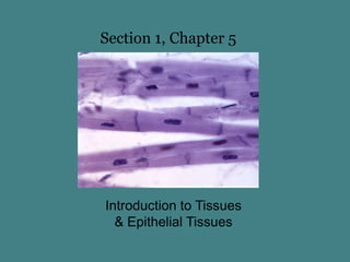
  • 1. Section 1, Chapter 5 Introduction to Tissues & Epithelial Tissues
  • 2. Tissues A Tissue is to a group of similar cells that carries out a specialized function. Histology is the science of tissues. examples of tissues
  • 3. There are four broad categories of tissues Muscular tissue – specialized to contract Nervous tissue – conducts, senses, and stores information Epithelial tissue – forms coverings (skin) & linings (blood vessels) Connective tissues – support, transport, protect
  • 4. Intercellular junctions Cells may be separated by a matrix or connected together by cellular junctions. 1. Tight Junction A tight junction fuses the cell membranes of adjacent cells together so molecules cannot move between the cells. Instead, they must move through the cells. Tight junctions prevent the passage of unwanted & harmful substances Examples: Blood Brain Barrier & Digestive Tract Cell 1 Interlocking membranes form tight junction between two cells Cell 2
  • 5. Cellular Junctions 2. Desmosome desmosomes form “spot welds” between cells Provides structural support Examples of desmosomes are in the epidermis of the skin Cells connected by desmosomes
  • 6. Cellular Junctions 3. Gap Junction Gap Junctions connect cells by protein ion channels that allow for cell-to-cell diffusion of ions Example of gap junctions are within the intercalated discs of cardiac muscle Intercalated discs of cardiac muscle are one type of gap junction.
  • 7. Types of Tissues: Epithelial Tissue Epithelial Tissue: Epithelial tissue lines cavities, such as the digestive & respiratory tracts It also forms coverings, such as the skin Functions include protection, secretion, absorption, excretion
  • 8. Epithelium: structure Epithelium is anchored to a layer of connective tissue, called the basement membrane. Epithelium has a basal surface that faces the basement membrane The free (apical) surface is exposed to the open space (lumen) Open space = lumen Basement membrane
  • 9. Characteristics of Epithelial Tissue Epithelium consists of tightly packed cells They lack a blood supply, instead they receive nutrients by diffusion Epithelium readily divide, so they are continually replaced = rapid healing
  • 10. Classifications of Epithelial Tissues Epithelium are classified by the layer of cells 1. Simple = Single layer of epithelial cells 2. Stratified = two or more layers of cells And they’re classified by the shape of cells 1. squamous = scale-like 2. cuboidal = cube-like shape 3. Columnar = tall cells
  • 11. Simple Squamous Epithelium Single layer of flattened cells Functions include diffusion, filtration, and secretion Locations include: air sacs of lungs (alveoli) lining of capillaries and the walls of blood vessels glomerulus of kidneys. surface view of simple squamous ep. alveoli of lung
  • 12. Simple Cuboidal Epithelium Single layer of cube-shaped cells Functions include secretion and absorption Locations include the lining and the ducts of some glands tubules within kidneys lumen cross-section of tubules in kidneys
  • 13. Simple Columnar Epithelium Single layer of elongated cells and their nuclei appear as a single row Functions include absorption, secretion, or transport Often contain goblet cells Goblet cells secrete mucus onto apical surface may be ciliated, or have microvilli, or have no modifications on apical surface. columnar cells goblet cell microvilli “brush border”
  • 14. Simple Columnar Epithelium Some simple columnar epithelium are covered with cilia on their free surface. Cilia beat constantly in a rhythmic motion to propel substances across the cell surface. Cilia propels the egg through uterine tube
  • 15. Simple Columnar Epithelium Some simple columnar epithelium are covered with microvili on their free surface. Microvilli are extensions of the cell membrane that increases the surface area of a cell. Example of microvilli include the intestinal epithelium, where they increase the amount of contact each cell makes with food. Microvilli greatly enhance absorption of nutrients.
  • 16. Pseudostratified Columnar Epithelium (PSCE) PSCE appear striated, but each cell touches the basement membrane. Usually ciliated Most PSCE contain goblet cells that secrete mucus Locations of PSCE include the lining of the respiratory tract: nasal cavity, pharynx, trachea, bronchi Functions: Mucus from goblet cells traps debris & cilia moves the mucus (and trapped debris) away from the lungs
  • 17. Stratified Squamous Epithelium Several layers of cells = thick tissue Primary function is to protect underlying tissues from abrasion Superficial cells are squamous but the deeper cells are cuboidal or columnar May be keratinized or non-keratinized
  • 18. Keratinized Stratified Squamous Epithelium Keratinized epithelium is found in the epidermis (outer layer) of skin Keratin is an insoluble protein that hardens and forms a protective coating. Cells accumulate keratin over time, so the older outermost cells of the epidermis are keratinized Stratified squamous epithelium of epidermis
  • 19. Non-keratinized Stratified Squamous Epithelium Non-keratinized (moist) stratified squamous epithelium lines the esophagus, oral cavity, vagina, and anal canal. Apical surface Stratified squamous epithelium of vagina
  • 20. Transitional Epithelium Transitional epithelium forms an expandable lining (Function: permit expansion) Locations include the inner layer of the urinary bladder & ureters The lining is several layers thick and the cells appear cuboidal when the urinary bladder is contracted, but only a few layers thick when the urinary bladder is distended and the cells appear squamous. empty urinary bladder distended urinary bladder
  • 21. Glandular Epithelium Glandular epithelium is specialized to produce & secrete substances Glandular epithelium form glands Exocrine Glands Exocrine glands secretes substances through ducts onto an open surface Examples includes goblet cells, sweat glands, and mammary glands Duct of a sweat gland
  • 22. Glandular Epithelium Endocrine Glands Endocrine glands secretes substances (hormones) directly into the blood stream. Examples includes the thyroid gland, pituitary gland, adrenal glands, ect. Follicles of thyroid gland are composed of simple cuboidal epithelium End of Section 1, Chapter 5