Diseases of the ruminant digestive tract
•
1 recomendación•1,002 vistas

The document summarizes the anatomy and functions of the ruminant digestive tract. It describes the four compartments of the ruminant stomach (rumen, reticulum, omasum, and abomasum) and explains their roles in fermenting feed, absorbing nutrients, and breaking down material. Key functions of the rumen like microbial fermentation of fibers into volatile fatty acids and belching of gases are also outlined. Common digestive disorders like bloat and acidosis are briefly discussed.

Recomendados
Más contenido relacionado
La actualidad más candente
La actualidad más candente (20)
Similar a Diseases of the ruminant digestive tract
Similar a Diseases of the ruminant digestive tract (20)
Más de karima Akool AlSalihi
Más de karima Akool AlSalihi (20)
Último
Último (20)
Diseases of the ruminant digestive tract
- 1. Diseases of the Ruminant digestive tract Dr Karima Al Salihi
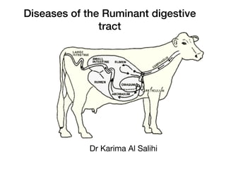
- 2. Anatomy of bovine digestive tract Quick facts • Ruminant stomachs have four compartments: the rumen, the reticulum, the omasum and the abomasum. • Rumen microbes ferment feed and produce volatile fatty acids, which is the cow’s main energy source. • Rumen microbes also produce B vitamins, vitamin K and amino acids.
- 3. The digestive tract of the adult cow The cow's digestive tract consists of the following. • Mouth • Esophagus • A four-compartment stomach, which includes ◦ The rumen (paunch) ◦ The reticulum (“honeycomb”) ◦ The omasum (“manyplies”) ◦ The abomasum (“true stomach”) • Small intestine • Large intestine
- 4. Stomach comportents 1. The rumen The rumen (on the left side of the animal) is the largest stomach compartment and consists of several sacs. It can hold 25 gallons or more of material depending on the size of the cow. Because of its size, the rumen acts as a storage or holding vat for feed. Aside from storage, the rumen is also a fermentation vat. The rumen’s environment favors the growth of microbes. These microbes digest or ferment feed within the rumen and make volatile fatty acids (VFAs). The rumen absorbs most of the VFAs from fermentation. A good blood supply to the rumen walls improves absorption of VFAs and other digestion products. Tiny projections (papillae) line the rumen, which increases the rumen’s surface area and the amount it can absorb.
- 5. 2. The reticulum The reticulum is a pouch-like structure in the forward area of the body, close to the heart. The tissues in the reticulum form a network similar to a honeycomb. A small tissue fold lies between the reticulum and rumen, but the two aren’t separate compartments. Together they’re called the rumino-reticulum. Heavy or dense feed and metal objects eaten by the cow drop into this compartment. Nails and other sharp objects may work into the tissue and cause “hardware disease.” You can use magnets to prevent disease or correct the problem through surgery. Leaving it untreated may lead to infection and possibly death. 3. The omasum The omasum is a globe-shaped structure containing leaves of tissue (like pages in a book). It absorbs water and other substances from digestive contents. Feed material (ingesta) between the leaves will be drier than ingesta found in the other compartments. 4. The abomasum The abomasum is the only compartment lined with glands. These glands release hydrochloric acid and digestive enzymes, needed to breakdown feeds. The abomasum is similar to a nonruminant stomach.
- 6. The small intestine • The small intestine consists of three sections: the duodenum, jejunum and ileum. • It measures about 20 times the length of the animal. • Secretions from the pancreas and gallbladder aid in digestion within the small intestine. • The small intestine completes most of the digestive process and absorbs many nutrients through villi (small finger-like projections). • From the villi the nutrients enter into the blood and lymphatic systems.
- 7. Cecum The cecum is the large area where the small and large intestine meet. The cecum breaks down some previously undigested fiber, but the exact importance of the cecum remains unknown.
- 8. Large intestine The large intestine is the last section of the tract that undigested feedstuffs pass through. Microbes digest some undigested feed here, but the main digestive function of the large intestine is to absorb water.
- 9. Digestive tract functions 1. Belching ( eructation) Digestion produces 30 to 50 quarts of gas per hour in the rumen. Carbon dioxide and methane are the main gases present. Cows must release this gas to avoid bloating. Under normal conditions, swelling from gas formation causes the cow to belch and release the gas. 2. Rumination Cows may spend 35 to 40 percent of each day ruminating (cud chewing). The amount of time spent ruminating depends on the diet. Little ruminating occurs when cows eat grain or finely ground rations. But when eating long hay, cows may ruminate for several hours. Mature cattle spend little time chewing while eating feed. Thus, during rest periods, cows regurgitate (bring up) soft feed wads (cud) to re-chew and break into smaller pieces. They also re- salivate the feed and re-swallow it. This process makes the feed easier for the microbes to digest.
- 10. 3. Feed movement in the rumen The rumen contracts and moves continuously. Healthy cows have one to two rumen contractions each minute. Poor rumen movement may indicate a sick animal. When the rumen contracts it • Mixes contents • Brings microbes in contact with new feedstuffs • Reduces flotation of solids • Moves materials out of the rumen
- 11. 4. Saliva production A cow’s salivary glands can make and add 50 to 80 quarts of saliva to the rumen daily. Saliva has several functions in cows. • It provides liquid for the microbes. • It recirculates nitrogen and minerals. • It buffers the rumen. ◦ Saliva keeps the rumen pH between 6.2 and 6.8 for best digestion of forage and feedstuffs.
- 12. 5. VomitingCattle rarely vomit. Sometimes certain feeds will induce vomiting. A few pasture plants, usually weeds, contain compounds called alkaloids that can cause vomiting. Work with a veterinarian if this problem continues.
- 13. 6. Energy feed digestion in the rumen Rumen microbes digest simple and complex carbohydrates (fiber) and convert them into VFAs. VFAs mainly consist of acetic, propionic and butyric acids and provide 50 to 70 percent of the cow’s energy.
- 14. Diet can affect the amounts of each VFA microbes produce. • High forage diets result in more acetic acid forming (60 to 70 percent of total) than propionic (15 to 20 percent) and butyric acids (5 to 15 percent). • More grain or finely ground forages can cause the amount of acetic acid to decline to 40 percent, while the amount of propionic acid may increase to 40 percent. Such changes in VFA production usually relate to a reduction in milk fat test. Microbes digest about 30 to 50 percent of the fiber units, cellulose and hemicellulose, in the rumen. Sixty percent or more of the starch is degraded depending on the amount fed and how fast ingested materials move through the rumen. Most sugars get completely digested within the rumen.
- 15. VFAs absorb into the bloodstream from the rumen and move to the body tissues including the udder. Once in the tissues, the cow uses VFAs as a source of energy for • Maintenance • Growth • Reproduction • Milk production
- 16. Protein and nonprotein nitrogen use in the rumen Not all consumed proteins get broken down in the rumen, (see figure 2). Through fermentation, protein is converted to ammonia, organic acids, amino acids and other products. About 40 to 75 percent of the natural protein in feed gets broken down.
- 18. The amount of breakdown depends on many factors including: • The ability of the protein to dissolve • How resistant the protein is to breakdown • How fast the feed passes through the rumen Aside from protein breakdown, nonprotein nitrogen (NPN) sources also provide ammonia. NPN sources include urea, ammonium salts, nitrates and other compounds. Many rumen microbes need ammonia to grow and build protein. Rumen microbes convert ammonia and organic acids into amino acids to use for building protein. Most of the extra ammonia absorbs into the bloodstream from the rumen. But small amounts may pass into the lower digestive tract and absorb there. Feed protein (not broken down in the rumen) and microbial protein pass to the abomasum and small intestine for digestion and absorption.
- 19. Vitamin Production Rumen microbes make vitamin K and all B vitamins. Microbes make enough of these vitamins for cattle growth and maintenance. Thus, under most conditions, cattle with healthy rumens don’t need added B vitamins or vitamin K in their diet. Cows under stress may need added niacin (B3) and thiamine (B1).
- 20. Fat digestion Most fat digestion and absorption occurs in the small intestine. Cows absorb more saturated fat than simple-stomach animals. This is because rumen microbes change unsaturated fatty acids to saturated fatty acids by adding hydrogen molecules. Feeding a lot of unsaturated fatty acids can be toxic to rumen bacteria, slow fiber digestion, and lower rumen pH.
- 21. Calf digestive system The rumen, reticulum and omasum remain undeveloped at birth and during the first few weeks of life. The calf’s largest stomach compartment is the abomasum. At this stage of life, the rumen doesn’t function and thus some feeds that mature cows can digest, calves can not.
- 22. Esopageal groove During nursing or bucket feeding milk bypasses the rumen and directly enters the abomasum via the esophageal groove. Reflex action (e.g. when the calf nurses) closes the groove to form a tube-like structure. This prevents milk or milk replacer from entering the rumen. If the calf drinks milk rapidly, some may overflow into the rumen.
- 23. Rumen development The rumen will remain undeveloped as long as the calf stays on milk. Once the calf begins eating grain and forage, a microbial population will develop in the rumen and reticulum. The end products from microbial fermentation are responsible for developing the rumen. Calves don’t need cud inoculation to start rumen development. Feeding grain with or without forage during the first few weeks of life will result in larger and heavier papillae growth in the rumen. Thus, the rumen will begin functioning like the adult’s when the calf is three months old.
- 24. Diseases of the Digestive System 1. Bloat ( Tympany). Bloat is the accumulation of free gas or froth in the rumen. Bloat is less common in small ruminants than in cattle, and it occurs less frequently in goats than in sheep. Bloat can occur from ingesting diets such as high-legume diets or cereal grains that promote the formation of froth in the rumen. Some diets, again such as those high in grain, promote the formation of free gas. Sometimes an animal will have an accumulation of free gas in the rumen because of a failure to eructate. CLINICAL SIGNS • Abdominal distension, especially apparent in the left paralumbar fossa • Signs of abdominal pain • Anxiety • Respiratory distress DIAGNOSIS • Observation of obvious abdominal distension • In the case of free gas bloat, a “ping” can be heard in the left paralumbar fossa • If an orogastric tube is passed, free gas can be heard (and smelled) escaping through the tube. Frothy bloat will result in froth being seen on the tube. TREATMENT • Bloat is an emergency situation and requires immediate attention • Passage of orogastric tube • Diocctyl sodium succinate (DSS), mineral oil, vegetable oil for frothy bloat • Trocarization of the rumen • Rumenotomy
- 25. RUMEN ACIDOSIS Rumen acidosis is caused by the rapid fermentation of highly digestible carbohydrates. The problem occurs more often with finely ground grains because the bacteria can more rapidly ferment the carbohydrate. The condition is more commonly seen in animals that have been fed a primarily forage-based diet, and then have received a large amount of concentrate feed. As the carbohydrates are digested, the pH declines. The decline in pH causes the normal resident microfauna and flora of the rumen to die. Water is drawn into the rumen, and the animal becomes dehydrated and may die. The acid buildup causes damage to the rumen epithelium. This allows the leakage of bacteria into the system, and the animal may become septic. CLINICAL SIGNS • Vary with the amount and type of feed • Signs first appear from 12 to 36 hours after ingestion of feed • Signs include shock, anorexia, weakness, depression • Severe dehydration • Signs of sepsis • Diarrhea DIAGNOSIS • Examination of rumen fluid shows the following: pH < 5.5 Protozoa reduced in number Large, Gram-positive rods TREATMENT • Treatment is aimed at reversing hypovolemia and shock, removing offending feedstuffs, and correcting the acidosis and sepsis • Intravenous (IV) fluids containing sodium bicarbonate • Flunixin meglumine • Rumenotomy to remove feedstuffs • Transfaunation of rumen microflora • Thiamine subcutaneously (SQ)
- 26. DIARRHEA Diarrhea involves loose to runny feces, and it is defined as the increase in the volume and frequency of defecation. Dehydration and electrolyte imbalances are the concerning sequelae to diarrhea, especially in very young kids and lambs. Animals of certain ages are more prone to specific infectious causes of diarrhea
- 28. Enterotoxigenic Escherichia coli Enterotoxigenic E. coli causes diarrhea mainly in neonatal lambs and, to a lesser extent, in kids. This type of bacteria has two main methods by which it causes disease. The first is by attachment and colonization of the intestinal villi. As these villi are destroyed, the intestine loses its absorptive capabilities. The second method is by production of an enterotoxin, which interferes with normal gut function. E. coli causes disease in young animals younger than 10 days, with 1 to 4 days of age being the most common age of onset. CLINICAL SIGNS • Usually presents as an outbreak, rather than in a single animal • Diarrhea • Dehydration • Recumbency • Many animals die before development of diarrhea DIAGNOSIS • Fecal culture and serotyping for K99 and F41 antigens • Histopathology from necropsy confirms TREATMENT • Fluid therapy: PO, IV, SQ • Antibiotics (amoxicillin, ampicillin, neomycin, trimethoprim/sulfa [TMS]) may be beneficial, but may also kill off beneficial gut flora • Flunixin meglumine