Esthetics with Veneers: A Review of Indications, Techniques and Maintenance
•Descargar como PPTX, PDF•
22 recomendaciones•4,767 vistas

The document provides an overview of esthetics with veneers. It discusses the definitions, history, indications and contraindications of veneers. It describes the processes of shade selection, tooth preparation including principles, rationale and types of preparation. It also discusses provisional restorations, cementation, maintenance and failures of veneers. Recent advancements discussed include feldspathic, lithium disilicate and minimally invasive veneers. In conclusion, veneers are a conservative treatment for improving aesthetics when done according to principles of preparation, cementation and maintenance.

Recomendados
Más contenido relacionado
La actualidad más candente
La actualidad más candente (20)
Similar a Esthetics with Veneers: A Review of Indications, Techniques and Maintenance
Similar a Esthetics with Veneers: A Review of Indications, Techniques and Maintenance (20)
Más de Naveed AnJum
Más de Naveed AnJum (11)
Último
Último (20)
Esthetics with Veneers: A Review of Indications, Techniques and Maintenance
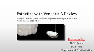
- 1. Esthetics with Veneers: A Review Presented by: Nabid Anjum PG IInd year Department of Prosthodontics Sowmya S, Sunitha S, Dhakshaini M R, Raghavendraswamy K N. Int J Dent Health Concern 2015;1:1-5.
- 2. CONTENTS • Introduction • Definitions • History • Indications and Contraindications • Shade selection • Tooth Preparation – Principles of tooth preparation – Rationale – Types of preparation – Armamentarium – Procedure
- 3. • Provisional Restoration • Cementation of Veneers • Maintenance • Failures of Restoration • Recent Advancements • Conclusion • References
- 4. INTRODUCTION • The oral region is a dynamic part of the face, with tooth and gingival display during functional lip movements creating an expression of aesthetics that is unique to an individual. • Esthetic dentistry is the fourth dimension in addition to other factors like biological, physiological and mechanical factors, all of which are to be combined for a successful result. • Since, esthetic dentistry has become an integral part of everyday practice in dental clinic. So with increasing patient demand, it has also become a challenging job for our profession. • Based on their strength, longevity, conservative nature, biocompatibility and aesthetics, veneers have been considered one of the most viable treatment modalities.
- 5. DEFINITIONS • Veneer: A thin sheet of material usually used as a finish. (GPT, 9th Edition) A veneer is a layer of tooth colored material that is applied to a tooth to restore localized or generalized defects and intrinsic discolorations. ( Sturdevant Art & Science of Dentistry ) • Laminate Veneer Restorations: A conservative esthetic restoration of anterior teeth to mask discoloration, restore malformed teeth, close diastemas & correct minor tooth alignment. (Mosby’s dental dictionary)
- 6. HISTORY • Dr. Charles L Pincus introduced the concept of veneering anterior teeth with laminates when approached by Hollywood directors in 1928. (HOLLYWOOD’S DENTIST) • Buonocore’s - Acid etching technique in 1955’s (increasing adhesion to enamel surface.) • Due to increasing aesthetic demand and the possibility of joining laminates to the tooth structure (particularly enamel), a new concept was introduced: minimally invasive restorative dentistry, which causes little damage to dental structures. • In this context, laminate veneer, also known as contact lenses, emerged. Shirley Temple, age 8, went before the cameras — no veneers. She lost her baby teeth, just like any child, but was never photographed with any teeth missing. Dr. Pincus placed "Hollywood Veneers" on Shirley's front teeth. They were only temporary, and had to be removed daily when eating, chewing or sleeping
- 7. INDICATIONS • Improve extreme discolorations such as tetracycline staining, fluorosis, devitalized teeth, and teeth darkened from age. • Repair chipped or fractured teeth.
- 8. • Closing of diastemas between teeth. • Ability to lengthen anterior teeth. • Improve the appearance of rotated or misaligned teeth. • Poor restorations.• Enamel defects
- 9. CONTRAINDICATIONS • If little or no enamel is present, full crown should be considered. • Bruxing or Clenching, or other para-functional habits • Severe crowding/Endodontically treated tooth • Poor oral hygiene • High caries rate • Certain types of occlusal problems such as Class III & end-to-end bites.
- 10. What are the clinical considerations ? • Esthetics and function • Anterior guidance and occlusal harmony • Physiologic contours • Emergence Profile
- 11. SHADE SELECTION: • The next important clinical parameter for the long-term success of veneer is shade selection procedure. • Based on the available literatures, a myriad of factors were available that influences the assessment of color of restoration. The factors under consideration are: Shade and optical properties of tooth laminate characteristics Dental shade matching devices Influence of polymerization Shade and thickness of resin cement
- 12. TYPES OF VENEER: Directly fabricated veneers: Composite Veneers Indirectly fabricated veneers Preformed laminates Laboratory fabricated veneers -Acrylic resin veneers -Microfill resin veneers -Porcelain veneers
- 13. TOOTH PREPARATION • Principles of tooth Preparation – • Rationale: Enamel preparation is done: i. To provide adequate space for porcelain opaquing and composite resin luting materials. ii. To remove convexities in the surface and provide a definitive path for insertion. iii. To assist veneer seating during placement and bonding the laminate. iv. To facilitate margin placement v. To provide adequate contour and colour without over contouring
- 14. • PROCEDURE: It involves the following steps: – Labial Reduction – Proximal reduction – Sulcular Extension – Incisal Reduction – Lingual Reduction LVS no. 1 – 0.5 mm reduction LVS no. 2 – 0.3 mm reduction
- 15. LABIAL REDUCTION • The thickness of the ceramic laminate should be 0.5 mm. • To achieve this, the labial preparation should achieve a uniform reduction of 0.3-0.5 mm, less gingivally and more incisally. • This involves:- Depth Cuts & Reducting Remaining Enamel Gently draw the diamond across the labial surface of the tooth from mesial to distal side.
- 16. PROXIMAL REDUCTION • Depth can often be as 0.8- 1 mm, since the enamel layer is thick towards proximal surface. • Done with round end tapered diamond, just continued into the proximal area (halfway). • It is ensured that the diamond is parallel with the long axis of the tooth. • Proximal reduction should stop just short of breaking the contact • Margin should be hidden within the embrasure area.
- 17. • Reasons to break contacts: present of pre-existing restoration diastema closure (will extend lingually) For proper contour If dentin exposure occurs at the periphery, such as the cervical region, it is advisable to prepare a little deeper into this area: • Use a layer of GIC can be used as a base. • The GIC will bond to dentin, and seal it as opposed to a dentin bonding agent, which may only adhere but not seal effectively. • Reasons to not break contacts: Simplifies try in ,no need to adjust contact Simplifies bonding and finishing Improve retention Improve aesthetics
- 18. SULCULAR EXTENSION AND MARGIN PLACEMENT • Routinely the margins are placed supragingivally. • When discoloration is excessive, the margins are extended subgingivally. • A rounded 0.3mm chamfer serves as an ideal margin for ceramic laminate veneer. ADVANTAGESOFSUPRAGINGIVALMARGIN
- 19. Conservative , Distinct. Provides increased bulk of porcelain giving adequate strength, avoids over contouring. Good marginal seal. Accuracy of fit – veneers is easily inserted at try-in and final placement Advantages of chamfer finish line
- 20. • WINDOW OR INTRA ENAMEL PREPARATION LABIALLY • FEATHERED INCISAL EDGE PREPARATION LABIALLY • INCISAL BEVEL PREPARATION • OVERLAPPED INCISAL EDGE PREPARATION INCISAL PREPARATION
- 22. LINGUAL REDUCTION • Any reduction of the incisal edge would necessitate some lingual enamel modification so that there is no butt joint at this incisal/lingual junction but rather a rounded chamfer. This modification will help to prevent the porcelain from shearing away from the incisal edge during function. • The round end tapered diamond is held parallel to the lingual surface with its end forming a slight chamfer 0.5 mm deep. • The lingual extension will also enhance the retention and increase the surface areas for bonding.
- 23. IMPRESSION MAKING GINGIVAL RETRACTION: If possible, retraction cord should be left during impression. IMPRESION MAKING: Usually 1 step procedure is preferred • Materials (light and heavy body) • Trays IMPRESSION MAKING : • Actual impression material can vary from polysulfide to polyether, but the vinyl polysiloxane injection method is the cleanest and easiest. Also because multiple pours are required for laboratory procedure.
- 24. PROVISIONAL RESTORATION • Provisional restoration for laminates may not be essential as there is no exposure of dentine (no sensitivity) and the proximal contacts are maintained (no drifting of adjacent teeth). • But most often it may be necessary for a patient to maintain their social engagements and if proximal contact is broken (wrap-around technique) Two Methods:- Direct Method (intraorally) Indirect Method (extraoral)
- 26. INDIRECT COMPOSITE RESIN/ACRYLIC RESIN VENEER
- 27. Laboratory procedure • Platinum foil backing • Refractory models • Direct castings • CAD –CAM machining
- 28. Platinum foil backing : • Thin layer of platinum foil is placed on the die .The porcelain is layered on the foil. Then the porcelain foil combination is removed from the die and fired in an oven . Before try-in ,the foil is removed, and the porcelain is etched .
- 29. Direct castings : • cast ceramic restorations are fabricated using the ‘lost wax’ technique. This eliminates the need for multiple firings but requires extrinsic staining for coloration. CAD/CAM Machining : • A model or video image of the preparation is required, and the restoration always requires modification of the surface porcelain to obtain proper esthetics.
- 30. CEMENTATION OF VENEERS • Second appointment : Remove temporary Evaluate fit and esthetics All veneers should be placed without bonding medium on teeth to assess the fit. Preparation of veneer: • Following cleaning of the veneer with a solvent such as acetone, it is etched with 10-15% hydrofluoric acid for 30 seconds to 1 minute according to the manufacturer’s instructions and the ceramic used. • A silane coupling agent is now applied to the fitting surface of the veneer and is allowed to remain for one minute. • It is then air-dried. The silane creates a chemical bond between composite cement and ceramic.
- 31. Aka Chemical coupling Agents used in silanization:- • 3-methacryloyloxypropyl-trimethoxysilane • Butylacrylate –acrylic acid copolymer Bond of the porcelain laminate to the tooth Silane greatly enhances the adhesive properties of the resin and thus increases bond strength. A salty-looking appearance should be observed. Once the silane is dried out, the choice of adhesive is applied over the whole interior surface.
- 32. Preparation of teeth: The teeth should definitely be kept clean and purified of blood, saliva or oral contaminants. • 37% phosphoric acid is applied on the prepared area. • It will create micromechanical porosities on the enamel. Coat the etched tooth surface with bonding agent of the light activated type, which is gently air dispersed into a thin, even layer. Light cure this evenly dispersed layer to seal the tooth surface
- 33. • Fill the laminate with the selected composite resin luting agent. • After 3-5 seconds of light curing, the excess luting resin that comes out of the margins has a jelly consistency and can be easily cleaned with an explorer. Place the laminate in position on the tooth rotating it about the incisal edge and toward the gingiva. Ensure that excess luting material extrudes from all peripheral aspects.
- 34. Use carbide finishing bur to remove excess cement. Use the LVS no. 8 bur to remove composite resin along the incisal margin. Clear the contacts with a extra fine metal strip to ensure they are free Polish the porcelain interface with diamond polishing paste. Wash and dry. Post operative viewCheck interproximal areas for clearance with dental floss
- 35. MAINTENANCE OF VENEERS • For 72-96 hours following insertion, patients should avoid highly coloured foods, tea or coffee, hard food and extreme temperatures. Routine scaling should be done, and ultrasonic scalers should be avoided. • Abrasive and highly fluoridated toothpastes should be avoided. • Excessive biting forces and nail biting and pencil chewing habits should be avoided. Soft acrylic mouth guard can be used during contact sports.
- 36. FAILURES OF RESTORATION • The survival probability of porcelain veneers according to the Kaplan - Meier survival estimation method was 97% at 5 years and 91% at 10½ years • Three Types: Mechanical : Poor positioning of incisal margin: less incisal thickness, margin too subgingival. Debonding: Use of expired cement Faulty veneer/tooth preparation during luting
- 37. Biological : Postoperative sensitivity Secondary caries Improper curing of cement, poor marginal adaptation. Marginal Microleakage – poor fit and extension. Aesthetic : Improper shade selection Gingival recession – over contour and improper subgingival placement
- 38. RECENT ADVANCEMENTS • The recent years there have been various advancements in dental laminates and veneers with the aim of overcoming the previous shortcomings and for a more conservative feasible approach. • The recent advancements are: Feldspathic teeth veneers Lithium disilicate teeth veneers Minimally invasive veneers or no prep veneers Zirconia Veneers (Prettau anterior, Zirkonzahn)
- 39. • These veneers contain many stacks of porcelain giving rise to multiple layers in the veneer. • Feldspars are naturally occurring aluminium silicate containing sodium or potassium. The feldspars contain fluoroapatite crystals improving the optical appearance of the tooth. • It has a polychromatic appearance and high translucency, hence closely resembles the natural tooth. Hence, it is of great esthetic value. • It is the highest quality cosmetic veneers. • A major concern with feldspathic porcelain veneers, however, was their strength, which was only approximately 70 MPa to 90 MPa. FELDSPATHIC VENEERS
- 40. LITHIUM DISILICATE VENEERS • They are the most widely used true glass ceramics. It is versatile and is stronger than other porcelain veneers . • It has high resistance to thermal shock thus managing the problem between two similar materials. • It is used for teeth which requires minimal reshaping. It can be used to correct the shape of a malformed tooth. • They can be conventionally cemented or adhesively bonded. • IPS Emax (Ivoclair vivadent) is an example of these veneers.
- 41. MINIMALLY INVASIVE / NO PREP VENEERS These veneers are ultrathin having a thickness similar to contact lenses of about 0.3-0.5 mm and hence get are called "contact lenses of teeth". These help in greatly conserving the tooth structure as previously used porcelain veneers needed a mandatory 0.5mm to 1 mm removal of tooth structure so that the thin layer of porcelain does not fracture. They consist of Lumineers, Durathin veneers and Vivaneers.
- 42. Lumineers: • They are exceptionally thin veneers (0.3mm) made of a special cerinate porcelain. • Cerinate is material made of feldspathic porcelain reinforced with leucite crystals. • They have high strength and resilience despite being exceptionally thin. • They can be showed according to the patients wishes and can be placed with minimal visits to the dentist. They can be placed within two visits to the dentist. Lumineers are a reversible procedure. • However the disadvantage of lumineers is that they have an opaque appearance interfering with the aesthetics of the patient.
- 43. Although Lumineers are most advantageous option, there are certain limitations to be considered: Lumineers can only be placed on teeth that are in good structural condition. The patient must have good oral hygiene, with no receding gums or signs of gum disease. Bleeding of the gums will interfere with the bonding process. Because there is very little or no tooth preparation, a small bump is likely to develop between the veneers and the gum.. The bump may create an irritation to the gum, and may increase the chances for staining and tooth decay.
- 44. The LUMINEERS Minimal Contouring Technique • It requires slight modification of the enamel but never touches dentin during LUMINEERS placement. Only 0.3 mm-0.5 mm enamel is removed, causing no sensitivity for the patient and therefore no need for any anesthesia. Add 5 coats of Tenure® A+B. Add 1 coat of Tenure S to the teeth. Note: Tooth surfaces must be shiny.
- 46. Componeers: • Pre-fabricated nano-hybrid composite enamel-shells • Attractive teeth and a new smile after only one visit • Very little removal of healthy tooth structure – 0.3mm • Individual, customized shaping of the front teeth. • Shine can be refreshed by polishing at any time • Unlike porcelain veneers, they can be easily repaired. Modeling MB5
- 47. Edelweiss Veneer System: • For the first time in the history of dental, it is now possible to work with prefabricated veneers made from nano-hybrid composite using modern laser technology.
- 48. • Low shrinkage due to nano-technology and high amount of filler-83 % • Good abrasion resistance • Very good physical and mechanical properties • Antibacterial surface due to zinc and fluorine particles in the filler • Easy polishing • Natural fluorescence and opalescence
- 50. CRITICAL ANALYSIS • Types of veneers were not highlighted. • Different types of tooth preparation for veneer have not been described. • There were no post operative photographs. • Advancements in veneers were not discussed.
- 51. CONCLUSION • Esthetic procedures have the ability to alter the entire appearance of the patient by providing them with a beautiful smile. The patient gains not only a positively improved appearance, but also a potential moral “boost” that acts positively on their mental health and self-esteem. • There are several types of veneers used commonly in practice today. Fired or pressed ceramic veneers are the most popular. Thin ceramic veneers bonded to acid-etched enamel have been suggested as the most acceptable, predictable type of veneer.
- 52. REFERENCES • Christensen, GJ. What is veneer? Resolving the confusion. JADA 2004:135;11,1574–76 • Lim CC. Case selection for porcelain veneers. Quintessence Int 1995;26:311-5. • Clyde JS, Gilmour A. Porcelain veneers: A preliminary review. Br Dent J 1988;164:9-14. • P. A. Brunton, A. Aminian, and N. H. F. Wilson. Tooth preparation techniques for porcelain laminate veneers B D J 2000;189:5 • Malone WF, Tylman SD, Koth DL. Tylman’s Theory and Practice of Fixed Prosthodontics. 8th ed. St Louis: Ishiyaku Euro-America; 1989
Notas del editor
- dental veneers are custom shells made from tooth colored materials that facilitate covering the front surface of the tooth and these are alternately known as dental laminates. Dental appearance has been judged to be an important indicator when assessing facial attractiveness with physical beauty being a significant factor in a person’s well-being.
- WE should have a proper knowledge regarding the emergence profile in a patients, the gingival contours and the anatomic variations of teeth. The esthetics should be evaluated with keeping in mind the variations in smile lines .the contours of the teeth should be maintained. The shade selection should be proper. Also adequate overbite and overjet should be kept in mind.any parafunctional habits if there should be considered,the smile architecture shouldbe kept in mind . High lip line are less favoured, a diagnosic mop up can also be done.
- early hours of appointment to avoid color fatigue, Clean the teeth and remove all stains and debris • Have patient’s mouth at dentist’s eye level , Use canine as reference , If there is confusion between two shades then it is always better to select a shade of lower chroma and higher value.
- Hence, preparation is needed mainly to • Get definite finish line • Provide space • Get fluoride-rich layer • Rough surface for better retention
- The facial surface should be reduced in two planes; one nearly parallel with the path of insertion, and one parallel with the incisal two- thirds of the facial surface of the tooth
- For the standard preparation, chamfer is placed at the height of gingival crest unless severe discoloration mandates a subgingival margin to gain extra veneer thickness. More success rate was seen with supragingival finish line because it: • Increases the area of enamel • Moisture control is better • Visual confirmation is excellent • Accessibility is good • Maintenance of hygiene is better
- One of the ways of protecting the proximal surface of the adjacent tooth is to place a metal matrix band in between. However, sometimes, poor placement of this metal band may injure the papilla.
- In the opinion of some authors incisal coverage in necessary in all cases to enhance the mechanical resistance of veneer, even though this involve the removal of 0.5-2.0 mm of intact incisal edge and may place the vulnerable cavosurface margin in an area of opposing tooth contact. Also, it was found that the window type of preparation was strongest compared with an overlapping and feathered design. Never end incisal edge where excursive movements of the mandible will cause shearing stresses across the junction of porcelain laminates and tooth.
- A single cord is used which remains in place when impression is being made and no extra hemostatic agent in the cord is needed because bleeding should be minimal with healthy gingivae.
- Materials used are tooth coloured acrylics and resin composites as in routine fixed prosthodontics. They are cemented with either a flowable luting resin or eugenol free cement.
- Each of the veneers is tried in individually beginning with the distal-most veneer, with the margins checked carefully. After ascertaining individual fit- place each laminate on one by one, until all are in place. Then check the collective fit and relationship of one laminate to another, especially in the contact areas.
- At this stage, the adhesive should not be light cured. As soon as the bonding is applied, the transparent composite luting agent is placed inside the veneer.
- The ceramic veneer bonded to tooth with composite resin cements produces two bonded interfaces. One between ceramic – composite resin cement and other between the tooth- composite resin interface. ... The light activated composite resin luting cement is preferred due to its longer working time and better colour stability. igh compressive and tensile srength – Ability to tint, opaque and characterize – Low viscosity – Low polymerization shrinkage – Good colour stability
- HOWEVER IN RECENT YEARS ZIRCONIA CERAMICS HAVE UNDERGONE MANY CHAGES IN ITS MICROSSTRUCTURE AND COMPOSITION TO INCREASE THEIR TRANSLUCENCY WITHOUT LOSING THEIR FRACTURE RESISTANCE. ITS MAIN DIFFICULTY ARE IN SITUATION OF LITTLE MECHANICAL RETENTION OF PREPARATION SINCE ZIRCONIA IS CHEMICALLY INERT AND CANNOT BE ETCHED BY HYDROFLUORIC ACID WHICH IMPLIES A LESS EFFECTIVE ADHESION COMPARED TO CERAMICS
- Feldspars are primarily composed of silicon oxide (60%–64%) and aluminum oxide (20%–23%), and are typically modified in different ways to create glass that can then be used in dental restorations.they are not strong due to their low mechanical properties as the flexural strength is from 60-70 MPA. With this material, it is possible to have a thickness of less than 0.5 mm, with or without preparation in the enamel. To preserve the health of the gingival tissues and prevent overcontouring, a slight 0.5 mm reduction of tooth surface is found to work best
- These veneers are exceptionally thin and are about 0.2 mm whereas the traditional veneers are usually about 0.5 mm thick. These veneers have gained popularity due to its good esthetic effects as it gives a natural translucency to the teeth closely resembling natural teeth. This is one of the advantages that durathinveneers have over lumineers as lumineers have an opaque appearance thus failing to give a natural effect
- The main difference is that Lumineers are made from a special patented cerinate porcelain that is very strong but much thinner than traditional laboratory-fabricated veneers. Their thickness is comparable to contact lenses. Therefore, anesthesia and temporaries are also not required.
- 1) The teeth must be free of decay. Any existing fillings must also be in good condition, along with the surrounding gum in the area where the Lumineers will be placed..
- Add an even layer of Ultra-Bond® Plus resin cement to the inner side of the LUMINEERS. Remove more excess cement with a probe. Light-cure each tooth for 3 seconds through the tray.