
Recomendados
Más contenido relacionado
La actualidad más candente
La actualidad más candente (20)
Similar a Brain and Spinal Cord
Similar a Brain and Spinal Cord (20)
Más de Dr. Neeta Gupta
Más de Dr. Neeta Gupta (20)
Último
Último (20)
Brain and Spinal Cord
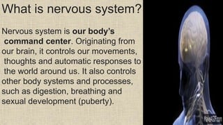
- 1. What is nervous system? Nervous system is our body’s command center. Originating from our brain, it controls our movements, thoughts and automatic responses to the world around us. It also controls other body systems and processes, such as digestion, breathing and sexual development (puberty).
- 2. Functionsof Nervous system: The 4 main functionsof the nervous system are: 1.Reception of general sensoryinformation (touch, pressure,temperature, pain, vibration) 2.Receiving and perceiving special sensations (taste, smell, vision, sounds) 3.Integration of sensoryinformation from different parts of the body and processing them 4.Responsegeneration
- 3. These major functions can again be dividedintotwo major categories. 1.Somatic functions,whichpeoplecan control voluntarily,such as blinking of the eyelids 2.Autonomicfunctions,whichpeoplecannotcontrol,such as the beating of the heart
- 4. Classification of Nervous System:
- 5. The brain is like a committee of experts. All the parts of the brain work together, but each part has its own special properties. The brain can be divided into three basic units: the forebrain, the midbrain, and the hindbrain.
- 6. Forebrain(Telencephalon): also called prosencephalon, it includes the telencephalon, which contains the cerebral hemispheres, and, under these, the diencephalon, which contains the thalamus, hypothalamus, epithalamus, and subthalamus. The forebrain is the largest and most highly developed part of the human brain: it consists primarily of the cerebrum. The cerebrum is split into two halves (hemispheres) by a deep fissure. Despite the split, the two cerebral hemispheres communicate with each other through a thick tract of nerve fibers that lies at the base of this fissure.
- 7. Although the two hemispheres seem to be mirror images of each other, they are different. For instance, the ability to form words seems to lie primarily in the left hemisphere, while the right hemisphere seems to control many abstract reasoning skills. the right cerebral hemisphere primarily controls the left side of the body and the left hemisphere primarily controls the right side. When one side of the brain is damaged, the opposite side of the body is affected. For example, a stroke in the right hemisphere of the brain can leave the left arm and leg paralyzed.
- 8. Cerebral cortex Cerebral cortex is the outer Most layer of brain. Its surface has many folds, giving it a wrinkled appearance. The folds consist of many deep grooves called sulci and raised areas called gyri.
- 9. Cortex is divided into four lobes: frontal, parietal, temporal and occipital. Each of these lobes is responsible for processing different types of information. Collectively, your cerebral cortex is responsible for the higher-level processes of the human brain, including language, memory, reasoning, thought, learning, decision-making, emotion, intelligence and personality.
- 10. Cerebral cortex and the cerebrum Cerebral cortex is the outer layer that lies on top of cerebrum. Cerebrum is the largest area of brain. Cerebrum divides brain into two halves called hemispheres. The hemispheres are attached by a bundle of nerve fibers called the corpus callosum. The corpus callosum allows two hemispheres to communicate with each other.
- 11. Diencephalon: Thalamus The thalamus is a small structure, located right above the brainstem responsible for relaying sensory information from the sense organs. It is responsible for transmitting motor information for movement and coordination. It is found in the limbic system within the cerebrum. This limbic system is mainly responsible for the formation of new memories and storing past experiences. Hypothalamus: The hypothalamus is a small and essential part of the brain, located precisely below the thalamus. controls appetite, peristalsis, the rate of heartbeat, and blood pressure and forms an axis with the pituitary gland which is the main link between the nervous and the endocrine systems.
- 12. Midbrain, also called mesencephalon, region of the developing vertebrate brain that is composed of the tectum and tegmentum. The midbrain serves important functions in motor movement, particularly movements of the eye, and in auditory and visual processing. a.The tectum (from Latin for “roof”) makes up the rear portion of the midbrain. Tectum controls eye movements, approach, and avoidance movements and is formed by two paired rounded swellings, the superior and Inferior colliculi.
- 13. The superior colliculus (SC) is a multisensory midbrain structure that integrates visual, auditory, and somatosensory spatial information to initiate orienting movements of the eyes and head toward salient objects in space. The superior colliculus receives input from the retina and the visual cortex and participates in a variety of visual reflexes, particularly the tracking of objects in the visual field.
- 14. The inferior colliculus is a part of the midbrain that serves as a main auditory(sound) center for the body. Its primary roles are signal integration, frequency recognition, and pitch discrimination. Bilateral damage to the inferior colliculi results in deafness, whereas unilateral damage may result in the inability to localize sound. The tegmentum is located in front of the tectum. It consists of fibre tracts and three regions distinguished by their colour—the red nucleus, the gray, and the substantia nigra. These regions are involved in the coordination of sensorimotor information.
- 15. Difference between tectum and tegmentum: The tectum is the dorsal part of the midbrain while the tegmentum is the ventral part of the midbrain. Tegmentum is located between the ventricular system and distinctive basal or ventral structures at each level. It forms the floor of the midbrain (mesencephalon) whereas the tectum forms the ceiling.
- 16. The hindbrain It is located at the lower back part of the brain and includes most of the brainstem (containing the medulla and pons), and the cerebellum. The metencephalon is the embryonic part of the hindbrain that differentiates into the pons and the cerebellum. The metencephalon grows into the cerebellum and pons of the adult brain, and the myelencephalon will become the medulla of the adult brain. The hindbrain is a conduit for information passing between the forebrain and the spinal cord.
- 17. Brainstem The brainstem is the structure that connects the cerebrum of the brain to the spinal cord and cerebellum. It is composed of four sections in descending order: the diencephalon, midbrain, pons, and medulla oblongata.
- 18. Pons is a part of brainstem, a structure that links brain to spinal cord. It handles unconscious processes and jobs, such as your sleep-wake cycle and breathing. It also contains several junction points for nerves that control muscles and carry information from senses in your head and face. Medulla oblongata: The bottom part of the brainstem helps regulate your breathing, heart rhythms, blood pressure and swallowing..
- 19. Whereas the pons is located in the upper part of the brainstem, the medulla oblongata is a structure located in the lower half of the brainstem. Just because the medulla oblongata is beneath the pons doesn't mean it's any less significant. In fact, they often work in tandem on issues such as breathing. The spinal cord The spinal cord extends downward from the base of your brain. It's made up of nerve cells and groups of nerves that carry messages between your brain and the rest of your body. Spinal cord injuries can result from damage to the vertebrae, ligaments or disks of the spinal column or to the spinal cord itself.
- 20. Functions of Spinal Cord .Control body movements It receives Signals from brain to other body parts and controls movements. It also directs autonomic (involuntary) functions like breathing rate as well as bowel and bladder function. •Reports senses to brain. It transits signals from other parts of body to the brain and helps brain to record and process sensations like pressure or pain.
- 21. •Manages reflexes. Spinal cord controls ome reflexes (involuntary movements) wihout involving your brain. For example, spinal cord manages patellar reflex (involuntarily moving your leg when someone taps your shin in a certain spot) relex action, is an involuntary, unplanned sequence or action and nearly instantaneous movement in response to a stimulus. A reflex is made possible by neural pathways called reflex arcs which can act on an impulse before that impulse reaches the brain.
- 22. The Ventral Root of the spinal nerve contains outgoing, efferent (meaning to "bear away from") fibers that carry information destined to control motor or glandular function. The cell bodies of these motor neurons are located in the ventral horns of the spinal cord's central grey region. Dorsal roots contain sensory axons which carry signals into the CNS. Ventral roots contains motor axons which carry signals from CNS-originating neurons to muscles and glands