SlideShare una empresa de Scribd logo
1 de 36
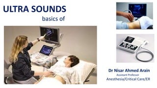
ULTRA SOUNDS
basics of
Dr Nisar Ahmed Arain
Assistant Professor
Anesthesia/Critical Care/ER
--1-What is the Ultrasound imaging
--2-Why Ultrasound is required
--3-Common uses of Ultrasound
--4-Small History of Ultrasound
--5-Properties of Ultrasound
--6-Equipment required for Ultrasound
--7-How does this procedure work
--8-Benefits and risks of Ultrasound
-OUTLINE
--Ultra sound imaging. Also called “sonography”
It involves “Exposing part of the body to high
frequency sound waves” to produce pictures
of the inside of the body
--Ultrasound examinations do not use Ionizing
radiations (as used in X Rays)
--Because Ultrasound images are captured in real
time, they can show the structure and movement
of the body’s internal organs, as well as blood
flowing through blood vessels
-GENERAL ULTRASOUND IMAGING
What is that
--1-Ultrasound (US) is the “Most widely used imaging
technology world wide
--2-It is popular due to “availability, Speed, low cost,
Patient friendless (No Radiation)
--3-Applied in “Obstetrics, Cardiology, Inner Medicine
and Urology”
--4-Ongoing research to improve image quality speed
and new application areas such as intraoperative
Navigation, and Tumor Therapy..
-WHY ULTRASOUND
Should be requested
--1-Ultrasound examinations can help to diagnose
a variety of conditions and to asses Organs
damage following illness
--2-Ultrasound is used to help Physicians to evaluate
symptoms such as
a-Pain
b-Swelling
c-Infection and
d-Hematuria (blood in urine)
-SOME COMMON USES
of the procedure
---Ultrasound is a useful way of examining many
of the body’s internal organs including but not
limited to the
-1-Heart and Blood vessels, including the abdominal
aorta and its major branches
-2-Liver
-3-Gall Bladder
-4- Spleen
-5-Pancreas
-6-Kindney
-7-Bladder
-8-Uterus, Ovaries, and unborn child (fetus) in pregnant
patients
-9-Eyes
-10-Thyroid and Parathyroid Glands
-11-Scrotum (testicles)
-12-Brain in infants
-13-Hips in infants
-SOME COMMON
USES
of the procedure
contd.
-ULTRASOUND
IS ALSO USED TO GUIDE
--1-Procedures such as Needle Biopsies
in which needles are used to Extract
sample cells from an abnormal area for
laboratory testing
--2-Image the Breasts and to guide Biopsy
of Breast cancer
--3-Diagnose a variety of Heart conditions
and to assess damage after a heart
attack or diagnose for Valvular
Heart disease
--1-Blockages to the blood flow (such as clots)
--2-Narrowing of vessels (which may be caused by a plaque)
--3-Tumors and congenital vascular malformations
IMPORTANT:-
With a knowledge about the speed and volume of blood flow
gained from a Doppler ultrasound image, the Physician can often
determine whether a patient is a good candidate for a procedure
like “Angioplasty”
DOPPLAR ULTRASOUND IMAGES CAN
HELP THE PHYSICIAN TO SEE AND EVALUATE
--Follow Fetal development
--Detect Pathologies
-APPLICATIONS IN OBSTETRICS
-Two Dimensional B Mode Ultrasound image of a fetus
-Three Dimensional image of the same fetus
5(Five) months after conception
-APPLICATIONS IN
OBSTETRICS
--1-Blood flow in vessels (Doppler US)
--2-Contraction, Rhythm
--3-Blood flow in the Heart (defects on
the wall muscle, valve defects
--4-Assessment of cardiac perfusion
-APPLICATIONS IN CARDIOLOGY
--1-Gallstone
--2-Perfusionn of Renal Transplant
-APPLICATIONS IN INNER MEDICINES
-Gallstone (Red arrow)within the Gallbladder
produces a bright surface ECHO and causes
a dark acoustic shadow (S)
-PERFUSION DOPPLAR IMAGE
OF A RENAL TRANSPLANT
--Visualization of Tendons, ligaments
--Investigations under movement is
possible
– simplifies the Detection of
RUPTURES, and OBSTRUCTIONS
-APPLICATIONS IN MUSCULOSKELETAL SYSTEM
The arrows show the large GAP of the
rupture of ACHILLES tendon
--Ultrasound Elastography is
often used to classify
Tumors and
--Malignant Tumors are
10 to 100 time stiffer
then the normal
tissue around
-APPLICATIONS OF
ULTRASOUND ELASTOGRAPHY
-The BAT use Ultrasound for navigation
-HISTORY
-Development of the B – mode Ultrasound image quality
-HISTORY
--1-Although Ultrasound is better known for its
diagnostic capabilities it was initially used
for therapy rather than diagnosis
--2-In the 1940’s Ultrasound was used to
perform services similar to that of radiation
or chemotherapy now
--3-Ultrasonic waves emit heat that can create
disruptive effects on animal tissue and
destroy malignant tissue.
-HISTORY contd.
-Common sound frequencies and frequency ranges
-COMMON SOUND
FREQUENCIES
Sound Frequency
Adultaudiblerange 15– 20’000Hz
Rangeforchildren'shearing Upto40’000Hz
Malespeakingvoice 100– 1’500Hz
Femalespeakingvoice 150‘2’500Hz
Standardpitch(ConcertA) 440Hz
Bat 50’000– 200’000Hz
MedicalUltrasound 2.5– 40MHz
Maximumsoundfrequency 600MHz
---Longitudinal mechanical waves
--Needs elastic medium
-Transducer needs to be in
contact with skin
--Component Resolution
-3 MHZ - > 1.1 mm
-10 MHZ - > 0.3 mm
--Wave velocity
-Fat - > 1450 m/s
-Muscle - > 1580 ms
-PHYSICS OF THE METHOD
-PRINCIPLES OF ULTRASOUND
-Its components
-Operations
-Applications
-ULRASOUND PARTS
-The basic Ultrasound Machine has the following parts
-ULTRASOUND MACINE
--1-Transducer Probe:-Probe that sends and receives the sound waves
--2-Central processing unit (CPU) :-Computer that does all of the calculations and
contains the electric power supplies for itself and the transducer probe
--3-Transducer Pulse Controls:-Changes the amplitude, frequency and duration of
the pulses emitted from the transducer probe
--4-Display:-Display the image from the Ultrasound data processed by the CPU
--5-Keyboard/Cursor:-Inputs data and takes measurements from the display
--6-Disc storage device(Hard, Floppy, CD) stores the acquired images
--7-Printer:-Prints the image from the displaced data
--1-Ultrasound scanners consist of a console
containing a computer and electronics
a video display screen and a transducer
that is used to do the scanning
--2-The transducer is a small hand – held
device that resembles a microphone
attached to the scanner by a cord
--3-The transducer sends out inaudible
high frequency sound waves into the
body and then listens for the returning
echoes from the tissues in the body
--4-The principles are similar to sonar
used by boats and submarines
-EQUIPMENT
--5-The Ultrasound image is immediately
visible on a video display screen that
looks like a computer or television screen
--6-The image is created based on the
amplitude (Strength) Frequency and
time it takes for the sound signal to
return from the area of the patient
being examined to the transducer and
the type of body structure the sound
travels through
-EQUIPMENT contd.
--1-Ultrasound imaging is based on the same
principles involved in the sonar used by Bats,
ships, Fishermen and the weather service.
--2-When a sound wave strikes an object, it
bounces back or Echoes
--3-By measuring these Echo waves, it is possible
to determine how far away the object is, its
shape, size and consistence (whether the object
Is solid, filled with fluid or both)
--4-In medicine, the Ultrasound is used to detect
changes in appearance of organs, tissues, and
vessels or detect abnormal masses such as
tumors
-HOW DOES THIS
PROCEEDURE WORK
--5-In an Ultrasound examination, a
Transducer both sends the sound
waves and receives/records the
Echoing waves
--6-When the transducer is pressed
against the skin, it directs small
pulses of inaudible, high frequency
sound waves into the body
--7-As the sound wave bounce off of
internal organ, fluids and tissues
the sensitive microphone in the
transducer records tiny Changes in
the sound’s pitch and direction
-HOW DOES THIS
PROCEEDURE WORK contd.
--8-These signature waves are instantly measured and
displayed by a computer, which in tern creates a
real – time picture on the monitor
--9-One or more frames of the moving pictures are
typically captured as still images
--10-Small loops of the moving “real time” images
may also be saved
--11-Dopplar Ultrasound, a special application of
Ultrasound, measures The direction and speed
of blood cells as they move through vessels
--12-The movement of blood cells causes a change
in pitch of the reflected sound waves
(called the Doppler effect)
--13-A computer collects and processes the sounds and
creates graphs or colour pictures that represent the
flow of blood through the blood vessels
-HOW DOES THIS
PROCEEDURE WORK contd
--1-For the most Ultrasound Examinations, the patient is
positioned lying Face – up on an examination table that
can be titled or moved
--2-A clear water – based gel is applied to the area of the
body being studied to help the transducer make secure
contact with the body and eliminate air pockets
between the transducer and the skin that can block the
sound waves from passing into your body
--3-The sonographer (Ultrasound technologist) or radiologist
then presses the transducer firmly against the skin in
various locations, sweeping over the area of interest or
angling the sound beam from a further location to better
see an area of concern
-HOW IS THE
PROCEDURE PERFORMED
--1-Dopplar sonography is performed using the same
transducer
--2-When the examination is complete, the patient may
be asked to dress and wait while the Ultrasound images
are reviewed
--3-In some Ultrasound studies, the transducer is attached
to a probe and inserted into a natural opening in the body.
These Examinations include:-
a-Trans-Esophageal-Echocardiogram:-The transducer is
inserted into the esophagus to obtain images of the Heart
b-Trans-Rectal-Ultrasound:-The transducer is inserted into a
man’s rectum to view the prostate
c-Trans-Vaginal Ultrasound:-The transducer is inserted into
a women’s vagina to view the Uterus and Ovaries
--4-Most Ultrasound examinations are completed within 30
minutes to an hours
-HOW IS THE PROCEDURE
PERFORMED contd.
BENEFITS
--1-Most Ultrasound scanning is non invasive ( no needles
or injections are required) and is usually painless
--2-Ultrasound is widely available, easy-to-use and less
expensive than other imaging methods
--3-Ultrasound imaging does not use any ionizing radiation
--4-Ultrasound imaging gives a clear picture of soft tissue
that do not show up well on X Ray images
--5-Ultrasound is the preferred imaging Modality for the
diagnosis and monitoring of pregnant women and their
unborn babies
--6-Ultrasound provides real time imaging, making it a good
tool for guiding Minimally invasive procedures such as
-Needle Biopsies and
-Needle Aspiration
RISKS
--1-For slandered diagnostic Ultrasound there are no known
harmful effects on humans (contd.)
-WHAT ARE THE
BENEFITS VS RISKS
--2-Unlike X Rays, Ultrasound involves only
sound waves
--3-There is no Radiation Danger
--4-However, sound waves can increase
body temperature
-This is known as cavitation
-Significant only for long exposures
time
-WHAT ARE THE BENEFITS
VS RISKS contd.
-WHAT ARE THE BENEFITS
VS RISKS contd.
--1-Many studies have been conducted
to determine the physiological effects
of Ultrasound cavitation
--2-No direct co-Relation have been found
between Ultrasound imaging and
cancer, low birth weight, dyslexia or
delayed speed of development
--3-Reliable data from Ultrasound
techniques is hard to come by
--4-Additional studies are ongoing
--5-Biggest risk is Misdiagnosis
--1-Improved clarity for use
in cancer diagnosis
--2-Increased therapeutic use
to correct blood clots and
kidney stones
--3-Portability and veterinary
use
--4-Joint and muscle treatment
through cavitation
-FUTURE OF ULTRASOUND
--1-Ultrasound waves are disrupted by air or gas: therefore
ultrasound is not an ideal imaging technique for air-filled
bowel or organs obscured by the bowel. In most cases,
-Barium exams(studies)
-CT Scanning and
-MRI
are the methods of choice in this setting
--2-Large patients are more difficult to image by Ultrasound
because greater amounts of tissue attenuates (weakens)
the sound waves as they pass deeper into the body
--3-Ultrasound has difficulty penetrating bone and, therefore,
can only see the outer surface of bony structures and not
what lies within (except in infants) For visualizing internal
structure of bones or certain joints, other imaging
Modalities such as MRI are typically used
-WHAT ARE THE
LIMITATIONS
OF GENERAL
ULTRASOUND
IMAGING
Ultasound

Más contenido relacionado

La actualidad más candente

Anaesthesia in robotic surgery
Anaesthesia in robotic surgeryAnaesthesia in robotic surgery
Anaesthesia in robotic surgerypankaj bhosale
 
Ultrasonography
UltrasonographyUltrasonography
UltrasonographyAJAZ KHAN
 
Chapter20 radiology and diagnostic imaging terminology
Chapter20 radiology and diagnostic imaging terminologyChapter20 radiology and diagnostic imaging terminology
Chapter20 radiology and diagnostic imaging terminologyRad Tech
 
Basic radiology points for homeo pathy
Basic radiology points for homeo pathyBasic radiology points for homeo pathy
Basic radiology points for homeo pathyBen Sakthivel
 
Medical Equipment Ultrasound Lecture 1
Medical Equipment Ultrasound Lecture 1Medical Equipment Ultrasound Lecture 1
Medical Equipment Ultrasound Lecture 1cairo university
 
MRI Procedure and Preparation
MRI Procedure and PreparationMRI Procedure and Preparation
MRI Procedure and PreparationMuhammad Zaid
 
Types of Ultrasound Probes
Types of Ultrasound ProbesTypes of Ultrasound Probes
Types of Ultrasound Probeshafsatalat1
 
Robotic surgery in ENT
Robotic surgery in ENTRobotic surgery in ENT
Robotic surgery in ENTJinu Iype
 
bone or marrow scan NMT
bone or marrow scan NMTbone or marrow scan NMT
bone or marrow scan NMTSUMAN GOWNDER
 
Anesthesia for robotic surgery
Anesthesia for robotic surgery Anesthesia for robotic surgery
Anesthesia for robotic surgery Aditya Kejriwal
 
Tele and robotic surgery
Tele and robotic surgeryTele and robotic surgery
Tele and robotic surgeryfathi neana
 
Radiology instruments
Radiology instrumentsRadiology instruments
Radiology instrumentsLourdes1506
 
Da vinci robotic surgical system
Da vinci robotic surgical systemDa vinci robotic surgical system
Da vinci robotic surgical systemOmer Khalil
 

La actualidad más candente (20)

Ultrasound
UltrasoundUltrasound
Ultrasound
 
Anaesthesia in robotic surgery
Anaesthesia in robotic surgeryAnaesthesia in robotic surgery
Anaesthesia in robotic surgery
 
Fluoroscopy ppt
Fluoroscopy pptFluoroscopy ppt
Fluoroscopy ppt
 
Ultrasonography
UltrasonographyUltrasonography
Ultrasonography
 
Chapter20 radiology and diagnostic imaging terminology
Chapter20 radiology and diagnostic imaging terminologyChapter20 radiology and diagnostic imaging terminology
Chapter20 radiology and diagnostic imaging terminology
 
Introduction to ultrasound
Introduction to ultrasoundIntroduction to ultrasound
Introduction to ultrasound
 
Radiology
RadiologyRadiology
Radiology
 
089 intravascular magnetic resonance imaging
089 intravascular magnetic resonance imaging089 intravascular magnetic resonance imaging
089 intravascular magnetic resonance imaging
 
Basic radiology points for homeo pathy
Basic radiology points for homeo pathyBasic radiology points for homeo pathy
Basic radiology points for homeo pathy
 
Medical Equipment Ultrasound Lecture 1
Medical Equipment Ultrasound Lecture 1Medical Equipment Ultrasound Lecture 1
Medical Equipment Ultrasound Lecture 1
 
Diagnostic imaging clinics
Diagnostic imaging clinicsDiagnostic imaging clinics
Diagnostic imaging clinics
 
MRI Procedure and Preparation
MRI Procedure and PreparationMRI Procedure and Preparation
MRI Procedure and Preparation
 
Types of Ultrasound Probes
Types of Ultrasound ProbesTypes of Ultrasound Probes
Types of Ultrasound Probes
 
Robotic surgery in ENT
Robotic surgery in ENTRobotic surgery in ENT
Robotic surgery in ENT
 
Robotic surgery
Robotic surgeryRobotic surgery
Robotic surgery
 
bone or marrow scan NMT
bone or marrow scan NMTbone or marrow scan NMT
bone or marrow scan NMT
 
Anesthesia for robotic surgery
Anesthesia for robotic surgery Anesthesia for robotic surgery
Anesthesia for robotic surgery
 
Tele and robotic surgery
Tele and robotic surgeryTele and robotic surgery
Tele and robotic surgery
 
Radiology instruments
Radiology instrumentsRadiology instruments
Radiology instruments
 
Da vinci robotic surgical system
Da vinci robotic surgical systemDa vinci robotic surgical system
Da vinci robotic surgical system
 

Similar a Ultasound

Ultrasound-1.pptx
Ultrasound-1.pptxUltrasound-1.pptx
Ultrasound-1.pptxcutefairy5
 
bams-4th-year-shalya-ultrasound-05-05-2020.pptx
bams-4th-year-shalya-ultrasound-05-05-2020.pptxbams-4th-year-shalya-ultrasound-05-05-2020.pptx
bams-4th-year-shalya-ultrasound-05-05-2020.pptxsaid462049
 
What is ultrasound
What is ultrasoundWhat is ultrasound
What is ultrasoundFetal Studio
 
ULTRASOUND BY DR.ABHIJEET MAJHI,MS .pptx
ULTRASOUND BY DR.ABHIJEET MAJHI,MS .pptxULTRASOUND BY DR.ABHIJEET MAJHI,MS .pptx
ULTRASOUND BY DR.ABHIJEET MAJHI,MS .pptxAbhijeet Majhi
 
Introduction to obs. ultrasound
Introduction to obs. ultrasoundIntroduction to obs. ultrasound
Introduction to obs. ultrasoundmagdy abdel
 
Sonography
SonographySonography
SonographyFLI
 
Ultrasound Basics.ppt
Ultrasound Basics.pptUltrasound Basics.ppt
Ultrasound Basics.pptghadaElbanby1
 
Its is useful for studying the basic concepts of ultrasound for medical imagi...
Its is useful for studying the basic concepts of ultrasound for medical imagi...Its is useful for studying the basic concepts of ultrasound for medical imagi...
Its is useful for studying the basic concepts of ultrasound for medical imagi...DilshanDillu1
 
3_2018_12_16!11_22_57_PM.pptx
3_2018_12_16!11_22_57_PM.pptx3_2018_12_16!11_22_57_PM.pptx
3_2018_12_16!11_22_57_PM.pptxKhaVuong2
 
Ultrasonography in Animals.pptxblba jhaha
Ultrasonography in Animals.pptxblba jhahaUltrasonography in Animals.pptxblba jhaha
Ultrasonography in Animals.pptxblba jhahaIzzatAftab
 
Ultrasonic applications
Ultrasonic applicationsUltrasonic applications
Ultrasonic applicationsOmar Salah
 

Similar a Ultasound (20)

Ultrasound-1.pptx
Ultrasound-1.pptxUltrasound-1.pptx
Ultrasound-1.pptx
 
ultrasonography.pptx
ultrasonography.pptxultrasonography.pptx
ultrasonography.pptx
 
Basics of Ultrasound
Basics of Ultrasound Basics of Ultrasound
Basics of Ultrasound
 
ultrasound JP.pptx
ultrasound JP.pptxultrasound JP.pptx
ultrasound JP.pptx
 
Basics of Ultrasound
Basics of UltrasoundBasics of Ultrasound
Basics of Ultrasound
 
bams-4th-year-shalya-ultrasound-05-05-2020.pptx
bams-4th-year-shalya-ultrasound-05-05-2020.pptxbams-4th-year-shalya-ultrasound-05-05-2020.pptx
bams-4th-year-shalya-ultrasound-05-05-2020.pptx
 
Ultrasound final.pptx
Ultrasound final.pptxUltrasound final.pptx
Ultrasound final.pptx
 
What is ultrasound
What is ultrasoundWhat is ultrasound
What is ultrasound
 
ULTRASOUND BY DR.ABHIJEET MAJHI,MS .pptx
ULTRASOUND BY DR.ABHIJEET MAJHI,MS .pptxULTRASOUND BY DR.ABHIJEET MAJHI,MS .pptx
ULTRASOUND BY DR.ABHIJEET MAJHI,MS .pptx
 
Us imaging
Us imagingUs imaging
Us imaging
 
Introduction to obs. ultrasound
Introduction to obs. ultrasoundIntroduction to obs. ultrasound
Introduction to obs. ultrasound
 
Sonography
SonographySonography
Sonography
 
Ultrasound Basics.ppt
Ultrasound Basics.pptUltrasound Basics.ppt
Ultrasound Basics.ppt
 
ultrasound JP.pptx
ultrasound JP.pptxultrasound JP.pptx
ultrasound JP.pptx
 
Its is useful for studying the basic concepts of ultrasound for medical imagi...
Its is useful for studying the basic concepts of ultrasound for medical imagi...Its is useful for studying the basic concepts of ultrasound for medical imagi...
Its is useful for studying the basic concepts of ultrasound for medical imagi...
 
3_2018_12_16!11_22_57_PM.pptx
3_2018_12_16!11_22_57_PM.pptx3_2018_12_16!11_22_57_PM.pptx
3_2018_12_16!11_22_57_PM.pptx
 
Ultrasonography in Animals.pptxblba jhaha
Ultrasonography in Animals.pptxblba jhahaUltrasonography in Animals.pptxblba jhaha
Ultrasonography in Animals.pptxblba jhaha
 
Ultrasonic applications
Ultrasonic applicationsUltrasonic applications
Ultrasonic applications
 
WIEB
WIEBWIEB
WIEB
 
Principle of Ultrasound.pptx
Principle of Ultrasound.pptxPrinciple of Ultrasound.pptx
Principle of Ultrasound.pptx
 

Más de Nisar Arain

Airway local blocks
Airway local blocksAirway local blocks
Airway local blocksNisar Arain
 
Endotracheal intubation
Endotracheal  intubationEndotracheal  intubation
Endotracheal intubationNisar Arain
 
Appropriate airway equipment and techniques.
Appropriate airway equipment and techniques.Appropriate airway equipment and techniques.
Appropriate airway equipment and techniques.Nisar Arain
 
Airway management
Airway managementAirway management
Airway managementNisar Arain
 
Theories of anesthesia
Theories of anesthesiaTheories of anesthesia
Theories of anesthesiaNisar Arain
 
Endotracheal intubation and laryngoscopy part 2
Endotracheal intubation and laryngoscopy part 2Endotracheal intubation and laryngoscopy part 2
Endotracheal intubation and laryngoscopy part 2Nisar Arain
 
Intubation part 1.
Intubation part 1.Intubation part 1.
Intubation part 1.Nisar Arain
 
Complications of artificial applications part 5
Complications of artificial applications part 5Complications of artificial applications part 5
Complications of artificial applications part 5Nisar Arain
 
Difficult airway management
Difficult airway managementDifficult airway management
Difficult airway managementNisar Arain
 
Airway management part 3.
Airway management part 3.Airway management part 3.
Airway management part 3.Nisar Arain
 
Airway assesment part 2
Airway assesment part 2Airway assesment part 2
Airway assesment part 2Nisar Arain
 
Airway assesment part 1
Airway assesment part 1Airway assesment part 1
Airway assesment part 1Nisar Arain
 
History of anesthesia.
History of anesthesia.History of anesthesia.
History of anesthesia.Nisar Arain
 
Local and regional anesthesia
Local and regional anesthesiaLocal and regional anesthesia
Local and regional anesthesiaNisar Arain
 
1d awareness problem during operation
1d awareness problem during operation1d awareness problem during operation
1d awareness problem during operationNisar Arain
 
1c geriatric patients
1c geriatric patients1c geriatric patients
1c geriatric patientsNisar Arain
 
1b general anesthesia
1b general anesthesia1b general anesthesia
1b general anesthesiaNisar Arain
 
1a respiratory system
1a respiratory system1a respiratory system
1a respiratory systemNisar Arain
 
Body temperature mechanism
Body temperature mechanismBody temperature mechanism
Body temperature mechanismNisar Arain
 

Más de Nisar Arain (20)

Airway local blocks
Airway local blocksAirway local blocks
Airway local blocks
 
Endotracheal intubation
Endotracheal  intubationEndotracheal  intubation
Endotracheal intubation
 
Appropriate airway equipment and techniques.
Appropriate airway equipment and techniques.Appropriate airway equipment and techniques.
Appropriate airway equipment and techniques.
 
Airway management
Airway managementAirway management
Airway management
 
Biophysics
BiophysicsBiophysics
Biophysics
 
Theories of anesthesia
Theories of anesthesiaTheories of anesthesia
Theories of anesthesia
 
Endotracheal intubation and laryngoscopy part 2
Endotracheal intubation and laryngoscopy part 2Endotracheal intubation and laryngoscopy part 2
Endotracheal intubation and laryngoscopy part 2
 
Intubation part 1.
Intubation part 1.Intubation part 1.
Intubation part 1.
 
Complications of artificial applications part 5
Complications of artificial applications part 5Complications of artificial applications part 5
Complications of artificial applications part 5
 
Difficult airway management
Difficult airway managementDifficult airway management
Difficult airway management
 
Airway management part 3.
Airway management part 3.Airway management part 3.
Airway management part 3.
 
Airway assesment part 2
Airway assesment part 2Airway assesment part 2
Airway assesment part 2
 
Airway assesment part 1
Airway assesment part 1Airway assesment part 1
Airway assesment part 1
 
History of anesthesia.
History of anesthesia.History of anesthesia.
History of anesthesia.
 
Local and regional anesthesia
Local and regional anesthesiaLocal and regional anesthesia
Local and regional anesthesia
 
1d awareness problem during operation
1d awareness problem during operation1d awareness problem during operation
1d awareness problem during operation
 
1c geriatric patients
1c geriatric patients1c geriatric patients
1c geriatric patients
 
1b general anesthesia
1b general anesthesia1b general anesthesia
1b general anesthesia
 
1a respiratory system
1a respiratory system1a respiratory system
1a respiratory system
 
Body temperature mechanism
Body temperature mechanismBody temperature mechanism
Body temperature mechanism
 

Último

Night 7k to 12k Navi Mumbai Call Girl Photo 👉 BOOK NOW 9833363713 👈 ♀️ night ...
Night 7k to 12k Navi Mumbai Call Girl Photo 👉 BOOK NOW 9833363713 👈 ♀️ night ...Night 7k to 12k Navi Mumbai Call Girl Photo 👉 BOOK NOW 9833363713 👈 ♀️ night ...
Night 7k to 12k Navi Mumbai Call Girl Photo 👉 BOOK NOW 9833363713 👈 ♀️ night ...aartirawatdelhi
 
Call Girls Vasai Virar Just Call 9630942363 Top Class Call Girl Service Avail...
Call Girls Vasai Virar Just Call 9630942363 Top Class Call Girl Service Avail...Call Girls Vasai Virar Just Call 9630942363 Top Class Call Girl Service Avail...
Call Girls Vasai Virar Just Call 9630942363 Top Class Call Girl Service Avail...GENUINE ESCORT AGENCY
 
Pondicherry Call Girls Book Now 9630942363 Top Class Pondicherry Escort Servi...
Pondicherry Call Girls Book Now 9630942363 Top Class Pondicherry Escort Servi...Pondicherry Call Girls Book Now 9630942363 Top Class Pondicherry Escort Servi...
Pondicherry Call Girls Book Now 9630942363 Top Class Pondicherry Escort Servi...GENUINE ESCORT AGENCY
 
Call Girls Service Jaipur {9521753030} ❤️VVIP RIDDHI Call Girl in Jaipur Raja...
Call Girls Service Jaipur {9521753030} ❤️VVIP RIDDHI Call Girl in Jaipur Raja...Call Girls Service Jaipur {9521753030} ❤️VVIP RIDDHI Call Girl in Jaipur Raja...
Call Girls Service Jaipur {9521753030} ❤️VVIP RIDDHI Call Girl in Jaipur Raja...Sheetaleventcompany
 
Independent Call Girls In Jaipur { 8445551418 } ✔ ANIKA MEHTA ✔ Get High Prof...
Independent Call Girls In Jaipur { 8445551418 } ✔ ANIKA MEHTA ✔ Get High Prof...Independent Call Girls In Jaipur { 8445551418 } ✔ ANIKA MEHTA ✔ Get High Prof...
Independent Call Girls In Jaipur { 8445551418 } ✔ ANIKA MEHTA ✔ Get High Prof...parulsinha
 
Call Girls Rishikesh Just Call 9667172968 Top Class Call Girl Service Available
Call Girls Rishikesh Just Call 9667172968 Top Class Call Girl Service AvailableCall Girls Rishikesh Just Call 9667172968 Top Class Call Girl Service Available
Call Girls Rishikesh Just Call 9667172968 Top Class Call Girl Service Availableperfect solution
 
Andheri East ) Call Girls in Mumbai Phone No 9004268417 Elite Escort Service ...
Andheri East ) Call Girls in Mumbai Phone No 9004268417 Elite Escort Service ...Andheri East ) Call Girls in Mumbai Phone No 9004268417 Elite Escort Service ...
Andheri East ) Call Girls in Mumbai Phone No 9004268417 Elite Escort Service ...Anamika Rawat
 
Call Girls Kolkata Kalikapur 💯Call Us 🔝 8005736733 🔝 💃 Top Class Call Girl Se...
Call Girls Kolkata Kalikapur 💯Call Us 🔝 8005736733 🔝 💃 Top Class Call Girl Se...Call Girls Kolkata Kalikapur 💯Call Us 🔝 8005736733 🔝 💃 Top Class Call Girl Se...
Call Girls Kolkata Kalikapur 💯Call Us 🔝 8005736733 🔝 💃 Top Class Call Girl Se...Namrata Singh
 
Premium Bangalore Call Girls Jigani Dail 6378878445 Escort Service For Hot Ma...
Premium Bangalore Call Girls Jigani Dail 6378878445 Escort Service For Hot Ma...Premium Bangalore Call Girls Jigani Dail 6378878445 Escort Service For Hot Ma...
Premium Bangalore Call Girls Jigani Dail 6378878445 Escort Service For Hot Ma...tanya dube
 
Independent Call Girls Service Mohali Sector 116 | 6367187148 | Call Girl Ser...
Independent Call Girls Service Mohali Sector 116 | 6367187148 | Call Girl Ser...Independent Call Girls Service Mohali Sector 116 | 6367187148 | Call Girl Ser...
Independent Call Girls Service Mohali Sector 116 | 6367187148 | Call Girl Ser...karishmasinghjnh
 
(Low Rate RASHMI ) Rate Of Call Girls Jaipur ❣ 8445551418 ❣ Elite Models & Ce...
(Low Rate RASHMI ) Rate Of Call Girls Jaipur ❣ 8445551418 ❣ Elite Models & Ce...(Low Rate RASHMI ) Rate Of Call Girls Jaipur ❣ 8445551418 ❣ Elite Models & Ce...
(Low Rate RASHMI ) Rate Of Call Girls Jaipur ❣ 8445551418 ❣ Elite Models & Ce...parulsinha
 
Call Girls in Delhi Triveni Complex Escort Service(🔝))/WhatsApp 97111⇛47426
Call Girls in Delhi Triveni Complex Escort Service(🔝))/WhatsApp 97111⇛47426Call Girls in Delhi Triveni Complex Escort Service(🔝))/WhatsApp 97111⇛47426
Call Girls in Delhi Triveni Complex Escort Service(🔝))/WhatsApp 97111⇛47426jennyeacort
 
Premium Call Girls In Jaipur {8445551418} ❤️VVIP SEEMA Call Girl in Jaipur Ra...
Premium Call Girls In Jaipur {8445551418} ❤️VVIP SEEMA Call Girl in Jaipur Ra...Premium Call Girls In Jaipur {8445551418} ❤️VVIP SEEMA Call Girl in Jaipur Ra...
Premium Call Girls In Jaipur {8445551418} ❤️VVIP SEEMA Call Girl in Jaipur Ra...parulsinha
 
Top Quality Call Girl Service Kalyanpur 6378878445 Available Call Girls Any Time
Top Quality Call Girl Service Kalyanpur 6378878445 Available Call Girls Any TimeTop Quality Call Girl Service Kalyanpur 6378878445 Available Call Girls Any Time
Top Quality Call Girl Service Kalyanpur 6378878445 Available Call Girls Any TimeCall Girls Delhi
 
Coimbatore Call Girls in Thudiyalur : 7427069034 High Profile Model Escorts |...
Coimbatore Call Girls in Thudiyalur : 7427069034 High Profile Model Escorts |...Coimbatore Call Girls in Thudiyalur : 7427069034 High Profile Model Escorts |...
Coimbatore Call Girls in Thudiyalur : 7427069034 High Profile Model Escorts |...chennailover
 
Top Rated Hyderabad Call Girls Erragadda ⟟ 9332606886 ⟟ Call Me For Genuine ...
Top Rated  Hyderabad Call Girls Erragadda ⟟ 9332606886 ⟟ Call Me For Genuine ...Top Rated  Hyderabad Call Girls Erragadda ⟟ 9332606886 ⟟ Call Me For Genuine ...
Top Rated Hyderabad Call Girls Erragadda ⟟ 9332606886 ⟟ Call Me For Genuine ...chandars293
 
Russian Call Girls Lucknow Just Call 👉👉7877925207 Top Class Call Girl Service...
Russian Call Girls Lucknow Just Call 👉👉7877925207 Top Class Call Girl Service...Russian Call Girls Lucknow Just Call 👉👉7877925207 Top Class Call Girl Service...
Russian Call Girls Lucknow Just Call 👉👉7877925207 Top Class Call Girl Service...adilkhan87451
 
Best Rate (Patna ) Call Girls Patna ⟟ 8617370543 ⟟ High Class Call Girl In 5 ...
Best Rate (Patna ) Call Girls Patna ⟟ 8617370543 ⟟ High Class Call Girl In 5 ...Best Rate (Patna ) Call Girls Patna ⟟ 8617370543 ⟟ High Class Call Girl In 5 ...
Best Rate (Patna ) Call Girls Patna ⟟ 8617370543 ⟟ High Class Call Girl In 5 ...Dipal Arora
 
Call Girls Service Jaipur {8445551418} ❤️VVIP BHAWNA Call Girl in Jaipur Raja...
Call Girls Service Jaipur {8445551418} ❤️VVIP BHAWNA Call Girl in Jaipur Raja...Call Girls Service Jaipur {8445551418} ❤️VVIP BHAWNA Call Girl in Jaipur Raja...
Call Girls Service Jaipur {8445551418} ❤️VVIP BHAWNA Call Girl in Jaipur Raja...parulsinha
 

Último (20)

Night 7k to 12k Navi Mumbai Call Girl Photo 👉 BOOK NOW 9833363713 👈 ♀️ night ...
Night 7k to 12k Navi Mumbai Call Girl Photo 👉 BOOK NOW 9833363713 👈 ♀️ night ...Night 7k to 12k Navi Mumbai Call Girl Photo 👉 BOOK NOW 9833363713 👈 ♀️ night ...
Night 7k to 12k Navi Mumbai Call Girl Photo 👉 BOOK NOW 9833363713 👈 ♀️ night ...
 
Call Girls Vasai Virar Just Call 9630942363 Top Class Call Girl Service Avail...
Call Girls Vasai Virar Just Call 9630942363 Top Class Call Girl Service Avail...Call Girls Vasai Virar Just Call 9630942363 Top Class Call Girl Service Avail...
Call Girls Vasai Virar Just Call 9630942363 Top Class Call Girl Service Avail...
 
Pondicherry Call Girls Book Now 9630942363 Top Class Pondicherry Escort Servi...
Pondicherry Call Girls Book Now 9630942363 Top Class Pondicherry Escort Servi...Pondicherry Call Girls Book Now 9630942363 Top Class Pondicherry Escort Servi...
Pondicherry Call Girls Book Now 9630942363 Top Class Pondicherry Escort Servi...
 
Call Girls Service Jaipur {9521753030} ❤️VVIP RIDDHI Call Girl in Jaipur Raja...
Call Girls Service Jaipur {9521753030} ❤️VVIP RIDDHI Call Girl in Jaipur Raja...Call Girls Service Jaipur {9521753030} ❤️VVIP RIDDHI Call Girl in Jaipur Raja...
Call Girls Service Jaipur {9521753030} ❤️VVIP RIDDHI Call Girl in Jaipur Raja...
 
Independent Call Girls In Jaipur { 8445551418 } ✔ ANIKA MEHTA ✔ Get High Prof...
Independent Call Girls In Jaipur { 8445551418 } ✔ ANIKA MEHTA ✔ Get High Prof...Independent Call Girls In Jaipur { 8445551418 } ✔ ANIKA MEHTA ✔ Get High Prof...
Independent Call Girls In Jaipur { 8445551418 } ✔ ANIKA MEHTA ✔ Get High Prof...
 
Call Girls Rishikesh Just Call 9667172968 Top Class Call Girl Service Available
Call Girls Rishikesh Just Call 9667172968 Top Class Call Girl Service AvailableCall Girls Rishikesh Just Call 9667172968 Top Class Call Girl Service Available
Call Girls Rishikesh Just Call 9667172968 Top Class Call Girl Service Available
 
Andheri East ) Call Girls in Mumbai Phone No 9004268417 Elite Escort Service ...
Andheri East ) Call Girls in Mumbai Phone No 9004268417 Elite Escort Service ...Andheri East ) Call Girls in Mumbai Phone No 9004268417 Elite Escort Service ...
Andheri East ) Call Girls in Mumbai Phone No 9004268417 Elite Escort Service ...
 
Call Girls Kolkata Kalikapur 💯Call Us 🔝 8005736733 🔝 💃 Top Class Call Girl Se...
Call Girls Kolkata Kalikapur 💯Call Us 🔝 8005736733 🔝 💃 Top Class Call Girl Se...Call Girls Kolkata Kalikapur 💯Call Us 🔝 8005736733 🔝 💃 Top Class Call Girl Se...
Call Girls Kolkata Kalikapur 💯Call Us 🔝 8005736733 🔝 💃 Top Class Call Girl Se...
 
Premium Bangalore Call Girls Jigani Dail 6378878445 Escort Service For Hot Ma...
Premium Bangalore Call Girls Jigani Dail 6378878445 Escort Service For Hot Ma...Premium Bangalore Call Girls Jigani Dail 6378878445 Escort Service For Hot Ma...
Premium Bangalore Call Girls Jigani Dail 6378878445 Escort Service For Hot Ma...
 
Independent Call Girls Service Mohali Sector 116 | 6367187148 | Call Girl Ser...
Independent Call Girls Service Mohali Sector 116 | 6367187148 | Call Girl Ser...Independent Call Girls Service Mohali Sector 116 | 6367187148 | Call Girl Ser...
Independent Call Girls Service Mohali Sector 116 | 6367187148 | Call Girl Ser...
 
(Low Rate RASHMI ) Rate Of Call Girls Jaipur ❣ 8445551418 ❣ Elite Models & Ce...
(Low Rate RASHMI ) Rate Of Call Girls Jaipur ❣ 8445551418 ❣ Elite Models & Ce...(Low Rate RASHMI ) Rate Of Call Girls Jaipur ❣ 8445551418 ❣ Elite Models & Ce...
(Low Rate RASHMI ) Rate Of Call Girls Jaipur ❣ 8445551418 ❣ Elite Models & Ce...
 
Call Girls in Delhi Triveni Complex Escort Service(🔝))/WhatsApp 97111⇛47426
Call Girls in Delhi Triveni Complex Escort Service(🔝))/WhatsApp 97111⇛47426Call Girls in Delhi Triveni Complex Escort Service(🔝))/WhatsApp 97111⇛47426
Call Girls in Delhi Triveni Complex Escort Service(🔝))/WhatsApp 97111⇛47426
 
Premium Call Girls In Jaipur {8445551418} ❤️VVIP SEEMA Call Girl in Jaipur Ra...
Premium Call Girls In Jaipur {8445551418} ❤️VVIP SEEMA Call Girl in Jaipur Ra...Premium Call Girls In Jaipur {8445551418} ❤️VVIP SEEMA Call Girl in Jaipur Ra...
Premium Call Girls In Jaipur {8445551418} ❤️VVIP SEEMA Call Girl in Jaipur Ra...
 
🌹Attapur⬅️ Vip Call Girls Hyderabad 📱9352852248 Book Well Trand Call Girls In...
🌹Attapur⬅️ Vip Call Girls Hyderabad 📱9352852248 Book Well Trand Call Girls In...🌹Attapur⬅️ Vip Call Girls Hyderabad 📱9352852248 Book Well Trand Call Girls In...
🌹Attapur⬅️ Vip Call Girls Hyderabad 📱9352852248 Book Well Trand Call Girls In...
 
Top Quality Call Girl Service Kalyanpur 6378878445 Available Call Girls Any Time
Top Quality Call Girl Service Kalyanpur 6378878445 Available Call Girls Any TimeTop Quality Call Girl Service Kalyanpur 6378878445 Available Call Girls Any Time
Top Quality Call Girl Service Kalyanpur 6378878445 Available Call Girls Any Time
 
Coimbatore Call Girls in Thudiyalur : 7427069034 High Profile Model Escorts |...
Coimbatore Call Girls in Thudiyalur : 7427069034 High Profile Model Escorts |...Coimbatore Call Girls in Thudiyalur : 7427069034 High Profile Model Escorts |...
Coimbatore Call Girls in Thudiyalur : 7427069034 High Profile Model Escorts |...
 
Top Rated Hyderabad Call Girls Erragadda ⟟ 9332606886 ⟟ Call Me For Genuine ...
Top Rated  Hyderabad Call Girls Erragadda ⟟ 9332606886 ⟟ Call Me For Genuine ...Top Rated  Hyderabad Call Girls Erragadda ⟟ 9332606886 ⟟ Call Me For Genuine ...
Top Rated Hyderabad Call Girls Erragadda ⟟ 9332606886 ⟟ Call Me For Genuine ...
 
Russian Call Girls Lucknow Just Call 👉👉7877925207 Top Class Call Girl Service...
Russian Call Girls Lucknow Just Call 👉👉7877925207 Top Class Call Girl Service...Russian Call Girls Lucknow Just Call 👉👉7877925207 Top Class Call Girl Service...
Russian Call Girls Lucknow Just Call 👉👉7877925207 Top Class Call Girl Service...
 
Best Rate (Patna ) Call Girls Patna ⟟ 8617370543 ⟟ High Class Call Girl In 5 ...
Best Rate (Patna ) Call Girls Patna ⟟ 8617370543 ⟟ High Class Call Girl In 5 ...Best Rate (Patna ) Call Girls Patna ⟟ 8617370543 ⟟ High Class Call Girl In 5 ...
Best Rate (Patna ) Call Girls Patna ⟟ 8617370543 ⟟ High Class Call Girl In 5 ...
 
Call Girls Service Jaipur {8445551418} ❤️VVIP BHAWNA Call Girl in Jaipur Raja...
Call Girls Service Jaipur {8445551418} ❤️VVIP BHAWNA Call Girl in Jaipur Raja...Call Girls Service Jaipur {8445551418} ❤️VVIP BHAWNA Call Girl in Jaipur Raja...
Call Girls Service Jaipur {8445551418} ❤️VVIP BHAWNA Call Girl in Jaipur Raja...
 

Ultasound

  • 1. ULTRA SOUNDS basics of Dr Nisar Ahmed Arain Assistant Professor Anesthesia/Critical Care/ER
  • 2. --1-What is the Ultrasound imaging --2-Why Ultrasound is required --3-Common uses of Ultrasound --4-Small History of Ultrasound --5-Properties of Ultrasound --6-Equipment required for Ultrasound --7-How does this procedure work --8-Benefits and risks of Ultrasound -OUTLINE
  • 3. --Ultra sound imaging. Also called “sonography” It involves “Exposing part of the body to high frequency sound waves” to produce pictures of the inside of the body --Ultrasound examinations do not use Ionizing radiations (as used in X Rays) --Because Ultrasound images are captured in real time, they can show the structure and movement of the body’s internal organs, as well as blood flowing through blood vessels -GENERAL ULTRASOUND IMAGING What is that
  • 4. --1-Ultrasound (US) is the “Most widely used imaging technology world wide --2-It is popular due to “availability, Speed, low cost, Patient friendless (No Radiation) --3-Applied in “Obstetrics, Cardiology, Inner Medicine and Urology” --4-Ongoing research to improve image quality speed and new application areas such as intraoperative Navigation, and Tumor Therapy.. -WHY ULTRASOUND Should be requested
  • 5. --1-Ultrasound examinations can help to diagnose a variety of conditions and to asses Organs damage following illness --2-Ultrasound is used to help Physicians to evaluate symptoms such as a-Pain b-Swelling c-Infection and d-Hematuria (blood in urine) -SOME COMMON USES of the procedure
  • 6. ---Ultrasound is a useful way of examining many of the body’s internal organs including but not limited to the -1-Heart and Blood vessels, including the abdominal aorta and its major branches -2-Liver -3-Gall Bladder -4- Spleen -5-Pancreas -6-Kindney -7-Bladder -8-Uterus, Ovaries, and unborn child (fetus) in pregnant patients -9-Eyes -10-Thyroid and Parathyroid Glands -11-Scrotum (testicles) -12-Brain in infants -13-Hips in infants -SOME COMMON USES of the procedure contd.
  • 7. -ULTRASOUND IS ALSO USED TO GUIDE --1-Procedures such as Needle Biopsies in which needles are used to Extract sample cells from an abnormal area for laboratory testing --2-Image the Breasts and to guide Biopsy of Breast cancer --3-Diagnose a variety of Heart conditions and to assess damage after a heart attack or diagnose for Valvular Heart disease
  • 8. --1-Blockages to the blood flow (such as clots) --2-Narrowing of vessels (which may be caused by a plaque) --3-Tumors and congenital vascular malformations IMPORTANT:- With a knowledge about the speed and volume of blood flow gained from a Doppler ultrasound image, the Physician can often determine whether a patient is a good candidate for a procedure like “Angioplasty” DOPPLAR ULTRASOUND IMAGES CAN HELP THE PHYSICIAN TO SEE AND EVALUATE
  • 9. --Follow Fetal development --Detect Pathologies -APPLICATIONS IN OBSTETRICS -Two Dimensional B Mode Ultrasound image of a fetus
  • 10. -Three Dimensional image of the same fetus 5(Five) months after conception -APPLICATIONS IN OBSTETRICS
  • 11. --1-Blood flow in vessels (Doppler US) --2-Contraction, Rhythm --3-Blood flow in the Heart (defects on the wall muscle, valve defects --4-Assessment of cardiac perfusion -APPLICATIONS IN CARDIOLOGY
  • 12. --1-Gallstone --2-Perfusionn of Renal Transplant -APPLICATIONS IN INNER MEDICINES -Gallstone (Red arrow)within the Gallbladder produces a bright surface ECHO and causes a dark acoustic shadow (S)
  • 13. -PERFUSION DOPPLAR IMAGE OF A RENAL TRANSPLANT
  • 14. --Visualization of Tendons, ligaments --Investigations under movement is possible – simplifies the Detection of RUPTURES, and OBSTRUCTIONS -APPLICATIONS IN MUSCULOSKELETAL SYSTEM The arrows show the large GAP of the rupture of ACHILLES tendon
  • 15. --Ultrasound Elastography is often used to classify Tumors and --Malignant Tumors are 10 to 100 time stiffer then the normal tissue around -APPLICATIONS OF ULTRASOUND ELASTOGRAPHY
  • 16. -The BAT use Ultrasound for navigation -HISTORY
  • 17. -Development of the B – mode Ultrasound image quality -HISTORY
  • 18. --1-Although Ultrasound is better known for its diagnostic capabilities it was initially used for therapy rather than diagnosis --2-In the 1940’s Ultrasound was used to perform services similar to that of radiation or chemotherapy now --3-Ultrasonic waves emit heat that can create disruptive effects on animal tissue and destroy malignant tissue. -HISTORY contd.
  • 19. -Common sound frequencies and frequency ranges -COMMON SOUND FREQUENCIES Sound Frequency Adultaudiblerange 15– 20’000Hz Rangeforchildren'shearing Upto40’000Hz Malespeakingvoice 100– 1’500Hz Femalespeakingvoice 150‘2’500Hz Standardpitch(ConcertA) 440Hz Bat 50’000– 200’000Hz MedicalUltrasound 2.5– 40MHz Maximumsoundfrequency 600MHz
  • 20. ---Longitudinal mechanical waves --Needs elastic medium -Transducer needs to be in contact with skin --Component Resolution -3 MHZ - > 1.1 mm -10 MHZ - > 0.3 mm --Wave velocity -Fat - > 1450 m/s -Muscle - > 1580 ms -PHYSICS OF THE METHOD
  • 21. -PRINCIPLES OF ULTRASOUND -Its components -Operations -Applications
  • 23. -The basic Ultrasound Machine has the following parts -ULTRASOUND MACINE --1-Transducer Probe:-Probe that sends and receives the sound waves --2-Central processing unit (CPU) :-Computer that does all of the calculations and contains the electric power supplies for itself and the transducer probe --3-Transducer Pulse Controls:-Changes the amplitude, frequency and duration of the pulses emitted from the transducer probe --4-Display:-Display the image from the Ultrasound data processed by the CPU --5-Keyboard/Cursor:-Inputs data and takes measurements from the display --6-Disc storage device(Hard, Floppy, CD) stores the acquired images --7-Printer:-Prints the image from the displaced data
  • 24. --1-Ultrasound scanners consist of a console containing a computer and electronics a video display screen and a transducer that is used to do the scanning --2-The transducer is a small hand – held device that resembles a microphone attached to the scanner by a cord --3-The transducer sends out inaudible high frequency sound waves into the body and then listens for the returning echoes from the tissues in the body --4-The principles are similar to sonar used by boats and submarines -EQUIPMENT
  • 25. --5-The Ultrasound image is immediately visible on a video display screen that looks like a computer or television screen --6-The image is created based on the amplitude (Strength) Frequency and time it takes for the sound signal to return from the area of the patient being examined to the transducer and the type of body structure the sound travels through -EQUIPMENT contd.
  • 26. --1-Ultrasound imaging is based on the same principles involved in the sonar used by Bats, ships, Fishermen and the weather service. --2-When a sound wave strikes an object, it bounces back or Echoes --3-By measuring these Echo waves, it is possible to determine how far away the object is, its shape, size and consistence (whether the object Is solid, filled with fluid or both) --4-In medicine, the Ultrasound is used to detect changes in appearance of organs, tissues, and vessels or detect abnormal masses such as tumors -HOW DOES THIS PROCEEDURE WORK
  • 27. --5-In an Ultrasound examination, a Transducer both sends the sound waves and receives/records the Echoing waves --6-When the transducer is pressed against the skin, it directs small pulses of inaudible, high frequency sound waves into the body --7-As the sound wave bounce off of internal organ, fluids and tissues the sensitive microphone in the transducer records tiny Changes in the sound’s pitch and direction -HOW DOES THIS PROCEEDURE WORK contd.
  • 28. --8-These signature waves are instantly measured and displayed by a computer, which in tern creates a real – time picture on the monitor --9-One or more frames of the moving pictures are typically captured as still images --10-Small loops of the moving “real time” images may also be saved --11-Dopplar Ultrasound, a special application of Ultrasound, measures The direction and speed of blood cells as they move through vessels --12-The movement of blood cells causes a change in pitch of the reflected sound waves (called the Doppler effect) --13-A computer collects and processes the sounds and creates graphs or colour pictures that represent the flow of blood through the blood vessels -HOW DOES THIS PROCEEDURE WORK contd
  • 29. --1-For the most Ultrasound Examinations, the patient is positioned lying Face – up on an examination table that can be titled or moved --2-A clear water – based gel is applied to the area of the body being studied to help the transducer make secure contact with the body and eliminate air pockets between the transducer and the skin that can block the sound waves from passing into your body --3-The sonographer (Ultrasound technologist) or radiologist then presses the transducer firmly against the skin in various locations, sweeping over the area of interest or angling the sound beam from a further location to better see an area of concern -HOW IS THE PROCEDURE PERFORMED
  • 30. --1-Dopplar sonography is performed using the same transducer --2-When the examination is complete, the patient may be asked to dress and wait while the Ultrasound images are reviewed --3-In some Ultrasound studies, the transducer is attached to a probe and inserted into a natural opening in the body. These Examinations include:- a-Trans-Esophageal-Echocardiogram:-The transducer is inserted into the esophagus to obtain images of the Heart b-Trans-Rectal-Ultrasound:-The transducer is inserted into a man’s rectum to view the prostate c-Trans-Vaginal Ultrasound:-The transducer is inserted into a women’s vagina to view the Uterus and Ovaries --4-Most Ultrasound examinations are completed within 30 minutes to an hours -HOW IS THE PROCEDURE PERFORMED contd.
  • 31. BENEFITS --1-Most Ultrasound scanning is non invasive ( no needles or injections are required) and is usually painless --2-Ultrasound is widely available, easy-to-use and less expensive than other imaging methods --3-Ultrasound imaging does not use any ionizing radiation --4-Ultrasound imaging gives a clear picture of soft tissue that do not show up well on X Ray images --5-Ultrasound is the preferred imaging Modality for the diagnosis and monitoring of pregnant women and their unborn babies --6-Ultrasound provides real time imaging, making it a good tool for guiding Minimally invasive procedures such as -Needle Biopsies and -Needle Aspiration RISKS --1-For slandered diagnostic Ultrasound there are no known harmful effects on humans (contd.) -WHAT ARE THE BENEFITS VS RISKS
  • 32. --2-Unlike X Rays, Ultrasound involves only sound waves --3-There is no Radiation Danger --4-However, sound waves can increase body temperature -This is known as cavitation -Significant only for long exposures time -WHAT ARE THE BENEFITS VS RISKS contd.
  • 33. -WHAT ARE THE BENEFITS VS RISKS contd. --1-Many studies have been conducted to determine the physiological effects of Ultrasound cavitation --2-No direct co-Relation have been found between Ultrasound imaging and cancer, low birth weight, dyslexia or delayed speed of development --3-Reliable data from Ultrasound techniques is hard to come by --4-Additional studies are ongoing --5-Biggest risk is Misdiagnosis
  • 34. --1-Improved clarity for use in cancer diagnosis --2-Increased therapeutic use to correct blood clots and kidney stones --3-Portability and veterinary use --4-Joint and muscle treatment through cavitation -FUTURE OF ULTRASOUND
  • 35. --1-Ultrasound waves are disrupted by air or gas: therefore ultrasound is not an ideal imaging technique for air-filled bowel or organs obscured by the bowel. In most cases, -Barium exams(studies) -CT Scanning and -MRI are the methods of choice in this setting --2-Large patients are more difficult to image by Ultrasound because greater amounts of tissue attenuates (weakens) the sound waves as they pass deeper into the body --3-Ultrasound has difficulty penetrating bone and, therefore, can only see the outer surface of bony structures and not what lies within (except in infants) For visualizing internal structure of bones or certain joints, other imaging Modalities such as MRI are typically used -WHAT ARE THE LIMITATIONS OF GENERAL ULTRASOUND IMAGING