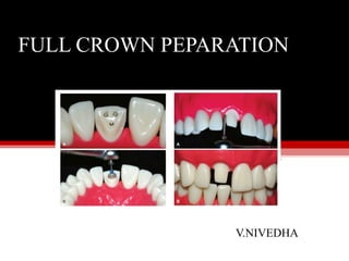
Full crown preparation
•Descargar como PPT, PDF•
303 recomendaciones•135,355 vistas

This document provides information on full crown tooth preparations, including definitions, biological and mechanical principles, and guidelines. It discusses the importance of margin location in relation to the biologic width to maintain gingival health. It also covers principles such as preservation of tooth structure, retention and resistance form, and considerations for different crest relationships to minimize risk of tissue recession. Guidelines are provided for preparation taper, height and diameter to enhance durability and resistance to dislodging forces.

Recomendados
Más contenido relacionado
La actualidad más candente
La actualidad más candente (20)
Similar a Full crown preparation
Similar a Full crown preparation (20)
Más de Nivedha Tina
Más de Nivedha Tina (16)
Último
Último (20)
Full crown preparation
- 2. CONTENTS • DEFINITION • PRINCIPLES OF TOOTH PREPARATION • REDUCTION GUIDE • FINISH LINES • BURS USED • TOOTH PREPARATION - All Metal - Porcelain fused to Metal - All Ceramic • COMMON ERRORS IN TOOTH PREPARATION • STRESSED PULP • SUMMARY & CONCLUSION • REFERENCES
- 3. DEFINITION According to the Glossary of Prosthodontic terms – 7 Full crown is described as “A restoration that covers all the coronal tooth surfaces (Mesial, Distal, Facial, Lingual and Occlusal)” • Tooth preparation may be defined as the mechanical treatment of dental disease or injury to hard tissue that restores a tooth to the original form (Tylman). • The mechanical preparation or the chemical treatment of the remaining tooth structure, which enables it to accommodate a restorative material without incurring mechanical or biological failure.(Marzouk) • The current focus is on conservative tooth preparation that is noninvasive and that minimally involves dentin.
- 4. REQIREMENTS OF TOOTH PREPARATION 1. Biologic considerations, which affect the health of the oral tissues 2. Mechanical considerations, which affect the integrity and durability of the restoration 3. Esthetic considerations, which affect the appearance of the patient
- 6. BIOLOGIC PRINCIPLES- SUMMIT Margin location is the most important biologic parameter in predictably maintaining gingival health. Even among patients receiving regular preventive dental care, subgingival margins are associated with increased probing depths and gingival inflammation. The critical factor in maintaining healthy gingival tissue is the relationship between margin location and supracrestal fiber attachment. If the restorative margins impinge on the supracrestal fiber attachment, chronic inflammation can result.
- 7. The term biologic width was given to this zone of connective tissue and epithelium by D. Walter Cohen (1962) in an unpublished presentation at Walter Reed Army Medical Center. This biologic zone was reintroduced to periodontics and restorative dentistry by Ing-ber et al The term biologic width describes a vertical measurement of 1.74 mm, the combined width of the connective and epithelial attachments. If the margin of a restoration violates the 2.04 mm biologic width, substantial gingival inflammation often results If the goal is to place a restorative margin in the sulcus without violating the biologic width, the base of the sulcus must be identified. However, this is extremely difficult. The periodontal literature indicates that the tip of the periodontal probe often penetrates the base of the sulcus and may extend into the connective tissue
- 8. BONE SOUNDING: The depth of penetration depends on the level of inflammation, the diameter of the probe, and the pressure used on the probe. Because the sulcus depth can be identified only histologically, the distance from the free gingival margin to the crest of the alveolar bone is the only predictable measurement available to determine intracrevicular margin location. At the crown preparation appointment, the entire dentogingival complex is measured. After the administration of local anesthesia, a periodontal probe is pushed through the sul-cus until resistance is felt .The probe is then angled away from the clinical crown while the tip is still touching the root surface and is pushed completely to the osseous crest . This process is called bone sounding, and measurements are taken on the midfacial aspect of the tooth and at both facioproximal line angles. If the probe is not angled correctly, there is a greater risk of the tip of the probe skipping past the thin facial plate of bone, resulting in an inaccurate measurement.
- 10. NORMAL CREST RELATIONSHIP In a normal crest relationship, the measurement from the free gingival margin to the osseous crest is 3.0 mm facially and 3.0 to 4.5 mm interproximally , which usually results in a gingival scallop of 3.0 to 4.0 mm and tissue levels that are stable in relation to the tooth. The normal crest is found on 85% of anterior teeth.In the normal crest relationship, restorative margins can be placed 0.5 to 1.0 mm into the sulcus facially and 0.5 to 2.5 mm interproximally. The apical limit of the restorative margin is 2.5 mm coronal to the osseous crest. The retraction technique is not critical in this crest relationship because the gingival level is stable. Typically, a normal crest relationship should yield no recession and no loss of papilla height following routine intervention
- 11. Research indicates that a normal crest relationship will reestablish itself even if the tissue is completely denuded, although it may take up to 3 years to return to its normal form
- 12. LOW CREST RELATIONSHIP A low crest relationship is the most difficult of all crest positions to manage and is found in 13% of anterior teeth. The relationship of the free gingival margin to the osseous crest is greater than 3.0 mm facially and greater than 4.5 mm interproximally . The gingival scallop does not mimic the osseous crest. Patients with low crests are at high risk for facial recession and loss of papilla height because of the increased distance from the alveolar crest to the gingival margin. The position of the soft tissues on the tooth is not stable in teeth with a low crest relationship and can easily be altered unintentionally duringtreatment.
- 13. If maintenance of the tissue levels is critical during restorative procedures, practitioners have two options. One option is to correct the low crest surgically before tooth preparation, creating a normal crest relationship and thus achieving predictability. This can be accomplished by reducing the tissue height with an internally beveled gingivectomy so that the gingival crest is 3 mm coronal to the osseous crest. However, if the position of the cementoenamel junction, root anatomy, gingival architecture, osseous support, or esthetic demands prevent proactive treatment, the second option is to take great care to avoid damage to the attachment during preparation and impression making. The finish line of the preparation should be located at or coronal to the free gingival margin, and there should be minimal, if any, tissue retraction during impression making. The patient should be warned of the possible tissue changes before the preparation begins and have an understanding of the treatment options if tissue loss does occur. While not predictable, thicker tissue seems more resistant to recession following intervention.
- 15. HIGH CREST RELATIONSHIP Patients with high crests are the least common (2%) and pose the greatest risk for violation of biologic width.Probing measurements are less than 3 mm facially or interproximally . The tissue levels are very stable, and the gingival scallop is flat, less than 3 mm. The high crest relationship sometimes occurs when excessive tissue covers the anatomical crown, such as in altered passive eruption or in patients with noncarious cervical lesions. However, it is most common adjacent to edentulous spaces where the gingival scallop has flattened. Margin location is determined by the demands of biologic width. High-crest teeth, by definition, will only allow an equigingival or supragingival restorative margin because of the short distance to the alveolar crest.
- 16. These teeth are at high risk for biologic width impingement with intracre-vicular margin placement. Gingival retraction for impressions should be minimal.
- 17. PULP PRESERVATION Preoperative radiographs and pulp testing are important steps in determining pulp vitality prior to tooth preparation. Unfortunately, pulp testing cannot identify degrees of health. Separately or cumulatively, the effects of large restorations, leaking restorations, caries lesions, deep cracks, pins, etc, increase the chances of pulpal necrosis after tooth preparation. In one study, irreversible pulpitis occurred in 5.7% of cases in which crowns were placed on vital teeth. Patients should be made aware of this risk preoperatively. If the tooth preparation involves an increase of heat to the tooth, pulpal necrosis can occur. In one in vivo study in primates, pulpal injury occurred in 15% of teeth with a 5.5°C rise in temperature.
- 18. An 11.1°C rise led to necrosis of the pulp in 60% of the teeth, and a 16.6°C rise caused necrosis of all the teeth tested. Temperature changes have been monitored during complete crown preparation. When a airwater spray coolant was used, a temperature decrease in the pulp chamber from 37°C to 25°C after 4 minutes of exposure occurred. However, when only air coolant was used, the pulpal temperature rose from 37°C to 48°C after 1 minute of continuous exposure. Therefore, continuous air-water coolant is a critical factor in maintaining pulpal health.
- 19. PRINCIPLES OF TOOTH PREPARATION (MECHANICAL) Preservation of tooth structure Retention & Resistance Structural durability Marginal integrity Preservation of the periodontium
- 20. PRESERVATION OF TOOTH STRUCTURE A. PRESERVATION/CONSERVATION Excessive reduction results a. Loss of retention and resistance (need area of structure) b. Thermal hypersensitivity (stripped insulation) c. Pulpal inflammation and necrosis (trauma) So We Do Necessary reduction. To preserve and to protect - we must properly design the preparation (Reduce enough for durability of the restorative material)
- 21. RETENTION AND RESISTANCE FORM Retention is the feature of a crown preparation that resists dislodgment in a vertical direction or along the path of placement. In 1926, Ward became the first practitioner to establish a standard for preparation taper. He prescribed 5% to 20% per inch or 3 to 12 degrees. Since then, recommendations have ranged from 3 to 5 degrees, to 6 degrees, to 10 to 14 degrees. Jørgensen indicated that there is a 50% reduction in retention when going from 5 to 10 degrees of taper. These tenets are heavily based on clinical empiricism and on two experiments in which crown and abutment analogs were pulled apart along their paths of insertion. The theoretical benefits of preparations with minimal convergence angles do not withstand scrutiny, and such preparations are difficult to produce in clinical practice..
- 22. Divergence from parallel might have to be as much as 12 degrees to be observed and produced clinically. Routine preparations in practice have been measured at between 15 and 36 degrees without apparent detrimental effect to the longevity of the restorations. For non adhesively cemented restorations, the minimum convergence value required clinically is unknown, although total convergence up to 20 degrees has been shown to be acceptable Today, crowns with greater taper may be cemented with adhesive resin cements, minimizing the need to prepare the crowns to minimal convergence angles
- 23. Magnitude of dislodging forces Forces that tend to remove a cemented restoration along its line of draw are small and rare. This can happen with sticky food and by pulling with floss under connector. The magnitude of these forces depends on stickiness of food, surface area of contact and texture of restoration being pulled. Surface area •Greater the surface area greater is the retention. Preparations on large teeth are more retentive than preparation on small teeth. Molar crowns are more retentive than premolar crowns of similar taper Surface area can be increased by adding boxes and grooves. However, they may be actually limiting freedom of movement than increasing surface area.
- 24. Freedom of displacement • Retention is improved by geometrically limiting the paths along which the restoration can be removed from the prepared tooth. Maximum retention is achieved when there is only one/ single path of removal • Grooves and proximal boxes can be used to increase retention by limiting the freedom of displacement in overtapered preparation and in the absence of oppos“ ing axial wall.
- 25. Stress concentration Retentive failure begins at junction of axial and occlusal surfaces where stress is concentrated. This then results in cohesive failure of the entire cemented area. Thus, rounding of line angles will reduce stress concentration and enhance retention. Type of restoration Preparations on different restoration designs have different retentive values when other factors are kept constant. Thus a complete crown is more retentive tha partialcoverage restorations
- 26. PATH OF INSERTION Definition: The specific direction in which prosthesis is placed on the abutment teeth (GPI'8). • Path of insertion is an imaginary line along which the restoration will be placed onto or removed from the preparation; also called path of placement. It is determined before beginning the preparation and the preparation is then planned to coincide with that imaginary line. In case of fixed partial dentures, it is important to keep the paths of all the abutments parallel to each other and the path of insertion to ensure a smooth fit of the restoration. • The path of insertion must be considered in two dimensions - faciolingually and mesiodistally.Faciolingual orientation can affect the aesthetics of the metal-ceramic crown or partial veneer crowns. For metal-ceramic crowns, it should be parallel to the long axis of the teeth. For partial veneer crowns, it should be parallel to the incisal half of the labial surface.The mesiodistal inclination should parallel the contact areas of adjacent teeth
- 27. To visually check a preparation for undercuts and taper, the centre of occlusal surface of the preparation is viewed with one eye from a distance of 30 em or 12 inch. If it is viewed with both eyes open, undercuts will be not be seen. In the mouth, a mouth mirror is held at an angle 0.5 inch above the preparation and preparation viewed with one eye . To view multiple abutments as in fixed partial dentures, the mirror is moved without changing the angulation from one abutment to another, after establishing a firm finger rest.
- 28. Roughness of the surfaces being cemented • The internal surface of casting is most effectively roughened by air-abrading with 50 microns of alumina. It has been seen that this increases retention by 64%. Acid- etching the fitting surface can also improve retention with certain luting agents. • But the tooth should not be roughened during preparation as this makes it difficult to make impressions and fabricate the prosthesis. Materials being cemented • More reactive the alloy better is retention. Hence, base metal alloys are better retained than gold alloys • Effect of retention to different core buildup materials is not consistent.
- 29. Types of luting agent In decreasing order, cement retention is best obtained with adhesive resin followed by glass ionomer, polycarboxylate, zinc phosphate and zinc oxide eugenol. However, the decision regarding which agent to use is also based on other factors.
- 30. RESISTANCE FORM: Resistance is the feature of a tooth preparation that enhances the restoration’s stability and resists dislodgment along an axis other than the path of placement. Most retention studies utilize conventional pull-type tests to evaluate preparations and/or cements. However, data on functional force vectors in the oral environment strongly suggest that these lift-off type forces are virtually nonexistent in the mouth. During chewing, teeth are subjected to alternating combinations of buccolingual and occlusogingival forces.These studies indicate that stresses that cause failure of an anterior restoration are repeated perpendicular or oblique forces. Therefore, as Caputo and Standlee concluded, “Resistance form is the most important factor that must be designed into any restoration if it is to succeed in function.” Resistance clinically is multifactoral. It is based on preparation taper, height, diameter, and cement type. Crowns generally loosen and fail by cleavage of the cement attachment without damaging the abutment or restoration. The cement
- 31. forces is unable to withstand load application. Attachment failure is a progressive phenomenon linked to increasing abutment taper. Increasing the preparation taper from 10 to 20 degrees creates a broader stress distribution and greater stress within the cement. The stress fields are consistently higher with greater taper and less surface area. The more parallel the walls of a preparation, the less stress on the cement lute irrespective of the preparation height. Taper becomes particularly important in teeth with a lower preparation surface area, such as an anterior tooth. Clinically, a minimal preparation taper decreases the damaging effects of occlusal stress on the cement attachment, improving a crown’s resistance even more than auxiliary preparation features like grooves or boxes. The height and diameter of the final preparation are also related to resistance. Resistance is increased by lengthening the axial walls of the preparation. The minimum height for resistance is one-half the diameter of the tooth.
- 32. This means that, on average, an anterior preparation must be 3.5 mm and a posterior preparation 4.0 mm in height. Of the total preparation height, the gingival 2.0 mm of the preparation must be on sound tooth structure to provide a proper ferrule, and the other 1.5 mm or more can be in either tooth structure or buildup material A ferrule is the marginal band of a crown that contacts tooth structure, providing protection from masticatory forces. In addition, the ferrule requires a dentin thickness of 1 mm from the external surface of the crown preparation to the wall of any endodontic preparation. In a comparison of in vivo restorations with and without 2 mm of remaining coronal tooth structure, the failure rate of the < 2 mm group was four times that of the control (26.20% and 6.67%, respectively)
- 34. Resistance is also affected by the mechanical properties of the cement. The limiting threshold of each crown is the cement’s resistance to fatigue under cyclic stress. The more stress that will be placed on the cement, because of a severe taper and/or lack of preparation height, the more resistant the cement must be. Resinmodified glass- ionomer cements are more resistant than conventional glass-ionomer cements, which are in turn more resistant than zinc phosphate cements. Most research on resistance and retention data is conducted on nonadhesively cemented crowns. Today, however, adhesive cements allow the placement of crowns that do not meet standard taper and length requirements. Yet, because the hydrodynamic nature of dentin bonding makes it unpredictable, it is suggested that all crown preparations meet minimum requirements
- 35. STRUCTURAL DURABILITY Structural durability is the relationship between occlusal stress and material strength. It ensures that a restoration does not deform or fracture under load. In a metal- ceramic crown, the minimum metal thickness under porcelain is 0.4 to 0.5 mm for gold alloys and 0.2 mm for base-metal alloys. If the metal is too thin, it will flex under load, resulting in possible porcelain fracture. The minimum porcelain thickness over metal is 0.9 mm (0.2 mm for the opaque material and 0.7 mm for body porcelain). Ceramists prefer a 1.3- to 1.5-mm reduction for the axial surfaces of metal-ceramic crowns and a 2.0-mm reduction incisally/occlusally. The greater the reduction, the easier it is to mask the opaque material in the gingival third of the crown with body porcelain
- 36. Most bonded all-ceramic crowns require a minimum thickness of 1.0 mm to provide esthetics and adequate strength. Cemented all ceramic crowns require an average circumferential tooth reduction of 1.5 mm for strength. OCCLUSAL REDUCTION:
- 37. FUNCTIONAL CUSP BEVEL Wide bevel on- Lingual inclines of the maxillary lingual cusps Buccal inclines of mandibular buccal cusps Adequate bulk of metal in area of heavy occlusal contact
- 38. LACK OF FUNCTIONAL CUSP BEVEL
- 39. MARGINAL INTEGRITY A completely closed margin is unattainable clinically. Even the finest margins are not sufficiently closed to prevent bacterial ingress. To place it in perspective, the width of a human hair is 50 μm; bacteria responsible for caries are 4 to 5 μm in diameter. Because bacteria are constantly passing under restoration margins, patient resistance to disease is more important than the marginal opening of crowns. What, then, is an acceptable marginal opening? One study reported that when the margin of an inlay or onlay could not be visualized, a marginal discrepancy of 119 μm was found to be acceptable. Björn et al reported that 83% of gold and 74% of porcelain crowns exhibited marginal defects; more than half were greater than 200 μm..
- 40. However, defective margins are to blame in only 10% of failed restorations. While all practitioners should strive for the finest margins possible, it is impossible to achieve a closed margin. The best possible margin enables the patient to floss and care for the restoration, minimizing cement dissolution and maximizing the patient’s natural resistance factors. Because 100 μm is the smallest detectable ledge, this can be used as a practical criterion for evaluating fit What finish line design provides the best marginal integrity? Traditionally, the margin design selected does not have a significant effect on marginal seal of metalceramic restorations. Shoulder ,shoulder-bevel , and Chamfer finish line preparations all allow acceptable marginal fit when complete seating is achieved. The shoulder-bevel margin is the least esthetic choice. The bevel should be used only with metal-ceramic crowns and is suited for structurally compromised teeth where ferrule extension is important. If porcelain is placed on a bevel, the cementation process may cause porcelain breakage.
- 41. In two studies, the geometry exhibiting the least marginal discrepancy after cementation was a shoulder preparation, which was significantly better than that of a shoulder bevel or chamfer. The shoulder finish line is thought to be better than the beveled shoulder because it allows the excess cement to escape more readily. The shoulder design exhibits less marginal distortion than a chamfer because of the crown’s thickness adjacent to the margin. Stress analysis of various margin finish lines showed that chamfer and internally rounded shoulder preparations had the lowest stress concentration when loaded vertically, reducing the risk of catastrophic ceramic failure under load. When utilizing metal-ceramic crowns, porcelain margins must provide an esthetic transition from tooth to crown, preventing the margin from becoming the visual focal point. Such margins are easier to fabricate and more predictable when they are fabricated on a 90-degree shoulder preparation
- 42. This is true for metal-ceramic and most all-ceramic systems. A chamfer requires minimal axial reduction and is appropriate for conservative all-ceramic restorations. It does not, however, provide an adequate reduction for metal-ceramic crowns. The opaque material used to mask the metal at the margin will show through the more translucent porcelain and will compromise the esthetics in the gingival third. A 90-degree shoulder between 1.0 and 1.5 mm in depth allows for a precise margin, maximum seating, and good esthetics.
- 44. The ideal finish line form and depth for all-ceramic systems vary between type of ceramic and fabrication technique. They all have two common elements: adequate axial depth and uniform smoothness. A study on the effect of preparation design on the marginal integrity of Procera crowns highlighted this fact. It demonstrated that a feather-edge finish line produced crowns with over twice the marginal opening of a chamfer or rounded shoulder finish line. Furthermore, the deeper axially the rounded shoulder, the tighter the marginal fit. Crowns created from IPS Empress CAD material exhibited similar results. The best finish line is a rounded axialgingival line angle with a butt-joint external cavosurface margin. Ideal cervical preparation taper is imperative with all-ceramic crowns. Ceramic systems cannot accurately reproduce the contours of auxiliary grooves or deep occlusal morphology, eliminating their use as resistance features. In addition, adequate occlusal/incisal reduction is more critical with all-ceramic than with metal-ceramic restorations. Inadequate space prevents the use of monoblock all- ceramic crowns and can compromise esthetics and strength.
- 45. PRESERVATION OF THE PERIODONTIUM: Margin placement Direct effect on ultimate success of restoration Margins should be as smooth as possible Placed in area that can be finished well by the dentist and kept clean by the patient Placed in enamel whenever possible Should be supragingival whenever possible
- 46. SUPRA GINGIVAL MARGINS: Supragingival margins Less potential for soft tissue damage Easily prepared and finished More easily kept clean Impressions are more easily made Restorations easily evaluated at recall appointments
- 47. SUBGINGIVAL MARGINS: Esthetics Existing caries, cervical erosion, or restorations extend subgingivally, and crown- lengthening is not indicated Proximal contact area extends to the gingival crest Additional retention is needed Margin of a metal-ceramic crown is to be hidden behind the labiogingival crest Root sensitivity cannot be controlled by more conservative procedures, such as the application of dentin bonding agents
- 48. Finish line should not be closer than 2mm to the alveolar crest Placement in this area causes gingival inflammation loss of alveolar crest height pocket formation
- 49. REDUCTION GUIDES for functional crown preparation When the teeth are severely worn or malpositioned or the vertical dimension is to be altered in the definitive restorations, the dentist will require a guide to allow proper reduction . Multiple reduction-matrix designs have been proposed in the literature. Most require the tooth to be prepared, after which the matrix is placed to confirm that the tooth was reduced adequately. The problem is that there is no recovery when the practitioner removes too much tooth structure. A more accurate guide would encourage the proper depth of reduction in all dimensions without the risk of blindly overreducing the tooth. Aesthetic preevaluative temporaries (APTs) were developed as a preoperative opportunity for the patient to evaluate the esthetics of veneer restorations. This same technique can be used as a guide for proper tooth reduction for an anterior crown.
- 50. The APT is fabricated from a properly contoured diagnostic wax-up .Condensation silicone is adapted directly on the cast. It is placed in a pressure pot at 20 psi for 15 minutes This will compress the silicone into the contours of the wax-up, producing a highly accurate matrix. A notch is made between the central incisors to be used as a seating guide intraorally. Deep cuts on the buccal aspect extending to the teeth or vent holes through to the teeth are made in the silicone, creating a spillway for the excess acrylic resin .Without this escape route, the acrylic resin will not flow, preventing the matrix from fully seating. If the matrix does not seat properly, it creates an error in the reduction. After the patient is anesthetized, the matrix is loaded with a provisional material (made from a bis-acrylic resin) and seated. After the material is allowed to set, the matrix is removed. The acrylic resin will remain on the teeth, engaging the embrasures .
- 51. There will be a variety of thicknesses of the acrylic resin depending on the amount of ideal tooth morphology missing. This is the proposed position and contour of the definitive restorations. Proper reduction cuts are made into the matrix, and then the remaining resin is removed Any bur cuts on the teeth indicate the location and depth of reduction that is necessary. If there is no mark on the tooth, it is already reduced enough for the definitive restoration ,although some preparation may be necessary to provide gingival margins and an appropriate path of draw.
- 53. FINISH LINES: Chamfer Is choice for most of the veneer cast metal restorations. It produces provides desired acute margins of the metal with sufficient thickness for strength.
- 54. It is an obtuse-angled finish line. • It is distinct. • Exhibits least stress. • Most conservative. • Indicated where metal forms the margin of a restoration, e.g. complete metal crown. Should not be given for porcelain restorations as the obtuse angle produces shearing forces which is not well tolerated by porcelain. • It is prepared using a round-end tapering diamond. This is produced by sinking in half the diamond into the tooth. The tip produces the chamfer while the sides give the necessary taper to the axial surface
- 55. HEAVY CHAMFER
- 56. SHOULDER Shoulder finish line: It provides an adequate bulk of the material at margins and is recommended for porcelain restorations.
- 58. RADIAL SHOULDER
- 59. SLOPING SHOULDER
- 60. KNIFE EDGE
- 62. BURS: Shape Use Round end tapered diamond 1.Depth orientation grooves 2.Occlusal reduction 3.Functional cusp Torpedo diamond 1.Axial reduction 2.Chamfer finish line Short needle 1.Initial interproximal axial reduction in posterior teeth Long needle 1.Initial proximal axial reduction in anterior teeth
- 63. Small wheel diamond 1. Lingual reduction in anterior teeth Tapered fissure bur (171L) 1.Seating groove 2.Proximal groove (posterior teeth 3.Smoothing and finishing 4.Occlusal and incisal bevels Tapered fissure burs (169L & 170L) 1.Initial groove alignment 2.Angles of proximal boxes 3.Smoothing and finishing 4.Occlusal and incisal bevels
- 64. End cutting bur Conventional shoulder finishing Torpedo bur 1.Axial wall finishing 2.Chamfer finishing Flame bur 1. Flare and bevel finishing
- 65. FULL VENEER CROWN ALL METAL PORCELAIN FUSED TO METAL ALL CERAMIC
- 66. COMPLETE CAST CROWN INDICATIONS On teeth that exhibit extensive coronal destruction On Non-esthetic zone teeth with extensive restorations Short clinical crowns Retainer for a long span fixed partial denture On endodontically treated teeth In cases of generalized attrition where vertical dimension is reduced.
- 67. CONTRAINDICATIONS If treatment objectives can be met with a more conservative restoration Extensively restored or cariously involved teeth within esthetic zone
- 68. ADVANTAGES • Greater retention and resistance than a more conservative restoration • Strength is superior to that of other restoration • Permits easy modification of the occlusion • Protects the coronal integrity of a natural tooth compromised by restorations
- 69. DISADVANTAGES It is no longer feasible to perform electric vitality testing of the abutment tooth
- 70. TOOTH PREPARATION ARMAMENTARIUM Handpiece Round ended tapered diamond 171L bur Torpedo diamond Torpedo bur Short needle diamond Red utility wax
- 71. Procedure of tooth preparation Place the round ended tapered diamond bur approx . 1mm deep in central , mesial , and distal fossae, and creating about 1.5 mm of clearance on the functional cusps and 1mm on the non-functional cusps.
- 72. Place these grooves in the buccal and lingual developmental grooves and in each triangular ridge extending from the cusp tip to the centre of its base.
- 73. On the non-functional cusp, the groove should parallel the intended cuspal inclination, on the functional it should be slightly flatter to ensure additional reduction of the functional cusp.
- 74. Place depth orientation grooves for a functional cusp bevel across the facial occlusal line angle of the mandibular premolar or molar, and across the lingual occlusal of a maxillary tooth
- 75. Round ended tapered diamond is used to give bevel parallel to the inward facing inclines of the opposing tooth, at a depth of 1.5mm, usually forming a 45 degree angle with the axial wall.
- 76. Complete the occlusal reduction in two steps. Half of the occlusal surface is reduced first so that the other half will be maintained as reference.
- 77. After completing the occlusal reduction the clearance can be checked by having the patient to close on a 2mm thick strip of red utility wax.
- 78. Alignment grooves for axial reduction With the help of narrow round end tapered diamond bur three alignment grooves are placed in each buccal and lingual wall.
- 79. When grooves are placed be sure that the shank of the diamond is parallel to the proposed path of withdrawal. when diamond with a 6 degree taper is used, an ideal axial convergence on the preparation wall will result.
- 80. Gingivally the resulting depth of preparation should be not more than half the width of the tip of diamond otherwise a lip of unsupported enamel will be created.
- 81. While doing axial reduction cut into the proximal area from both sides until a few mm of interproximal area remains.
- 82. Now a thin diamond is worked through the proximal area in an occlusogingival or buccolingual sawing motion carefully avoiding the adjacent teeth.
- 83. • Once sufficient room is created the torpedo diamond is introduced to plane the walls while simultaneously forming a chamfer as the interproximal gingival finish line.
- 84. All the axial surfaces are smoothened with a torpedo carbide finishing bur which will also finish the chamfer finish line. Special care should be taken in rounding the corners from the buccal or lingual surfaces to the proximal surfaces.
- 85. The final step is the placement of a seating groove. It will prevent the rotational tendencies during cementation and will help the casting to guide in place.
- 86. PORCELAIN FUSED TO METAL CROWN Indications • On teeth that require complete coverage and where significant aesthetic demands are placed • If all ceramic crown is contraindicated
- 87. Contraindications • Patient with active caries or untreated periodontal disease • In young patients with large pulp chambers • Whenever a more conservative retainer is technically feasible. • If there is intact buccal wall
- 88. Advantages Superior esthetics as compared to cast metal crowns Disadvantages • Requires significant tooth reduction • For esthetics, margin is placed subgingivally . . . . • Subject to fracture . . . . • Difficult to obtain accurate occlusion in glazed porcelain • Shade selection can be difficult.
- 89. Posterior porcelain fused to metal crown preparation Lab knife with no. 25 blade Silicone putty with accelerator Handpiece Flat ended tapered diamond Short needle diamond Torpedo diamond Torpedo bur Radial fissure bur Biangle chisel Armamentarium
- 90. PREPARATION Silicone putty is adapted to the facial , lingual, and occlusal surfaces of the tooth to be prepared.
- 91. After polymerization silicone is cut half into faciolingual midline of the tooth. the more conventional facial index is fabricated by cutting along the facial cusps of the teeth, the occlusal portion is discarded and gingival portion can be used as an index.
- 92. Occlusal reduction • Round end tapered diamond is used to give depth orientation grooves. In the areas of ceramic coverage reduction should be 1.5 to 2mm
- 93. Depth orientation grooves for functional cusp bevel is provided on the lingual inclines of the maxillary lingual cusps and facial inclines of the mandibular facial cusps. they provide a uniform bulk of restorative material. the depth is 1.5 mm if covered by metal and 2mm if veneered by porcelain.
- 94. Axial reduction Cut three vertical grooves in the occlusal portion of the facial surface. These are placed with full diameter of the instrument fading out in the area where the facial surface is most curved.
- 95. Now align the bur along the gingival component and place atleast 2 more grooves near the line angles of the tooth. the tip of diamond should be supragingival at this time. Now remove the facial surface with the flat end tapered diamond
- 96. If facial reduction is less than1.2mm for a base metal crown or 1.4mm for a noble metal ceramic crown . . . . . . . . .
- 97. Proximal axial reduction is begun with short needle diamond Proximal surface planed with needle diamond.
- 98. The lingual axial wall is reduced with a torpedo diamond. produce adequate reduction on the lingual and proximal wall to create a distinct chamfer finish line.
- 99. At the lingual most extension of the proximal contact the transition from a deeper facial reduction to the relative shallower lingual reduction results in vertical wall or “wing” of tooth structure. If the wing is not lingual to the proximal contact . . . . Besides conserving the tooth structure the wings add to the torque resistance to the preparation.
- 100. Finishing is done with – 170 bur on the occlusal surface - end cutting bur on the facial surface
- 101. • The facial silicone and midsaggital silicone putty index shows uniform reduction on the facial surface and occlusogingivally.
- 102. ALL CERAMIC CROWN INDICATIONS • In areas with a high esthetic requirement where a more conservative restoration will be inadequate. CONTRAINDICATIONS • Where a more conservative restoration can be used • Not indicated in molar tooth because of increased occlusal load and reduced aesthetic demand.
- 103. ADVANTAGES Superior aesthetics DISADVANTAGES Reduced strength Proximal and lingual reduction are less conservative Not effective as retainers for fixed partial dentures Wear has been observed on the natural teeth opposing all ceramic restoration If inadequate tooth preparation goes uncorrected can result in fracture.
- 104. All ceramic crown tooth preparation A combination of facial and lingual index is made by adapting silicone putty to the facial, lingual ,and occlusal surface of the posterior teeth. This will provide an accurate reference for both facial and lingual reduction
- 105. OCCLUSAL REDUCTION Use large round end tapered diamond to place depth orientation grooves on the occlusal surface. The final occlusal preparation should be between 1.5mm to 2mm.
- 106. Remove the tooth structure remaining between the grooves following inclined planes of occlusal surface.
- 107. Use the same round ended tapered diamond to produce depth orientation grooves for the functional cusp bevel Create the functional cusp bevel to insure that the facial incline of the facial cusp will have the same porcelain thickness as the lingual incline..
- 108. Check the occlusal reduction by asking the patient to bite on a 1.5mm leaf of thickness gauge
- 109. Make depth orientation grooves on the facial and lingual surfaces to insure that adequate reduction of tooth with a minimum thickness of 1mm at gingival finish line 1.5mm at slightly less at mid-crown Remove the remaining tooth structure between the grooves with the help of large round end tapered diamond so that shoulder with a rounded internal line angle can be formed.
- 110. Now use this short needle diamond to begin the proximal axial reduction. As more space is created the needle diamond can be brushed across to produce more reduction.
- 111. Use round end tapered diamond to blend the proximal axial reduction and shoulder of facial and lingual surfaces. Finishing of preparation- round end tapered diamond bur..
- 112. Now use the horizontal and mid Sagittal index to reveal the amount of facial and lingual and occlusal reduction
- 113. STRESSED PULP T he term “stressed pulp” describes a vital dental pulp that has been subjected to repeated damage, including operative trauma, accidents, or other pathologic changes. The stressed pulp condition is a clinical concept and not a histologic entity.
- 114. Operative procedures and materials that cause pulpal injury and may lead to pulpal stress include the following: Deep tooth preparation (where less than 2 mm of remaining dentin covers the pulp) High-speed tooth cutting without coolant Application of topical medications on cavity preparations’ Continuous air-drying of preparation Cement with high acidity Direct pulp capping Local anesthesia of long duration (while full-crown preparation is made) Unlilled resin restoration Unbased restoration allowing for thermal conductivity
- 115. The following diseases, conditions, and treatment cause pulpal injury and may lead to pulpal stress: Chronic bruxism Chronic caries Chronic periodontal disease’ Chronic trauma from occlusion’ Chronic occlusal attrition and erosion Cracks in tooth structure Radiation therapy Systemic diseases of oral manifestation Diabetes, vitamin C deficiency, leukemia, endocrine disturbances
- 116. The ability of the pulp to recover from pathologic and operative trauma is related to: Type of injury. Mild injury is believed to be of no significance, and repair can be expected. Duration of injury. A primary injury of short duration provides the best chance for recovery. Thickness of remaining dentin. Remaining dentin between the cavity floor or crown preparation and the pulp is necessary to protect the pulp. If the remaining dentin is less than 2 mm thick,4 trauma to the pulp becomes more damaging and the recovery chances are reduced. Physiologic age of the tooth. The pulp chamber and apical foramen size should be large enough to allow adequate vascular flow to the tooth. A receded pulp chamber or pulp chamber filled with calcification stones or reparative dentin may not have a good reparative potential due to its deprivation of vascular and cellular elements.“’ Host factors. Patient age and systemic health play an important role in pulp recovery potentials. Young, healthy patients have better healing responses
- 117. MANAGEMENT: Endodontics should be considered strongly before beginning restorative therapy, orthodontics, or other traumatic procedures on teeth with stressed pulps.
- 118. COMMON ERRORS IN TOOTH PREPARATION Insufficient occlusal reduction Lack of uniform reduction on buccal surface comprising esthetics Minimal axial reduction on the buccal and lingual surfaces Over reduction of teeth and violation of biologic width Excessive taper of proximal surfaces Variable shoulder width Undercuts on the distolingual surface of the preparation Rough tooth preparations.
- 119. SUMMARY
- 122. CONCLUSION The removal of all morphologic form of the tooth is a radical treatment and restoring it properly can be difficult. The full veneer crown is a restoration that replaces lost tooth structure and imparts some measure of structural support to the tooth. Hence, one must be able to judge correctly the type of restoration required for each individual tooth and try to follow the guidelines for the respective tooth preparation.
- 123. REFERENCES Shillingburg(1981) Fundamentals of Fixed Prosthodontics 2nd edition Quintessence Shillingburg (1987) Fundamentals of Tooth Preparation for Cast metal and Porcelain restoration. Quintessence Pub. Co. C.J.Goodacre –designing tooth preparation for optimal success.DCNA 2004;48:359-385. Textbook of Prosthodontics - V Rangarajan & T V Padmanabhan - 2nd Edition (2017) SUMMIT 4 TH EDITION