SlideShare una empresa de Scribd logo
1 de 14
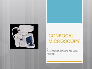
CONFOCAL
MICROSCOPY
By:
Noor Munirah bt Awang Abu Bakar
P82498
Confocal microscopy-Outline
Introduction: What is confocal microscopy
Clinical use of confocal microscopy
Type of confocal microscopy available
Confoscan 4
Optic of confocal microscopy
Anatomy of cornea layer
Intro: Confocal microscopy
 A non-invasive histological imaging technique to demonstrate the
characteristic of corneal and conjunctival anatomy in vivo at the
cellular level.
 Use reflected light from the living tissue. Therefore, it is an in vivo
imaging method of the living cornea.
 Record normal corneal innervation and cell distribution, as well as
changes associated with age, contact lens wear and systemic disease
such as diabetes.
(Elisabeth, 2008)
Clinical use
 Observation and characterization of the corneal layers and cells
 Detection and Management of Corneal Dystrophies & Ectasies
 Detection and Management of Pathologic Infectious Conditions
 Pre and post surgical evaluation (PRK, LASIK and LASEK, flap
evaluations and Radial Keratotomy)
 Monitoring contact lens induced corneal changes
 Diagnosing Peripheral Neuropathies
 Penetrating keratoplasty
Type of confocal microscope
 Four types of confocal microscopes are commercially available:
1. Confocal laser scanning microscopes
use multiple mirrors (typically 2 or 3 scanning linearly along the x and the
y axis) to scan the laser across the sample and "descan" the image across a
fixed pinhole and detector.
Example: Confoscan 4 by NIDEK
2. Spinning-disk (Nipkow disk) confocal microscopes
3. Microlens enhanced or dual spinning disk confocal microscopes
4. Programmable array microscopes (PAM)
Confoscan 4
 The only instrument that combines confocal microscopy, endothelial
microscopy and accurate pachymetry in one compact unit.
 High Precision
 Wider measurement area (up to 1000 cells/exam)
 Full cornea, endothelium or epithelium scan
 Fully automatic cell count and endothelial density measurement
 Confocal Microscope with 40X Probe or 20X probe
 Gel immersion exam
 Examination time below 15 sec.
 Fully non-contact (12 mm working distance in air)
 High quality imaging through corneal haze and opacities
 ±5 microns instrumental accuracy
Optic of confocal microscopy
1. It uses focused light or laser beam.
2. A bright light beam is projected and focused through an objective lens to the cornea.
3. Then reflected light spot is collected from the illuminated tissue area by objective lens.
4. With the help of beam splitter, reflected light is separated from the light mixture and directed to the
detection unit.
5. Reflected light reaches to the detection apparatus by passing a pinhole.
6. The pin-hole at the entrance of detector apparatus filters the light coming from outside the intended
focal point. This filtration helps grabbing sharper and clearer images than conventional light microscopy.
7. Detector transcodes the reflected light into electrical signal and records to the storage media.
Anatomy of corneal layer
 Cornea is a transparent and avascular layer of
the eye that cover front part of the eye.
 Funtion: To refract or bend and focusing light
that enters the eye.
 Consists of 5 layers
 Corneal epithelium
 Bowman’s layer
 Corneal stroma
 Descemet’s membrane
 Corneal endothelium
Anatomy of corneal layer –
1. Epithelium
Superficialcells
• Depth 0 μm,
~50mm in diameter
• Density ~850
cells/mm2
• Bright cell borders
and a dark nucleus
& cytoplasm are
readily visualized
• Shape: often
hexagonal
Wingcells
• Depth 20 μm,
~20mm in diameter
• Density ~5,000
cells/mm2
• Bright cell borders
and a dark
cytoplasm
• Shape: minimal
variation shape &
size
Basalcells
• Depth 30 μm,
~10µm in diameter
• Density ~9,000
cells/mm2
• Bright cell borders
and nucleus not
visible
• Shape: minimal
variation shape &
size
Anatomy of corneal layer
No Corneal layer Confocal image
2. Bowman’s layer
• An acellular layer
• 8-14 µm thick
• Composed of randomly arranged collagen fibers (finer than
those found in corneal stroma, continuous with anterior
stroma.
• Resistant to trauma.
• Cannot be regenerated if destroyed
3. Stroma layer
• Thickness: 0.5 mm,
• 90 % of total corneal thickness
• Consists of collagen fibrils (lamellae) and cells embedded in
hydrated matrix of proteoglycans (ground substance).
• Lamellae arranged parallelly (200 – 250 layers)
• Cells: keratocytes
< Anterior stroma
Posterior stroma>
Anatomy of corneal layer
No Corneal layer Confocal image
4. Descemet membrane
• Represents the basement membrane of the endothelium
• Made up of collagen and glycoprotein
• Thickness :10-12 µm
• Very resistant to chemical agents, trauma, infection and pathological
processes, can regenerate, when destroyed.
• Maintains integrity of the eyeball,
• Images with CM-hazy appearance and no cellular structures can be
identified.
• Normal Descemet’s membrane is not visible in young subjects, becomes
more visible with increasing age (Hollingsworth, 2001)
5. Endothelial
• Single layer of flat hexagonal cells, Mosaic pattern
• Bright cell bodies with dark cell borders
• 4-6 mm thick, 20µm in diameter
• Cell density: 2400-3000cells/mm2
• Contains active pump mechanism
• Best evaluated by specular microscopy
Example
Fuch’s
dystrophy
Keratoconus with no
CL wear
Keratoconus associated with
RGP wear
Conclusion
 The introduction of in vivo confocal microscopy into the research and
clinical practice is a revolutionary approach for both inherited or
acquired corneal diseases.
 Able to demonstrate the characteristic corneal and conjunctival
anatomy at the cellular level.
 It allowed the clinician in vivo microscopic analysis of
 affected corneas like corneal guttata and Fuch dystrophy
 wound healing response after refractive surgery
 screening of unaffected family members.
References:
 Elisabeth M. Messmer,E.M. Confocal Microscopy: When Is it Helpful to Diagnose Corneal and
Conjunctival Disease?.Rev Ophthalmol. 2008;3(2):177-192.
 Hollingsworth J, Perez-Gomez I, Mutalib HA, et al. A population study of the normal cornea using
an in vivo, slit-scanning confocal microscope. Optometry and Vision Science. 2001;78:706–11.
 Umit Yolcu, Omer Faruk Sahin and Fatih C. Gundogan (2014). Imaging in Ophthalmology,
Ophthalmology - Current Clinical and Research Updates, Associate Prof. Pinakin Davey (Ed.), ISBN:
978-953-51-1721-6, InTech, DOI: 10.5772/58314. Available from:
http://www.intechopen.com/books/ophthalmology-current-clinical-and-research-updates/imaging-in-
ophthalmology

Más contenido relacionado

La actualidad más candente

Corneal topography wavefront analysis
Corneal topography wavefront analysisCorneal topography wavefront analysis
Corneal topography wavefront analysisikramdr01
 
Principles of optical coherence tomography
Principles of optical coherence tomographyPrinciples of optical coherence tomography
Principles of optical coherence tomographyJagdish Dukre
 
OPTICAL COHERENCE TOMOGRAPHY ANGIOGRAPHY
OPTICAL COHERENCE TOMOGRAPHY  ANGIOGRAPHY  OPTICAL COHERENCE TOMOGRAPHY  ANGIOGRAPHY
OPTICAL COHERENCE TOMOGRAPHY ANGIOGRAPHY Shruti Laddha
 
OCTA Optical Coherence Tomography angiography
OCTA Optical Coherence Tomography angiographyOCTA Optical Coherence Tomography angiography
OCTA Optical Coherence Tomography angiographyMohammad Abusamak
 
Electrophysiology of the Eye
Electrophysiology of the EyeElectrophysiology of the Eye
Electrophysiology of the EyeVedant Murkey
 
OCT – Introduction and Macular disorders
OCT – Introduction and Macular disordersOCT – Introduction and Macular disorders
OCT – Introduction and Macular disordersSajjan Sangai
 
Optical coherence tomography(OCT) --macula
Optical coherence tomography(OCT) --maculaOptical coherence tomography(OCT) --macula
Optical coherence tomography(OCT) --maculaAkshay Nayak
 
Wound healing of cornea
Wound healing of cornea Wound healing of cornea
Wound healing of cornea Panit Cherdchu
 
Electrophysiology of retina
Electrophysiology of retinaElectrophysiology of retina
Electrophysiology of retinaGanesh Gaikwad
 
Abberation by suraj chhetri
Abberation by suraj chhetriAbberation by suraj chhetri
Abberation by suraj chhetriSuraj Chhetri
 
Optical Coherence Tomography
Optical Coherence TomographyOptical Coherence Tomography
Optical Coherence TomographyTushya Parkash
 

La actualidad más candente (20)

Confocal microscopy
Confocal microscopyConfocal microscopy
Confocal microscopy
 
Corneal topography wavefront analysis
Corneal topography wavefront analysisCorneal topography wavefront analysis
Corneal topography wavefront analysis
 
Principles of optical coherence tomography
Principles of optical coherence tomographyPrinciples of optical coherence tomography
Principles of optical coherence tomography
 
OPTICAL COHERENCE TOMOGRAPHY ANGIOGRAPHY
OPTICAL COHERENCE TOMOGRAPHY  ANGIOGRAPHY  OPTICAL COHERENCE TOMOGRAPHY  ANGIOGRAPHY
OPTICAL COHERENCE TOMOGRAPHY ANGIOGRAPHY
 
Cofocal microscope
Cofocal microscopeCofocal microscope
Cofocal microscope
 
OCTA Optical Coherence Tomography angiography
OCTA Optical Coherence Tomography angiographyOCTA Optical Coherence Tomography angiography
OCTA Optical Coherence Tomography angiography
 
Electrophysiology of the Eye
Electrophysiology of the EyeElectrophysiology of the Eye
Electrophysiology of the Eye
 
OCT – Introduction and Macular disorders
OCT – Introduction and Macular disordersOCT – Introduction and Macular disorders
OCT – Introduction and Macular disorders
 
Confocal microscope
Confocal microscopeConfocal microscope
Confocal microscope
 
Confocal microscopy
Confocal microscopyConfocal microscopy
Confocal microscopy
 
Optical coherence tomography(OCT) --macula
Optical coherence tomography(OCT) --maculaOptical coherence tomography(OCT) --macula
Optical coherence tomography(OCT) --macula
 
FUNDUS AUTOFLUORESCENCE
FUNDUS  AUTOFLUORESCENCEFUNDUS  AUTOFLUORESCENCE
FUNDUS AUTOFLUORESCENCE
 
Imaging in glaucoma
Imaging in glaucomaImaging in glaucoma
Imaging in glaucoma
 
Wound healing of cornea
Wound healing of cornea Wound healing of cornea
Wound healing of cornea
 
OCT
OCTOCT
OCT
 
Principles of Confocal Microscopy
Principles of Confocal MicroscopyPrinciples of Confocal Microscopy
Principles of Confocal Microscopy
 
B SCAN
B SCAN B SCAN
B SCAN
 
Electrophysiology of retina
Electrophysiology of retinaElectrophysiology of retina
Electrophysiology of retina
 
Abberation by suraj chhetri
Abberation by suraj chhetriAbberation by suraj chhetri
Abberation by suraj chhetri
 
Optical Coherence Tomography
Optical Coherence TomographyOptical Coherence Tomography
Optical Coherence Tomography
 

Similar a Confocal microscopy

Assessment of corneal endothelium
Assessment of corneal endotheliumAssessment of corneal endothelium
Assessment of corneal endotheliumReshma Shaji
 
Assessment of corneal endothelium
Assessment of corneal endotheliumAssessment of corneal endothelium
Assessment of corneal endotheliumanudeep kannegolla
 
Assessment of corneal endothelium
Assessment of corneal endotheliumAssessment of corneal endothelium
Assessment of corneal endotheliumdrvasant162
 
Optical Coherence Tomography (OCT)
Optical Coherence Tomography (OCT)Optical Coherence Tomography (OCT)
Optical Coherence Tomography (OCT)ShrutiDagar1
 
ELECTRON MICROSCOPE-1.pptx
ELECTRON MICROSCOPE-1.pptxELECTRON MICROSCOPE-1.pptx
ELECTRON MICROSCOPE-1.pptxOpenPhone
 
Oct in post seg disorders
Oct in post seg disordersOct in post seg disorders
Oct in post seg disordersDinesh Madduri
 
Optical coherence tomography in multiple sclerosis
Optical coherence tomography in multiple sclerosisOptical coherence tomography in multiple sclerosis
Optical coherence tomography in multiple sclerosisAhmed Mamdouh
 
slit lamp photography.pdf
slit lamp photography.pdfslit lamp photography.pdf
slit lamp photography.pdfMusabFathallah
 
Examination of the cornea by Dr. Iddi Ndyabawe
Examination of the cornea by Dr. Iddi NdyabaweExamination of the cornea by Dr. Iddi Ndyabawe
Examination of the cornea by Dr. Iddi NdyabaweIddi Ndyabawe
 
Cornea- Anatomy & Physiology.pptx
Cornea- Anatomy & Physiology.pptxCornea- Anatomy & Physiology.pptx
Cornea- Anatomy & Physiology.pptxAyurgyan2077
 
Keratoprosthesis
KeratoprosthesisKeratoprosthesis
Keratoprosthesispeterroy90
 
OCT , Laser therapy for DR , Vitrectomy
OCT , Laser therapy for DR , VitrectomyOCT , Laser therapy for DR , Vitrectomy
OCT , Laser therapy for DR , VitrectomyAl Amin
 
Optical Coherence Tomography(OCT) in posterior segment diseases
Optical Coherence Tomography(OCT) in posterior segment diseasesOptical Coherence Tomography(OCT) in posterior segment diseases
Optical Coherence Tomography(OCT) in posterior segment diseasesShagil Khan
 
MACULAR FUNCTION TEST PRESENTATION VERY IMP
MACULAR FUNCTION TEST PRESENTATION VERY IMPMACULAR FUNCTION TEST PRESENTATION VERY IMP
MACULAR FUNCTION TEST PRESENTATION VERY IMPVidhiMadrecha
 

Similar a Confocal microscopy (20)

Assessment of corneal endothelium
Assessment of corneal endotheliumAssessment of corneal endothelium
Assessment of corneal endothelium
 
Assessment of corneal endothelium
Assessment of corneal endotheliumAssessment of corneal endothelium
Assessment of corneal endothelium
 
Assessment of corneal endothelium
Assessment of corneal endotheliumAssessment of corneal endothelium
Assessment of corneal endothelium
 
Optical Coherence Tomography (OCT)
Optical Coherence Tomography (OCT)Optical Coherence Tomography (OCT)
Optical Coherence Tomography (OCT)
 
ELECTRON MICROSCOPE-1.pptx
ELECTRON MICROSCOPE-1.pptxELECTRON MICROSCOPE-1.pptx
ELECTRON MICROSCOPE-1.pptx
 
Oct in post seg disorders
Oct in post seg disordersOct in post seg disorders
Oct in post seg disorders
 
Oct introduction
Oct  introductionOct  introduction
Oct introduction
 
Optical coherence tomography in multiple sclerosis
Optical coherence tomography in multiple sclerosisOptical coherence tomography in multiple sclerosis
Optical coherence tomography in multiple sclerosis
 
slit lamp photography.pdf
slit lamp photography.pdfslit lamp photography.pdf
slit lamp photography.pdf
 
Examination of the cornea by Dr. Iddi Ndyabawe
Examination of the cornea by Dr. Iddi NdyabaweExamination of the cornea by Dr. Iddi Ndyabawe
Examination of the cornea by Dr. Iddi Ndyabawe
 
Cornea- Anatomy & Physiology.pptx
Cornea- Anatomy & Physiology.pptxCornea- Anatomy & Physiology.pptx
Cornea- Anatomy & Physiology.pptx
 
Keratoprosthesis
KeratoprosthesisKeratoprosthesis
Keratoprosthesis
 
OCT , Laser therapy for DR , Vitrectomy
OCT , Laser therapy for DR , VitrectomyOCT , Laser therapy for DR , Vitrectomy
OCT , Laser therapy for DR , Vitrectomy
 
Sl exam pt i
Sl exam pt iSl exam pt i
Sl exam pt i
 
OCT
OCTOCT
OCT
 
Catract
CatractCatract
Catract
 
Histo tool.pptx
Histo tool.pptxHisto tool.pptx
Histo tool.pptx
 
Histological tools
Histological toolsHistological tools
Histological tools
 
Optical Coherence Tomography(OCT) in posterior segment diseases
Optical Coherence Tomography(OCT) in posterior segment diseasesOptical Coherence Tomography(OCT) in posterior segment diseases
Optical Coherence Tomography(OCT) in posterior segment diseases
 
MACULAR FUNCTION TEST PRESENTATION VERY IMP
MACULAR FUNCTION TEST PRESENTATION VERY IMPMACULAR FUNCTION TEST PRESENTATION VERY IMP
MACULAR FUNCTION TEST PRESENTATION VERY IMP
 

Más de Noor Munirah Aab

Biometry: Iol calculation
Biometry: Iol calculation Biometry: Iol calculation
Biometry: Iol calculation Noor Munirah Aab
 
Pseudophakic Bullous Keratopathy case
Pseudophakic Bullous Keratopathy case Pseudophakic Bullous Keratopathy case
Pseudophakic Bullous Keratopathy case Noor Munirah Aab
 
Case study: Sixth Nerve Palsy (Optometric Management)
Case study: Sixth Nerve Palsy (Optometric Management)Case study: Sixth Nerve Palsy (Optometric Management)
Case study: Sixth Nerve Palsy (Optometric Management)Noor Munirah Aab
 
AION Anterior Ischemic Optic Neuropathy
AION Anterior Ischemic Optic NeuropathyAION Anterior Ischemic Optic Neuropathy
AION Anterior Ischemic Optic NeuropathyNoor Munirah Aab
 
Fixation characteristics and abnormalities
Fixation characteristics and abnormalitiesFixation characteristics and abnormalities
Fixation characteristics and abnormalitiesNoor Munirah Aab
 
Contact lens for Keratoconus case study
Contact lens for Keratoconus case studyContact lens for Keratoconus case study
Contact lens for Keratoconus case studyNoor Munirah Aab
 
Ill-sustained accommodation
Ill-sustained accommodationIll-sustained accommodation
Ill-sustained accommodationNoor Munirah Aab
 
Contact lens complication
Contact lens complication Contact lens complication
Contact lens complication Noor Munirah Aab
 

Más de Noor Munirah Aab (13)

Biometry: Iol calculation
Biometry: Iol calculation Biometry: Iol calculation
Biometry: Iol calculation
 
Pseudophakic Bullous Keratopathy case
Pseudophakic Bullous Keratopathy case Pseudophakic Bullous Keratopathy case
Pseudophakic Bullous Keratopathy case
 
Holladay2
Holladay2Holladay2
Holladay2
 
Bacterial keratitis
Bacterial keratitis Bacterial keratitis
Bacterial keratitis
 
Case study: Sixth Nerve Palsy (Optometric Management)
Case study: Sixth Nerve Palsy (Optometric Management)Case study: Sixth Nerve Palsy (Optometric Management)
Case study: Sixth Nerve Palsy (Optometric Management)
 
Sixth nerve palsy
Sixth nerve palsySixth nerve palsy
Sixth nerve palsy
 
Scleral lenses
Scleral lensesScleral lenses
Scleral lenses
 
AION Anterior Ischemic Optic Neuropathy
AION Anterior Ischemic Optic NeuropathyAION Anterior Ischemic Optic Neuropathy
AION Anterior Ischemic Optic Neuropathy
 
Fixation characteristics and abnormalities
Fixation characteristics and abnormalitiesFixation characteristics and abnormalities
Fixation characteristics and abnormalities
 
Contact lens for Keratoconus case study
Contact lens for Keratoconus case studyContact lens for Keratoconus case study
Contact lens for Keratoconus case study
 
Ill-sustained accommodation
Ill-sustained accommodationIll-sustained accommodation
Ill-sustained accommodation
 
Pediatric contact lens
Pediatric contact lensPediatric contact lens
Pediatric contact lens
 
Contact lens complication
Contact lens complication Contact lens complication
Contact lens complication
 

Último

Chandigarh Call Girls 👙 7001035870 👙 Genuine WhatsApp Number for Real Meet
Chandigarh Call Girls 👙 7001035870 👙 Genuine WhatsApp Number for Real MeetChandigarh Call Girls 👙 7001035870 👙 Genuine WhatsApp Number for Real Meet
Chandigarh Call Girls 👙 7001035870 👙 Genuine WhatsApp Number for Real Meetpriyashah722354
 
Jaipur Call Girls 9257276172 Call Girl in Jaipur Rajasthan
Jaipur Call Girls 9257276172 Call Girl in Jaipur RajasthanJaipur Call Girls 9257276172 Call Girl in Jaipur Rajasthan
Jaipur Call Girls 9257276172 Call Girl in Jaipur Rajasthanindiancallgirl4rent
 
Ozhukarai Call Girls 👙 6297143586 👙 Genuine WhatsApp Number for Real Meet
Ozhukarai Call Girls 👙 6297143586 👙 Genuine WhatsApp Number for Real MeetOzhukarai Call Girls 👙 6297143586 👙 Genuine WhatsApp Number for Real Meet
Ozhukarai Call Girls 👙 6297143586 👙 Genuine WhatsApp Number for Real MeetCall Girls Service
 
❤️♀️@ Jaipur Call Girl Agency ❤️♀️@ Manjeet Russian Call Girls Service in Jai...
❤️♀️@ Jaipur Call Girl Agency ❤️♀️@ Manjeet Russian Call Girls Service in Jai...❤️♀️@ Jaipur Call Girl Agency ❤️♀️@ Manjeet Russian Call Girls Service in Jai...
❤️♀️@ Jaipur Call Girl Agency ❤️♀️@ Manjeet Russian Call Girls Service in Jai...Gfnyt.com
 
Punjab❤️Call girls in Mohali ☎️7435815124☎️ Call Girl service in Mohali☎️ Moh...
Punjab❤️Call girls in Mohali ☎️7435815124☎️ Call Girl service in Mohali☎️ Moh...Punjab❤️Call girls in Mohali ☎️7435815124☎️ Call Girl service in Mohali☎️ Moh...
Punjab❤️Call girls in Mohali ☎️7435815124☎️ Call Girl service in Mohali☎️ Moh...Sheetaleventcompany
 
Call Girl Raipur 📲 9999965857 whatsapp live cam sex service available
Call Girl Raipur 📲 9999965857 whatsapp live cam sex service availableCall Girl Raipur 📲 9999965857 whatsapp live cam sex service available
Call Girl Raipur 📲 9999965857 whatsapp live cam sex service availablegragmanisha42
 
💚😋Chandigarh Escort Service Call Girls, ₹5000 To 25K With AC💚😋
💚😋Chandigarh Escort Service Call Girls, ₹5000 To 25K With AC💚😋💚😋Chandigarh Escort Service Call Girls, ₹5000 To 25K With AC💚😋
💚😋Chandigarh Escort Service Call Girls, ₹5000 To 25K With AC💚😋Sheetaleventcompany
 
❤️♀️@ Jaipur Call Girls ❤️♀️@ Jaispreet Call Girl Services in Jaipur QRYPCF ...
❤️♀️@ Jaipur Call Girls ❤️♀️@ Jaispreet Call Girl Services in Jaipur QRYPCF  ...❤️♀️@ Jaipur Call Girls ❤️♀️@ Jaispreet Call Girl Services in Jaipur QRYPCF  ...
❤️♀️@ Jaipur Call Girls ❤️♀️@ Jaispreet Call Girl Services in Jaipur QRYPCF ...Gfnyt.com
 
Call Girls Service Chandigarh Gori WhatsApp ❤7710465962 VIP Call Girls Chandi...
Call Girls Service Chandigarh Gori WhatsApp ❤7710465962 VIP Call Girls Chandi...Call Girls Service Chandigarh Gori WhatsApp ❤7710465962 VIP Call Girls Chandi...
Call Girls Service Chandigarh Gori WhatsApp ❤7710465962 VIP Call Girls Chandi...Niamh verma
 
VIP Call Girl Sector 25 Gurgaon Just Call Me 9899900591
VIP Call Girl Sector 25 Gurgaon Just Call Me 9899900591VIP Call Girl Sector 25 Gurgaon Just Call Me 9899900591
VIP Call Girl Sector 25 Gurgaon Just Call Me 9899900591adityaroy0215
 
(Ajay) Call Girls in Dehradun- 8854095900 Escorts Service 50% Off with Cash O...
(Ajay) Call Girls in Dehradun- 8854095900 Escorts Service 50% Off with Cash O...(Ajay) Call Girls in Dehradun- 8854095900 Escorts Service 50% Off with Cash O...
(Ajay) Call Girls in Dehradun- 8854095900 Escorts Service 50% Off with Cash O...indiancallgirl4rent
 
VIP Call Girls Noida Jhanvi 9711199171 Best VIP Call Girls Near Me
VIP Call Girls Noida Jhanvi 9711199171 Best VIP Call Girls Near MeVIP Call Girls Noida Jhanvi 9711199171 Best VIP Call Girls Near Me
VIP Call Girls Noida Jhanvi 9711199171 Best VIP Call Girls Near Memriyagarg453
 
Krishnagiri call girls Tamil aunty 7877702510
Krishnagiri call girls Tamil aunty 7877702510Krishnagiri call girls Tamil aunty 7877702510
Krishnagiri call girls Tamil aunty 7877702510Vipesco
 
Russian Call Girls Kota * 8250192130 Service starts from just ₹9999 ✅
Russian Call Girls Kota * 8250192130 Service starts from just ₹9999 ✅Russian Call Girls Kota * 8250192130 Service starts from just ₹9999 ✅
Russian Call Girls Kota * 8250192130 Service starts from just ₹9999 ✅gragmanisha42
 
VIP Call Girl Sector 32 Noida Just Book Me 9711199171
VIP Call Girl Sector 32 Noida Just Book Me 9711199171VIP Call Girl Sector 32 Noida Just Book Me 9711199171
VIP Call Girl Sector 32 Noida Just Book Me 9711199171Call Girls Service Gurgaon
 
Call Girl Raipur 📲 9999965857 ヅ10k NiGhT Call Girls In Raipur
Call Girl Raipur 📲 9999965857 ヅ10k NiGhT Call Girls In RaipurCall Girl Raipur 📲 9999965857 ヅ10k NiGhT Call Girls In Raipur
Call Girl Raipur 📲 9999965857 ヅ10k NiGhT Call Girls In Raipurgragmanisha42
 
Call Girls Service In Goa 💋 9316020077💋 Goa Call Girls By Russian Call Girl...
Call Girls Service In Goa  💋 9316020077💋 Goa Call Girls  By Russian Call Girl...Call Girls Service In Goa  💋 9316020077💋 Goa Call Girls  By Russian Call Girl...
Call Girls Service In Goa 💋 9316020077💋 Goa Call Girls By Russian Call Girl...russian goa call girl and escorts service
 
Call Girls Thane Just Call 9907093804 Top Class Call Girl Service Available
Call Girls Thane Just Call 9907093804 Top Class Call Girl Service AvailableCall Girls Thane Just Call 9907093804 Top Class Call Girl Service Available
Call Girls Thane Just Call 9907093804 Top Class Call Girl Service AvailableDipal Arora
 
VIP Call Girls Sector 67 Gurgaon Just Call Me 9711199012
VIP Call Girls Sector 67 Gurgaon Just Call Me 9711199012VIP Call Girls Sector 67 Gurgaon Just Call Me 9711199012
VIP Call Girls Sector 67 Gurgaon Just Call Me 9711199012Call Girls Service Gurgaon
 
Jodhpur Call Girls 📲 9999965857 Jodhpur best beutiful hot girls full satisfie...
Jodhpur Call Girls 📲 9999965857 Jodhpur best beutiful hot girls full satisfie...Jodhpur Call Girls 📲 9999965857 Jodhpur best beutiful hot girls full satisfie...
Jodhpur Call Girls 📲 9999965857 Jodhpur best beutiful hot girls full satisfie...seemahedar019
 

Último (20)

Chandigarh Call Girls 👙 7001035870 👙 Genuine WhatsApp Number for Real Meet
Chandigarh Call Girls 👙 7001035870 👙 Genuine WhatsApp Number for Real MeetChandigarh Call Girls 👙 7001035870 👙 Genuine WhatsApp Number for Real Meet
Chandigarh Call Girls 👙 7001035870 👙 Genuine WhatsApp Number for Real Meet
 
Jaipur Call Girls 9257276172 Call Girl in Jaipur Rajasthan
Jaipur Call Girls 9257276172 Call Girl in Jaipur RajasthanJaipur Call Girls 9257276172 Call Girl in Jaipur Rajasthan
Jaipur Call Girls 9257276172 Call Girl in Jaipur Rajasthan
 
Ozhukarai Call Girls 👙 6297143586 👙 Genuine WhatsApp Number for Real Meet
Ozhukarai Call Girls 👙 6297143586 👙 Genuine WhatsApp Number for Real MeetOzhukarai Call Girls 👙 6297143586 👙 Genuine WhatsApp Number for Real Meet
Ozhukarai Call Girls 👙 6297143586 👙 Genuine WhatsApp Number for Real Meet
 
❤️♀️@ Jaipur Call Girl Agency ❤️♀️@ Manjeet Russian Call Girls Service in Jai...
❤️♀️@ Jaipur Call Girl Agency ❤️♀️@ Manjeet Russian Call Girls Service in Jai...❤️♀️@ Jaipur Call Girl Agency ❤️♀️@ Manjeet Russian Call Girls Service in Jai...
❤️♀️@ Jaipur Call Girl Agency ❤️♀️@ Manjeet Russian Call Girls Service in Jai...
 
Punjab❤️Call girls in Mohali ☎️7435815124☎️ Call Girl service in Mohali☎️ Moh...
Punjab❤️Call girls in Mohali ☎️7435815124☎️ Call Girl service in Mohali☎️ Moh...Punjab❤️Call girls in Mohali ☎️7435815124☎️ Call Girl service in Mohali☎️ Moh...
Punjab❤️Call girls in Mohali ☎️7435815124☎️ Call Girl service in Mohali☎️ Moh...
 
Call Girl Raipur 📲 9999965857 whatsapp live cam sex service available
Call Girl Raipur 📲 9999965857 whatsapp live cam sex service availableCall Girl Raipur 📲 9999965857 whatsapp live cam sex service available
Call Girl Raipur 📲 9999965857 whatsapp live cam sex service available
 
💚😋Chandigarh Escort Service Call Girls, ₹5000 To 25K With AC💚😋
💚😋Chandigarh Escort Service Call Girls, ₹5000 To 25K With AC💚😋💚😋Chandigarh Escort Service Call Girls, ₹5000 To 25K With AC💚😋
💚😋Chandigarh Escort Service Call Girls, ₹5000 To 25K With AC💚😋
 
❤️♀️@ Jaipur Call Girls ❤️♀️@ Jaispreet Call Girl Services in Jaipur QRYPCF ...
❤️♀️@ Jaipur Call Girls ❤️♀️@ Jaispreet Call Girl Services in Jaipur QRYPCF  ...❤️♀️@ Jaipur Call Girls ❤️♀️@ Jaispreet Call Girl Services in Jaipur QRYPCF  ...
❤️♀️@ Jaipur Call Girls ❤️♀️@ Jaispreet Call Girl Services in Jaipur QRYPCF ...
 
Call Girls Service Chandigarh Gori WhatsApp ❤7710465962 VIP Call Girls Chandi...
Call Girls Service Chandigarh Gori WhatsApp ❤7710465962 VIP Call Girls Chandi...Call Girls Service Chandigarh Gori WhatsApp ❤7710465962 VIP Call Girls Chandi...
Call Girls Service Chandigarh Gori WhatsApp ❤7710465962 VIP Call Girls Chandi...
 
VIP Call Girl Sector 25 Gurgaon Just Call Me 9899900591
VIP Call Girl Sector 25 Gurgaon Just Call Me 9899900591VIP Call Girl Sector 25 Gurgaon Just Call Me 9899900591
VIP Call Girl Sector 25 Gurgaon Just Call Me 9899900591
 
(Ajay) Call Girls in Dehradun- 8854095900 Escorts Service 50% Off with Cash O...
(Ajay) Call Girls in Dehradun- 8854095900 Escorts Service 50% Off with Cash O...(Ajay) Call Girls in Dehradun- 8854095900 Escorts Service 50% Off with Cash O...
(Ajay) Call Girls in Dehradun- 8854095900 Escorts Service 50% Off with Cash O...
 
VIP Call Girls Noida Jhanvi 9711199171 Best VIP Call Girls Near Me
VIP Call Girls Noida Jhanvi 9711199171 Best VIP Call Girls Near MeVIP Call Girls Noida Jhanvi 9711199171 Best VIP Call Girls Near Me
VIP Call Girls Noida Jhanvi 9711199171 Best VIP Call Girls Near Me
 
Krishnagiri call girls Tamil aunty 7877702510
Krishnagiri call girls Tamil aunty 7877702510Krishnagiri call girls Tamil aunty 7877702510
Krishnagiri call girls Tamil aunty 7877702510
 
Russian Call Girls Kota * 8250192130 Service starts from just ₹9999 ✅
Russian Call Girls Kota * 8250192130 Service starts from just ₹9999 ✅Russian Call Girls Kota * 8250192130 Service starts from just ₹9999 ✅
Russian Call Girls Kota * 8250192130 Service starts from just ₹9999 ✅
 
VIP Call Girl Sector 32 Noida Just Book Me 9711199171
VIP Call Girl Sector 32 Noida Just Book Me 9711199171VIP Call Girl Sector 32 Noida Just Book Me 9711199171
VIP Call Girl Sector 32 Noida Just Book Me 9711199171
 
Call Girl Raipur 📲 9999965857 ヅ10k NiGhT Call Girls In Raipur
Call Girl Raipur 📲 9999965857 ヅ10k NiGhT Call Girls In RaipurCall Girl Raipur 📲 9999965857 ヅ10k NiGhT Call Girls In Raipur
Call Girl Raipur 📲 9999965857 ヅ10k NiGhT Call Girls In Raipur
 
Call Girls Service In Goa 💋 9316020077💋 Goa Call Girls By Russian Call Girl...
Call Girls Service In Goa  💋 9316020077💋 Goa Call Girls  By Russian Call Girl...Call Girls Service In Goa  💋 9316020077💋 Goa Call Girls  By Russian Call Girl...
Call Girls Service In Goa 💋 9316020077💋 Goa Call Girls By Russian Call Girl...
 
Call Girls Thane Just Call 9907093804 Top Class Call Girl Service Available
Call Girls Thane Just Call 9907093804 Top Class Call Girl Service AvailableCall Girls Thane Just Call 9907093804 Top Class Call Girl Service Available
Call Girls Thane Just Call 9907093804 Top Class Call Girl Service Available
 
VIP Call Girls Sector 67 Gurgaon Just Call Me 9711199012
VIP Call Girls Sector 67 Gurgaon Just Call Me 9711199012VIP Call Girls Sector 67 Gurgaon Just Call Me 9711199012
VIP Call Girls Sector 67 Gurgaon Just Call Me 9711199012
 
Jodhpur Call Girls 📲 9999965857 Jodhpur best beutiful hot girls full satisfie...
Jodhpur Call Girls 📲 9999965857 Jodhpur best beutiful hot girls full satisfie...Jodhpur Call Girls 📲 9999965857 Jodhpur best beutiful hot girls full satisfie...
Jodhpur Call Girls 📲 9999965857 Jodhpur best beutiful hot girls full satisfie...
 

Confocal microscopy

  • 2. Confocal microscopy-Outline Introduction: What is confocal microscopy Clinical use of confocal microscopy Type of confocal microscopy available Confoscan 4 Optic of confocal microscopy Anatomy of cornea layer
  • 3. Intro: Confocal microscopy  A non-invasive histological imaging technique to demonstrate the characteristic of corneal and conjunctival anatomy in vivo at the cellular level.  Use reflected light from the living tissue. Therefore, it is an in vivo imaging method of the living cornea.  Record normal corneal innervation and cell distribution, as well as changes associated with age, contact lens wear and systemic disease such as diabetes. (Elisabeth, 2008)
  • 4. Clinical use  Observation and characterization of the corneal layers and cells  Detection and Management of Corneal Dystrophies & Ectasies  Detection and Management of Pathologic Infectious Conditions  Pre and post surgical evaluation (PRK, LASIK and LASEK, flap evaluations and Radial Keratotomy)  Monitoring contact lens induced corneal changes  Diagnosing Peripheral Neuropathies  Penetrating keratoplasty
  • 5. Type of confocal microscope  Four types of confocal microscopes are commercially available: 1. Confocal laser scanning microscopes use multiple mirrors (typically 2 or 3 scanning linearly along the x and the y axis) to scan the laser across the sample and "descan" the image across a fixed pinhole and detector. Example: Confoscan 4 by NIDEK 2. Spinning-disk (Nipkow disk) confocal microscopes 3. Microlens enhanced or dual spinning disk confocal microscopes 4. Programmable array microscopes (PAM)
  • 6. Confoscan 4  The only instrument that combines confocal microscopy, endothelial microscopy and accurate pachymetry in one compact unit.  High Precision  Wider measurement area (up to 1000 cells/exam)  Full cornea, endothelium or epithelium scan  Fully automatic cell count and endothelial density measurement  Confocal Microscope with 40X Probe or 20X probe  Gel immersion exam  Examination time below 15 sec.  Fully non-contact (12 mm working distance in air)  High quality imaging through corneal haze and opacities  ±5 microns instrumental accuracy
  • 7. Optic of confocal microscopy 1. It uses focused light or laser beam. 2. A bright light beam is projected and focused through an objective lens to the cornea. 3. Then reflected light spot is collected from the illuminated tissue area by objective lens. 4. With the help of beam splitter, reflected light is separated from the light mixture and directed to the detection unit. 5. Reflected light reaches to the detection apparatus by passing a pinhole. 6. The pin-hole at the entrance of detector apparatus filters the light coming from outside the intended focal point. This filtration helps grabbing sharper and clearer images than conventional light microscopy. 7. Detector transcodes the reflected light into electrical signal and records to the storage media.
  • 8. Anatomy of corneal layer  Cornea is a transparent and avascular layer of the eye that cover front part of the eye.  Funtion: To refract or bend and focusing light that enters the eye.  Consists of 5 layers  Corneal epithelium  Bowman’s layer  Corneal stroma  Descemet’s membrane  Corneal endothelium
  • 9. Anatomy of corneal layer – 1. Epithelium Superficialcells • Depth 0 μm, ~50mm in diameter • Density ~850 cells/mm2 • Bright cell borders and a dark nucleus & cytoplasm are readily visualized • Shape: often hexagonal Wingcells • Depth 20 μm, ~20mm in diameter • Density ~5,000 cells/mm2 • Bright cell borders and a dark cytoplasm • Shape: minimal variation shape & size Basalcells • Depth 30 μm, ~10µm in diameter • Density ~9,000 cells/mm2 • Bright cell borders and nucleus not visible • Shape: minimal variation shape & size
  • 10. Anatomy of corneal layer No Corneal layer Confocal image 2. Bowman’s layer • An acellular layer • 8-14 µm thick • Composed of randomly arranged collagen fibers (finer than those found in corneal stroma, continuous with anterior stroma. • Resistant to trauma. • Cannot be regenerated if destroyed 3. Stroma layer • Thickness: 0.5 mm, • 90 % of total corneal thickness • Consists of collagen fibrils (lamellae) and cells embedded in hydrated matrix of proteoglycans (ground substance). • Lamellae arranged parallelly (200 – 250 layers) • Cells: keratocytes < Anterior stroma Posterior stroma>
  • 11. Anatomy of corneal layer No Corneal layer Confocal image 4. Descemet membrane • Represents the basement membrane of the endothelium • Made up of collagen and glycoprotein • Thickness :10-12 µm • Very resistant to chemical agents, trauma, infection and pathological processes, can regenerate, when destroyed. • Maintains integrity of the eyeball, • Images with CM-hazy appearance and no cellular structures can be identified. • Normal Descemet’s membrane is not visible in young subjects, becomes more visible with increasing age (Hollingsworth, 2001) 5. Endothelial • Single layer of flat hexagonal cells, Mosaic pattern • Bright cell bodies with dark cell borders • 4-6 mm thick, 20µm in diameter • Cell density: 2400-3000cells/mm2 • Contains active pump mechanism • Best evaluated by specular microscopy
  • 12. Example Fuch’s dystrophy Keratoconus with no CL wear Keratoconus associated with RGP wear
  • 13. Conclusion  The introduction of in vivo confocal microscopy into the research and clinical practice is a revolutionary approach for both inherited or acquired corneal diseases.  Able to demonstrate the characteristic corneal and conjunctival anatomy at the cellular level.  It allowed the clinician in vivo microscopic analysis of  affected corneas like corneal guttata and Fuch dystrophy  wound healing response after refractive surgery  screening of unaffected family members.
  • 14. References:  Elisabeth M. Messmer,E.M. Confocal Microscopy: When Is it Helpful to Diagnose Corneal and Conjunctival Disease?.Rev Ophthalmol. 2008;3(2):177-192.  Hollingsworth J, Perez-Gomez I, Mutalib HA, et al. A population study of the normal cornea using an in vivo, slit-scanning confocal microscope. Optometry and Vision Science. 2001;78:706–11.  Umit Yolcu, Omer Faruk Sahin and Fatih C. Gundogan (2014). Imaging in Ophthalmology, Ophthalmology - Current Clinical and Research Updates, Associate Prof. Pinakin Davey (Ed.), ISBN: 978-953-51-1721-6, InTech, DOI: 10.5772/58314. Available from: http://www.intechopen.com/books/ophthalmology-current-clinical-and-research-updates/imaging-in- ophthalmology