SlideShare una empresa de Scribd logo
1 de 2
Department of Geology, GSS College,Belgaum.
Petrological Microscope and its parts
A mineral or a rock is studied under microscope by making a thin section out of it. To
make a thin section glass slide, mineral/rock fragment, canada balsam and section making
tools are required. The mineral/rock fragment is taken and is fixed to the glass slide with
the help of the canada balsam. Fixing of mineral fragment to the glass slide involves -
cooking of canada balsam, where few drops of canada balsam on glass slide is heated
until it gives thread when the sharp pin is put into it and pulled. The mineral section is
placed on it and uniformly pressed to remove air bubbles trapped between the mineral
fragments, Canada balsam and the glass slide. The mineral section now is subjected to
grinding on a section-cutting wheel using emery powder. Once the section is thin
(0.03mm) enough, few drops of Canada balsam is placed on it and heated to the required
temperature and a glass cover slip is placed on top of it. It is uniformly pressed to remove
trapped air bubbles. Once it is done a section is clean and it is ready for the study.
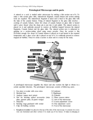
A petrological microscope magnifies the object and also restricts the light to vibrate in a
certain specified direction. The petrological microscope consists of following parts-
1. Eye piece or ocular with cross wires 8.Condenser
2. Bertrand lens 9. Diaphragm
3. Analyser (upper nicol prism) 10. Polarizer
4. Slot for inserting optical accessories (mica
plate, gypsum plate, & quartz wedge)
11. Plane concave mirror
12. Arm of the microscope
5. Objective 13. Coarse adjustment screw
6. Rotating stage graduated with vernier 14. Fine adjustment screw
7. Clips for holding sections 15. Foot of the microscope
1. Eyepiece or ocular:It is placed at the top of the tube of the microscope. It contains two cross
wire one horizontal and other vertical. It is made up of two lenses. It helps in enlarging or
Department of Geology, GSS College,Belgaum.
magnifying image of the mineral section. The ocular contains a lens, which is usually a 10x
lens. The magnification of your thin section is the product of the magnification of the ocular
and the objective. For example, if you are looking at a section using the 4x objective, and
your ocular is 10x then the total magnification is 40x.
2. Bertrand lens: It is placed in the tube of the microscope. It can be brought in and out of the
path of rays. This is used when examining interference figures and while studying the
proprties of minerals like sign of the mineral and finding out biaxial and uniaxial nature of
the mineral under conoscopic condition.
3. Analyser (Nicol Prism): This is similar to polarizer made up of transparent calcite crystal
called as Iceland spar in the form of a prism. Two rhombohedral parts of a calcite cut
diagonally, are joined by means of Canada balsam. This can be put into or out of the path of
rays of light as and when required. Below the analyser there is a slot or opening in which
optical accessories like mica plate, gypsum plate and quartz wedge are inserted.
4. Objective: This is placed at the other end of the tube of the microscope and can be removed
easily whenever required. Obectives are used for magnification. There are three types of
objectives- low power, high power and medium. Low power objectives magnify or enlarge 12
to 14 times of the original diameter. Medium and high power objectives magnify 40 to 80
times the diameter.
5. Tube of the microscope: Ocular, Bertrand lens, analyser and objective are fixed in the tube
of the microscope. This tube can be raised or lowered as and when focusing is required by
means of screw.
6. Rotating stage: This is provided just below the objective, which is graduated with vernier
scale. This can be rotated either clockwise or anticlock wise. For holding the thin section on
the stage it is provided with 2 clips. The stage can be fixed by operating a screw.
7. Clips for holding the section: These are two steel clips, which are fitted on the stage of the
microscope. Their one end is fixed to the stage and the other is moveable. These clips hold
the section in place.
8. Condenser: It is a lens, which condenses the rays and illuminates a particular point of the
mineral or rock section on the stage and is fixed below the stage of the microscope.
9. Iris Diaphragm: It is made up of metal piece. It works as a shutter and controls the amount
of light that falls on the object.
10. Polarizer: It is similar to analyser in construction. It is fixed at the lower end of sub stage and
is rotatable. It is also called as nicol prism.When both polarizer and analyser is in the path of
rays they are said to be under cross nicols (under analysed light). With the help of both the
nicol prisms (analyser & polarizer) the light can be made to vibrate in any specified plane.
11. Mirror: This is placed below the sub stage and can be tilted. It is a Plano-concave mirror.
Light can be reflected and made to fall on thin section. Plane mirror is used for studying
sections under low power objective. Below this there is a base which is the heaviest part of
the microscope.
12. Arm of the microscope: One end of the arm of the microscope is attached to the base with
the help of the fastening screw. The other end is attached to the tube of the microscope. The
arm can be moved in back to front for inclined position.
13. Coarse adjustment screws: Rotating clockwise or anti clockwise can bring the object into
rough focus.
14. Fine adjustment screws: By operating these screws the objective studied under investigation
is brought under sharp focus.
15. Subs-tage: The sub-stage arrangement consists of condenser, diaphragm, and polarizer. This
sub-stage can be raised or lowered by operating screws.

Más contenido relacionado

La actualidad más candente

Stereographic projection
Stereographic projection Stereographic projection
Stereographic projection PurvaPandey3
 
Extinction angle and Types of Extinction in Minerals.pdf
Extinction angle and Types of Extinction in Minerals.pdfExtinction angle and Types of Extinction in Minerals.pdf
Extinction angle and Types of Extinction in Minerals.pdfAasishGiri
 
Amphibole group of minerals
Amphibole group of mineralsAmphibole group of minerals
Amphibole group of mineralsRomit Kumar
 
Gravity & magnetic methods in geology
Gravity & magnetic methods in geologyGravity & magnetic methods in geology
Gravity & magnetic methods in geologyMd Asif Hasan
 
Lead and zinc deposits of india
Lead and zinc deposits of indiaLead and zinc deposits of india
Lead and zinc deposits of indiaPramoda Raj
 
Mineralogy and crystallography notes notes
Mineralogy and crystallography notes notes Mineralogy and crystallography notes notes
Mineralogy and crystallography notes notes Pramoda Raj
 
AN INTRODUCTION TO ORE MICROSCOPY
AN INTRODUCTION TO ORE MICROSCOPY AN INTRODUCTION TO ORE MICROSCOPY
AN INTRODUCTION TO ORE MICROSCOPY krishanunath1
 
Stereographic projection crystallography
Stereographic projection crystallographyStereographic projection crystallography
Stereographic projection crystallographyShivam Jain
 
Geological criteria for ore prospecting
Geological criteria for ore prospectingGeological criteria for ore prospecting
Geological criteria for ore prospectingPramoda Raj
 
MEMBERS OF AMPHIBOLE GROUP
MEMBERS OF AMPHIBOLE GROUPMEMBERS OF AMPHIBOLE GROUP
MEMBERS OF AMPHIBOLE GROUPparag sonwane
 
Singhbhum Craton
Singhbhum CratonSinghbhum Craton
Singhbhum CratonAkash Naik
 
Phanerozoic Stratigraphy of India
Phanerozoic Stratigraphy of IndiaPhanerozoic Stratigraphy of India
Phanerozoic Stratigraphy of IndiaRajkumarGhosh14
 
METAMORPHIC DIFFERENTIATION
METAMORPHIC DIFFERENTIATIONMETAMORPHIC DIFFERENTIATION
METAMORPHIC DIFFERENTIATIONGOPAL JI GUPTA
 
The rubidium strontium dating
The rubidium strontium dating The rubidium strontium dating
The rubidium strontium dating Avnee Kashyap
 
Foliation and lineation
Foliation and lineationFoliation and lineation
Foliation and lineationSaad Raja
 
Metamorphic facies
Metamorphic faciesMetamorphic facies
Metamorphic faciesAbid Ali
 

La actualidad más candente (20)

Stereographic projection
Stereographic projection Stereographic projection
Stereographic projection
 
Extinction angle and Types of Extinction in Minerals.pdf
Extinction angle and Types of Extinction in Minerals.pdfExtinction angle and Types of Extinction in Minerals.pdf
Extinction angle and Types of Extinction in Minerals.pdf
 
Anorthosite
Anorthosite Anorthosite
Anorthosite
 
Amphibole group of minerals
Amphibole group of mineralsAmphibole group of minerals
Amphibole group of minerals
 
Gravity & magnetic methods in geology
Gravity & magnetic methods in geologyGravity & magnetic methods in geology
Gravity & magnetic methods in geology
 
Ophiolite
OphioliteOphiolite
Ophiolite
 
Lead and zinc deposits of india
Lead and zinc deposits of indiaLead and zinc deposits of india
Lead and zinc deposits of india
 
Mineralogy and crystallography notes notes
Mineralogy and crystallography notes notes Mineralogy and crystallography notes notes
Mineralogy and crystallography notes notes
 
AN INTRODUCTION TO ORE MICROSCOPY
AN INTRODUCTION TO ORE MICROSCOPY AN INTRODUCTION TO ORE MICROSCOPY
AN INTRODUCTION TO ORE MICROSCOPY
 
Stereographic projection crystallography
Stereographic projection crystallographyStereographic projection crystallography
Stereographic projection crystallography
 
Geological criteria for ore prospecting
Geological criteria for ore prospectingGeological criteria for ore prospecting
Geological criteria for ore prospecting
 
Brachiopoda
Brachiopoda Brachiopoda
Brachiopoda
 
MEMBERS OF AMPHIBOLE GROUP
MEMBERS OF AMPHIBOLE GROUPMEMBERS OF AMPHIBOLE GROUP
MEMBERS OF AMPHIBOLE GROUP
 
Singhbhum Craton
Singhbhum CratonSinghbhum Craton
Singhbhum Craton
 
Phanerozoic Stratigraphy of India
Phanerozoic Stratigraphy of IndiaPhanerozoic Stratigraphy of India
Phanerozoic Stratigraphy of India
 
METAMORPHIC DIFFERENTIATION
METAMORPHIC DIFFERENTIATIONMETAMORPHIC DIFFERENTIATION
METAMORPHIC DIFFERENTIATION
 
The rubidium strontium dating
The rubidium strontium dating The rubidium strontium dating
The rubidium strontium dating
 
Foliation and lineation
Foliation and lineationFoliation and lineation
Foliation and lineation
 
Paleozoic stratigraphy
Paleozoic stratigraphy Paleozoic stratigraphy
Paleozoic stratigraphy
 
Metamorphic facies
Metamorphic faciesMetamorphic facies
Metamorphic facies
 

Similar a Petro microscope

Polarising microscope
Polarising microscope Polarising microscope
Polarising microscope parag sonwane
 
Slit lamp presentation 2015
Slit lamp presentation 2015Slit lamp presentation 2015
Slit lamp presentation 2015amanimohsen1
 
Microbiology lab 2
Microbiology lab 2Microbiology lab 2
Microbiology lab 2MBBS IMS MSU
 
Microbiology lab 2
Microbiology lab 2Microbiology lab 2
Microbiology lab 2MBBS IMS MSU
 
topic 4 Parts of Marine Sextant.pptx
topic 4 Parts of Marine Sextant.pptxtopic 4 Parts of Marine Sextant.pptx
topic 4 Parts of Marine Sextant.pptxAltair8
 
To Study Principles of Microscopy: Light Microscope, Phase Contrast Microsco...
To Study Principles of Microscopy: Light Microscope, Phase Contrast  Microsco...To Study Principles of Microscopy: Light Microscope, Phase Contrast  Microsco...
To Study Principles of Microscopy: Light Microscope, Phase Contrast Microsco...Om Prakash
 
Microscope part 2 BY DR. C. P. ARYA (B.Sc. B.D.S.; M.D.S.; P.M.S.; R.N.T.C.P.)
Microscope part 2 BY DR. C. P. ARYA (B.Sc. B.D.S.; M.D.S.; P.M.S.; R.N.T.C.P.)Microscope part 2 BY DR. C. P. ARYA (B.Sc. B.D.S.; M.D.S.; P.M.S.; R.N.T.C.P.)
Microscope part 2 BY DR. C. P. ARYA (B.Sc. B.D.S.; M.D.S.; P.M.S.; R.N.T.C.P.)DR. C. P. ARYA
 
Microscopes Applied
Microscopes AppliedMicroscopes Applied
Microscopes Appliedbeckydaw
 
Introduction - Bright Field Microscope.pptx
Introduction - Bright Field Microscope.pptxIntroduction - Bright Field Microscope.pptx
Introduction - Bright Field Microscope.pptxYuktiSharma55
 
Instrumentaion - MICROSCOPE
Instrumentaion - MICROSCOPEInstrumentaion - MICROSCOPE
Instrumentaion - MICROSCOPENayana Mohanan
 
Microscope and types of microscope
Microscope and types of microscopeMicroscope and types of microscope
Microscope and types of microscopeRimsha Pahore
 
Science7.q2.l1 2 final (1)
Science7.q2.l1 2 final (1)Science7.q2.l1 2 final (1)
Science7.q2.l1 2 final (1)MyraAbon
 
LAB-2 of hshaiiananaiaihana maiuhvwbj.pdf
LAB-2 of hshaiiananaiaihana maiuhvwbj.pdfLAB-2 of hshaiiananaiaihana maiuhvwbj.pdf
LAB-2 of hshaiiananaiaihana maiuhvwbj.pdfssuserdf9205
 

Similar a Petro microscope (20)

Polarising microscope
Polarising microscope Polarising microscope
Polarising microscope
 
Slit lamp presentation 2015
Slit lamp presentation 2015Slit lamp presentation 2015
Slit lamp presentation 2015
 
Parts of the microscope
Parts of the microscopeParts of the microscope
Parts of the microscope
 
Cunanan, mark bryan
Cunanan, mark bryanCunanan, mark bryan
Cunanan, mark bryan
 
Telescope
TelescopeTelescope
Telescope
 
Quality Assurance and Inspection OF CL.pptx
Quality Assurance and Inspection OF CL.pptxQuality Assurance and Inspection OF CL.pptx
Quality Assurance and Inspection OF CL.pptx
 
Microbiology lab 2
Microbiology lab 2Microbiology lab 2
Microbiology lab 2
 
Microbiology lab 2
Microbiology lab 2Microbiology lab 2
Microbiology lab 2
 
topic 4 Parts of Marine Sextant.pptx
topic 4 Parts of Marine Sextant.pptxtopic 4 Parts of Marine Sextant.pptx
topic 4 Parts of Marine Sextant.pptx
 
To Study Principles of Microscopy: Light Microscope, Phase Contrast Microsco...
To Study Principles of Microscopy: Light Microscope, Phase Contrast  Microsco...To Study Principles of Microscopy: Light Microscope, Phase Contrast  Microsco...
To Study Principles of Microscopy: Light Microscope, Phase Contrast Microsco...
 
Microscope part 2 BY DR. C. P. ARYA (B.Sc. B.D.S.; M.D.S.; P.M.S.; R.N.T.C.P.)
Microscope part 2 BY DR. C. P. ARYA (B.Sc. B.D.S.; M.D.S.; P.M.S.; R.N.T.C.P.)Microscope part 2 BY DR. C. P. ARYA (B.Sc. B.D.S.; M.D.S.; P.M.S.; R.N.T.C.P.)
Microscope part 2 BY DR. C. P. ARYA (B.Sc. B.D.S.; M.D.S.; P.M.S.; R.N.T.C.P.)
 
MICROSCOPE.pptx
MICROSCOPE.pptxMICROSCOPE.pptx
MICROSCOPE.pptx
 
Microscopes Applied
Microscopes AppliedMicroscopes Applied
Microscopes Applied
 
Introduction - Bright Field Microscope.pptx
Introduction - Bright Field Microscope.pptxIntroduction - Bright Field Microscope.pptx
Introduction - Bright Field Microscope.pptx
 
Microscope
MicroscopeMicroscope
Microscope
 
Instrumentaion - MICROSCOPE
Instrumentaion - MICROSCOPEInstrumentaion - MICROSCOPE
Instrumentaion - MICROSCOPE
 
Telescope
TelescopeTelescope
Telescope
 
Microscope and types of microscope
Microscope and types of microscopeMicroscope and types of microscope
Microscope and types of microscope
 
Science7.q2.l1 2 final (1)
Science7.q2.l1 2 final (1)Science7.q2.l1 2 final (1)
Science7.q2.l1 2 final (1)
 
LAB-2 of hshaiiananaiaihana maiuhvwbj.pdf
LAB-2 of hshaiiananaiaihana maiuhvwbj.pdfLAB-2 of hshaiiananaiaihana maiuhvwbj.pdf
LAB-2 of hshaiiananaiaihana maiuhvwbj.pdf
 

Más de Pramod Hanamgond

Más de Pramod Hanamgond (7)

Sediment texture
Sediment textureSediment texture
Sediment texture
 
Toposheet interpretation
Toposheet interpretationToposheet interpretation
Toposheet interpretation
 
Volcano
VolcanoVolcano
Volcano
 
Geology Introduction
Geology IntroductionGeology Introduction
Geology Introduction
 
Brunton compas
Brunton compasBrunton compas
Brunton compas
 
Attitude of beds
Attitude of bedsAttitude of beds
Attitude of beds
 
Geotectonic event details
Geotectonic event detailsGeotectonic event details
Geotectonic event details
 

Último

PSYCHOSOCIAL NEEDS. in nursing II sem pptx
PSYCHOSOCIAL NEEDS. in nursing II sem pptxPSYCHOSOCIAL NEEDS. in nursing II sem pptx
PSYCHOSOCIAL NEEDS. in nursing II sem pptxSuji236384
 
Proteomics: types, protein profiling steps etc.
Proteomics: types, protein profiling steps etc.Proteomics: types, protein profiling steps etc.
Proteomics: types, protein profiling steps etc.Silpa
 
LUNULARIA -features, morphology, anatomy ,reproduction etc.
LUNULARIA -features, morphology, anatomy ,reproduction etc.LUNULARIA -features, morphology, anatomy ,reproduction etc.
LUNULARIA -features, morphology, anatomy ,reproduction etc.Silpa
 
POGONATUM : morphology, anatomy, reproduction etc.
POGONATUM : morphology, anatomy, reproduction etc.POGONATUM : morphology, anatomy, reproduction etc.
POGONATUM : morphology, anatomy, reproduction etc.Silpa
 
Thyroid Physiology_Dr.E. Muralinath_ Associate Professor
Thyroid Physiology_Dr.E. Muralinath_ Associate ProfessorThyroid Physiology_Dr.E. Muralinath_ Associate Professor
Thyroid Physiology_Dr.E. Muralinath_ Associate Professormuralinath2
 
CURRENT SCENARIO OF POULTRY PRODUCTION IN INDIA
CURRENT SCENARIO OF POULTRY PRODUCTION IN INDIACURRENT SCENARIO OF POULTRY PRODUCTION IN INDIA
CURRENT SCENARIO OF POULTRY PRODUCTION IN INDIADr. TATHAGAT KHOBRAGADE
 
Bhiwandi Bhiwandi ❤CALL GIRL 7870993772 ❤CALL GIRLS ESCORT SERVICE In Bhiwan...
Bhiwandi Bhiwandi ❤CALL GIRL 7870993772 ❤CALL GIRLS  ESCORT SERVICE In Bhiwan...Bhiwandi Bhiwandi ❤CALL GIRL 7870993772 ❤CALL GIRLS  ESCORT SERVICE In Bhiwan...
Bhiwandi Bhiwandi ❤CALL GIRL 7870993772 ❤CALL GIRLS ESCORT SERVICE In Bhiwan...Monika Rani
 
TransientOffsetin14CAftertheCarringtonEventRecordedbyPolarTreeRings
TransientOffsetin14CAftertheCarringtonEventRecordedbyPolarTreeRingsTransientOffsetin14CAftertheCarringtonEventRecordedbyPolarTreeRings
TransientOffsetin14CAftertheCarringtonEventRecordedbyPolarTreeRingsSérgio Sacani
 
Grade 7 - Lesson 1 - Microscope and Its Functions
Grade 7 - Lesson 1 - Microscope and Its FunctionsGrade 7 - Lesson 1 - Microscope and Its Functions
Grade 7 - Lesson 1 - Microscope and Its FunctionsOrtegaSyrineMay
 
development of diagnostic enzyme assay to detect leuser virus
development of diagnostic enzyme assay to detect leuser virusdevelopment of diagnostic enzyme assay to detect leuser virus
development of diagnostic enzyme assay to detect leuser virusNazaninKarimi6
 
Phenolics: types, biosynthesis and functions.
Phenolics: types, biosynthesis and functions.Phenolics: types, biosynthesis and functions.
Phenolics: types, biosynthesis and functions.Silpa
 
Porella : features, morphology, anatomy, reproduction etc.
Porella : features, morphology, anatomy, reproduction etc.Porella : features, morphology, anatomy, reproduction etc.
Porella : features, morphology, anatomy, reproduction etc.Silpa
 
Cyanide resistant respiration pathway.pptx
Cyanide resistant respiration pathway.pptxCyanide resistant respiration pathway.pptx
Cyanide resistant respiration pathway.pptxSilpa
 
Reboulia: features, anatomy, morphology etc.
Reboulia: features, anatomy, morphology etc.Reboulia: features, anatomy, morphology etc.
Reboulia: features, anatomy, morphology etc.Silpa
 
Factory Acceptance Test( FAT).pptx .
Factory Acceptance Test( FAT).pptx       .Factory Acceptance Test( FAT).pptx       .
Factory Acceptance Test( FAT).pptx .Poonam Aher Patil
 
Asymmetry in the atmosphere of the ultra-hot Jupiter WASP-76 b
Asymmetry in the atmosphere of the ultra-hot Jupiter WASP-76 bAsymmetry in the atmosphere of the ultra-hot Jupiter WASP-76 b
Asymmetry in the atmosphere of the ultra-hot Jupiter WASP-76 bSérgio Sacani
 
CYTOGENETIC MAP................ ppt.pptx
CYTOGENETIC MAP................ ppt.pptxCYTOGENETIC MAP................ ppt.pptx
CYTOGENETIC MAP................ ppt.pptxSilpa
 
The Mariana Trench remarkable geological features on Earth.pptx
The Mariana Trench remarkable geological features on Earth.pptxThe Mariana Trench remarkable geological features on Earth.pptx
The Mariana Trench remarkable geological features on Earth.pptxseri bangash
 
THE ROLE OF BIOTECHNOLOGY IN THE ECONOMIC UPLIFT.pptx
THE ROLE OF BIOTECHNOLOGY IN THE ECONOMIC UPLIFT.pptxTHE ROLE OF BIOTECHNOLOGY IN THE ECONOMIC UPLIFT.pptx
THE ROLE OF BIOTECHNOLOGY IN THE ECONOMIC UPLIFT.pptxANSARKHAN96
 

Último (20)

PSYCHOSOCIAL NEEDS. in nursing II sem pptx
PSYCHOSOCIAL NEEDS. in nursing II sem pptxPSYCHOSOCIAL NEEDS. in nursing II sem pptx
PSYCHOSOCIAL NEEDS. in nursing II sem pptx
 
Proteomics: types, protein profiling steps etc.
Proteomics: types, protein profiling steps etc.Proteomics: types, protein profiling steps etc.
Proteomics: types, protein profiling steps etc.
 
LUNULARIA -features, morphology, anatomy ,reproduction etc.
LUNULARIA -features, morphology, anatomy ,reproduction etc.LUNULARIA -features, morphology, anatomy ,reproduction etc.
LUNULARIA -features, morphology, anatomy ,reproduction etc.
 
+971581248768>> SAFE AND ORIGINAL ABORTION PILLS FOR SALE IN DUBAI AND ABUDHA...
+971581248768>> SAFE AND ORIGINAL ABORTION PILLS FOR SALE IN DUBAI AND ABUDHA...+971581248768>> SAFE AND ORIGINAL ABORTION PILLS FOR SALE IN DUBAI AND ABUDHA...
+971581248768>> SAFE AND ORIGINAL ABORTION PILLS FOR SALE IN DUBAI AND ABUDHA...
 
POGONATUM : morphology, anatomy, reproduction etc.
POGONATUM : morphology, anatomy, reproduction etc.POGONATUM : morphology, anatomy, reproduction etc.
POGONATUM : morphology, anatomy, reproduction etc.
 
Thyroid Physiology_Dr.E. Muralinath_ Associate Professor
Thyroid Physiology_Dr.E. Muralinath_ Associate ProfessorThyroid Physiology_Dr.E. Muralinath_ Associate Professor
Thyroid Physiology_Dr.E. Muralinath_ Associate Professor
 
CURRENT SCENARIO OF POULTRY PRODUCTION IN INDIA
CURRENT SCENARIO OF POULTRY PRODUCTION IN INDIACURRENT SCENARIO OF POULTRY PRODUCTION IN INDIA
CURRENT SCENARIO OF POULTRY PRODUCTION IN INDIA
 
Bhiwandi Bhiwandi ❤CALL GIRL 7870993772 ❤CALL GIRLS ESCORT SERVICE In Bhiwan...
Bhiwandi Bhiwandi ❤CALL GIRL 7870993772 ❤CALL GIRLS  ESCORT SERVICE In Bhiwan...Bhiwandi Bhiwandi ❤CALL GIRL 7870993772 ❤CALL GIRLS  ESCORT SERVICE In Bhiwan...
Bhiwandi Bhiwandi ❤CALL GIRL 7870993772 ❤CALL GIRLS ESCORT SERVICE In Bhiwan...
 
TransientOffsetin14CAftertheCarringtonEventRecordedbyPolarTreeRings
TransientOffsetin14CAftertheCarringtonEventRecordedbyPolarTreeRingsTransientOffsetin14CAftertheCarringtonEventRecordedbyPolarTreeRings
TransientOffsetin14CAftertheCarringtonEventRecordedbyPolarTreeRings
 
Grade 7 - Lesson 1 - Microscope and Its Functions
Grade 7 - Lesson 1 - Microscope and Its FunctionsGrade 7 - Lesson 1 - Microscope and Its Functions
Grade 7 - Lesson 1 - Microscope and Its Functions
 
development of diagnostic enzyme assay to detect leuser virus
development of diagnostic enzyme assay to detect leuser virusdevelopment of diagnostic enzyme assay to detect leuser virus
development of diagnostic enzyme assay to detect leuser virus
 
Phenolics: types, biosynthesis and functions.
Phenolics: types, biosynthesis and functions.Phenolics: types, biosynthesis and functions.
Phenolics: types, biosynthesis and functions.
 
Porella : features, morphology, anatomy, reproduction etc.
Porella : features, morphology, anatomy, reproduction etc.Porella : features, morphology, anatomy, reproduction etc.
Porella : features, morphology, anatomy, reproduction etc.
 
Cyanide resistant respiration pathway.pptx
Cyanide resistant respiration pathway.pptxCyanide resistant respiration pathway.pptx
Cyanide resistant respiration pathway.pptx
 
Reboulia: features, anatomy, morphology etc.
Reboulia: features, anatomy, morphology etc.Reboulia: features, anatomy, morphology etc.
Reboulia: features, anatomy, morphology etc.
 
Factory Acceptance Test( FAT).pptx .
Factory Acceptance Test( FAT).pptx       .Factory Acceptance Test( FAT).pptx       .
Factory Acceptance Test( FAT).pptx .
 
Asymmetry in the atmosphere of the ultra-hot Jupiter WASP-76 b
Asymmetry in the atmosphere of the ultra-hot Jupiter WASP-76 bAsymmetry in the atmosphere of the ultra-hot Jupiter WASP-76 b
Asymmetry in the atmosphere of the ultra-hot Jupiter WASP-76 b
 
CYTOGENETIC MAP................ ppt.pptx
CYTOGENETIC MAP................ ppt.pptxCYTOGENETIC MAP................ ppt.pptx
CYTOGENETIC MAP................ ppt.pptx
 
The Mariana Trench remarkable geological features on Earth.pptx
The Mariana Trench remarkable geological features on Earth.pptxThe Mariana Trench remarkable geological features on Earth.pptx
The Mariana Trench remarkable geological features on Earth.pptx
 
THE ROLE OF BIOTECHNOLOGY IN THE ECONOMIC UPLIFT.pptx
THE ROLE OF BIOTECHNOLOGY IN THE ECONOMIC UPLIFT.pptxTHE ROLE OF BIOTECHNOLOGY IN THE ECONOMIC UPLIFT.pptx
THE ROLE OF BIOTECHNOLOGY IN THE ECONOMIC UPLIFT.pptx
 

Petro microscope

  • 1. Department of Geology, GSS College,Belgaum. Petrological Microscope and its parts A mineral or a rock is studied under microscope by making a thin section out of it. To make a thin section glass slide, mineral/rock fragment, canada balsam and section making tools are required. The mineral/rock fragment is taken and is fixed to the glass slide with the help of the canada balsam. Fixing of mineral fragment to the glass slide involves - cooking of canada balsam, where few drops of canada balsam on glass slide is heated until it gives thread when the sharp pin is put into it and pulled. The mineral section is placed on it and uniformly pressed to remove air bubbles trapped between the mineral fragments, Canada balsam and the glass slide. The mineral section now is subjected to grinding on a section-cutting wheel using emery powder. Once the section is thin (0.03mm) enough, few drops of Canada balsam is placed on it and heated to the required temperature and a glass cover slip is placed on top of it. It is uniformly pressed to remove trapped air bubbles. Once it is done a section is clean and it is ready for the study. A petrological microscope magnifies the object and also restricts the light to vibrate in a certain specified direction. The petrological microscope consists of following parts- 1. Eye piece or ocular with cross wires 8.Condenser 2. Bertrand lens 9. Diaphragm 3. Analyser (upper nicol prism) 10. Polarizer 4. Slot for inserting optical accessories (mica plate, gypsum plate, & quartz wedge) 11. Plane concave mirror 12. Arm of the microscope 5. Objective 13. Coarse adjustment screw 6. Rotating stage graduated with vernier 14. Fine adjustment screw 7. Clips for holding sections 15. Foot of the microscope 1. Eyepiece or ocular:It is placed at the top of the tube of the microscope. It contains two cross wire one horizontal and other vertical. It is made up of two lenses. It helps in enlarging or
  • 2. Department of Geology, GSS College,Belgaum. magnifying image of the mineral section. The ocular contains a lens, which is usually a 10x lens. The magnification of your thin section is the product of the magnification of the ocular and the objective. For example, if you are looking at a section using the 4x objective, and your ocular is 10x then the total magnification is 40x. 2. Bertrand lens: It is placed in the tube of the microscope. It can be brought in and out of the path of rays. This is used when examining interference figures and while studying the proprties of minerals like sign of the mineral and finding out biaxial and uniaxial nature of the mineral under conoscopic condition. 3. Analyser (Nicol Prism): This is similar to polarizer made up of transparent calcite crystal called as Iceland spar in the form of a prism. Two rhombohedral parts of a calcite cut diagonally, are joined by means of Canada balsam. This can be put into or out of the path of rays of light as and when required. Below the analyser there is a slot or opening in which optical accessories like mica plate, gypsum plate and quartz wedge are inserted. 4. Objective: This is placed at the other end of the tube of the microscope and can be removed easily whenever required. Obectives are used for magnification. There are three types of objectives- low power, high power and medium. Low power objectives magnify or enlarge 12 to 14 times of the original diameter. Medium and high power objectives magnify 40 to 80 times the diameter. 5. Tube of the microscope: Ocular, Bertrand lens, analyser and objective are fixed in the tube of the microscope. This tube can be raised or lowered as and when focusing is required by means of screw. 6. Rotating stage: This is provided just below the objective, which is graduated with vernier scale. This can be rotated either clockwise or anticlock wise. For holding the thin section on the stage it is provided with 2 clips. The stage can be fixed by operating a screw. 7. Clips for holding the section: These are two steel clips, which are fitted on the stage of the microscope. Their one end is fixed to the stage and the other is moveable. These clips hold the section in place. 8. Condenser: It is a lens, which condenses the rays and illuminates a particular point of the mineral or rock section on the stage and is fixed below the stage of the microscope. 9. Iris Diaphragm: It is made up of metal piece. It works as a shutter and controls the amount of light that falls on the object. 10. Polarizer: It is similar to analyser in construction. It is fixed at the lower end of sub stage and is rotatable. It is also called as nicol prism.When both polarizer and analyser is in the path of rays they are said to be under cross nicols (under analysed light). With the help of both the nicol prisms (analyser & polarizer) the light can be made to vibrate in any specified plane. 11. Mirror: This is placed below the sub stage and can be tilted. It is a Plano-concave mirror. Light can be reflected and made to fall on thin section. Plane mirror is used for studying sections under low power objective. Below this there is a base which is the heaviest part of the microscope. 12. Arm of the microscope: One end of the arm of the microscope is attached to the base with the help of the fastening screw. The other end is attached to the tube of the microscope. The arm can be moved in back to front for inclined position. 13. Coarse adjustment screws: Rotating clockwise or anti clockwise can bring the object into rough focus. 14. Fine adjustment screws: By operating these screws the objective studied under investigation is brought under sharp focus. 15. Subs-tage: The sub-stage arrangement consists of condenser, diaphragm, and polarizer. This sub-stage can be raised or lowered by operating screws.