SlideShare una empresa de Scribd logo
1 de 40
An Overview of Oral Mucous Membrane-II
• Parts of oral mucous
membrane
– Covering epithelium
– Underlying connective
tissue
– Submucosa
– Periosteum, muscle
• Three main types
– Masticatory mucosa
– Lining mucosa
– Specialized mucosa
courtesy :www.medscape.com/viewarticle/472915
•Two patterns of maturation
a) Keratinized
b) Non keratinized
Richard B. Presland, Ph.D.; Richard J. Jurevic, D.D.S.. Making Sense of the Epithelial Barrier:What Molecular Biology and
Genetics Tell Us About the Functions of Oral Mucosal and Epidermal Tissues. Journal of Dental Education April 2002.
Epithelial-Connective Tissue Interface
Basement membrane /Basal lamina
• Light microscope- 1-2µm thick is seen on the
lamina propria side of the junction
• Electron microscope- layer appears thinner
Basement membrane
Richard B. Presland, Ph.D.; Richard J. Jurevic, D.D.S.. Making Sense of the Epithelial Barrier:What Molecular Biology and
Genetics Tell Us About the Functions of Oral Mucosal and Epidermal Tissues. Journal of Dental Education April 2002.
• Using the electron microscope the following
layers of the basement membrane can be
distinguished:
Lamina rara externa - 10 to 50nm thick
Lamina densa -20 to 300nm, mostly 50 nm.
Lamina rara interna is only about 10 nm thick
Lamina fibroreticularis has a thickness of 200
- 500 nm. Rich in basotubules, 10 nm wide
microfibrillike structures.
Kimberley Brown. Epithelial Cells: Surface Domains & Junctions. Cell and Tissue Biology 2004
Structure of Hemidesmosome
Courtesy: xtal.cicancer.org/research.html
Protein Alternate terms Site
Keratin 5 Basal layer of stratified epithelia
Keratin 14 Basal layer of stratified epithelia
Plectin/HD1 Intracellular
IFAP300 Intracellular
P200 Intracellular
BPAg1 BP230 or dystonin Intracellular
BPAg2 BP180 or TYPE-XVII Transmembrane
collagen
a6b4 integrin Transmembrane
Laminin 5 Epiligrin or nicein or
kalinin BMZ
Laminin 6 BMZ
Ladinin LAD-1 BMZ
Uncein BMZ
Type-VII collagen BMZ
Type-IV collagen BMZ
Main hemidesmosome components
J Bagan, LL Muzio, C Scully.Mucosal Diseases Series.Number III.Mucous Membrane Pemphigoid.Oral diseases 2005;11:197-218
• Basement membrane (basal lamina) is a tough elastic structure
– Type IV and VII collagens
– heparan sulphate (HS) (perlecan)
– chondroitin sulphate (ChS)
– proteoglycans (versican), and
– glycoproteins
laminin
nidogen (entactin)
thrombospondin
tenascin
fibronectin
David F. Wilson et al. Oral cancer: Role of the basement membrane in invasion. Australian Dental Journal 1999;44:(2):93-97
Lamina lucida
• Glycoprotein layer
• It contains type IV
collagen
• It contains laminin and
entactin.
• Laminin and type IV
collagen promote
epithelial cell growth. Courtesy: www.ncbi.nlm.nih.gov/bookshelf/br.fcgi
• Laminins
– Laminin is a large, triple
chain molecule.
– α, β, γ subunits
• Integrins
– Family of transmembrane
proteins consisting of 2
subunits- α and β.
• Plectin and BP 230
– Members of the plakin
family
Courtesy: myworldatlarge.wordpress.com
Lamina densa
• Layer of finely
granular/filamentous
material that run
parallel to the basal cell
membranes of the
epithelial cells.
• Inserted into the lamina
densa are small loops of
finely banded fibrils
called anchoring fibrils.
REGIONAL VARIATIONS OF ORAL
MUCOSA
Lip
• Skin on its outer surface
• Labial mucosa on its inner
surface
• Vermilion zone/ Red zone/
Transitional zone
• Striated muscles in their core-
muscles of facial expression
• Minor mucous salivary glands
• Skin
– Keratinized layer of epidermis
on a bed of connective tissue
– Sweat glands, sebaceous glands
and bases of hair follicles
Section of skin of lip. A=keratinized epidermis,
B=shaft of hair, C=sebaceous gland, D=dermis.
Masson’s trichrome;x30
Fig.14.31
B.K.B.BERKOVITZ.ORAL ANTOMY, HISTOLOGY AND EMBRYOLOGY. FOURTH EDITION:MOSBY ELSEVIER;2009.CHAP.14;223-252.
Vermilion
• Lacks the appendages of skin
• Angles of the mouth-
sebaceous glands
• Constant moistening by the
tongue- lacks mucous glands
• Vermilion zone
– Epithelium is keratinized, thin
and translucent
– Connective tissue papilla are
long and narrow
• Intermediate zone- lacks a
granular layer and has a thick
parakeratinized layer
Section of red zone of lip. Keratinized epithelium
& lamina propria
H&E x80
Fig: 14.32
B.K.B.BERKOVITZ.ORAL ANTOMY, HISTOLOGY AND EMBRYOLOGY. FOURTH EDITION:MOSBY ELSEVIER;2009.CHAP.14;223-252.
Labial Mucosa
• Thick, non keratinized
epithelium
• Lamina propria is wide
with short and irregular
papillae
• Sub mucosa- Minor
salivary glands Section of labial mucosa. A=non-keratinized oral
epithellium, B=lamina propria, C=minor salivary gland
in submucosa, D=fibres of orbicularis oris
H&E; x15
Fig: 14.33
B.K.B.BERKOVITZ.ORAL ANTOMY, HISTOLOGY AND EMBRYOLOGY. FOURTH EDITION:MOSBY ELSEVIER;2009.CHAP.14;223-252.
Cheek
• Epithelium- non keratinized
• Lamina propria- dense with
short, irregular papillae
• Submucosa- minor mucous
salivary glands
• Linea alba- keratinized,
white line coincident with
the occlusal plane
• Fordyce’s spots- Sebaceous
glands appear as small
yellow patches
Section of buccal mucosa. B=non keratinized
oral epithelium, C=lamina propria, D=minor
salivary glands in submucosa, A=fibres of
buccinator muscle.
H&E;X15
Fig: 14.34
B.K.B.BERKOVITZ.ORAL ANTOMY, HISTOLOGY AND EMBRYOLOGY. FOURTH EDITION:MOSBY ELSEVIER;2009.CHAP.14;223-252.
Alveolar Mucosa
• Thin, non keratinized
epithelium
• Lamina propria shows
poorly developed
dermal papillae
• Sub mucosa houses
many minor mucous
salivary glands
Section of lining oral epithelium from the alveolar
mucosa. Absence of keratin in the superficial
layer
H&E;x80
Fig: 14.13
B.K.B.BERKOVITZ.ORAL ANTOMY, HISTOLOGY AND EMBRYOLOGY. FOURTH EDITION:MOSBY ELSEVIER;2009.CHAP.14;223-252.
Gingiva
• Two recognized regions
– Attached gingiva
– Free gingiva
– Gingival margin
– Gingival sulcus
• Mucogingival junction
Free gingival groove
courtesy :www.medscape.com/viewarticle/1893
Attached Gingiva
• Keratinized
• No sub mucosa
• Lamina propria-bound
directly to the bone
Crevicular(sulcular) epithelium
• Epithelium on the inner
surface of the gingiva
• Non keratinized
Section of parakeratinized oral epithelium from the
gingiva. The superficial layer stain more heavily for
keratin but nuclei are retained
H&E; x100
Fig: 14.12
B.K.B.BERKOVITZ.ORAL ANTOMY, HISTOLOGY AND EMBRYOLOGY. FOURTH EDITION:MOSBY ELSEVIER;2009.CHAP.14;223-252.
Junctional epithelium
• Epithelial collar that surrounds the
tooth and extends from the region of
the cementum-enamel junction to
the bottom of the gingival crevice
• Two zones:
– A single layer of cuboidal cells- stratum
germinativum
– Several layers of flattened cells-
stratum spinosum
• Features of the junctional epithelium
– Hemidesmosomes and basal lamina-
Attachment apparatus/ Epithelial
attachment
– Internal basal lamina
– External basal lamina
Demineralized section showing the
dentogingival junction .A=region of attached
gingiva covered by masticatory epithelium,
B=region of free gingiva covered externally by
masticatory epithelium,C=non-keratinized
crevicular epithelium, D=non-keratinized
junctional epithelium
H&E; x30
Fig: 14.42
B.K.B.BERKOVITZ.ORAL ANTOMY, HISTOLOGY AND EMBRYOLOGY. FOURTH EDITION:MOSBY ELSEVIER;2009.CHAP.14;223-252.
Cont…
Internal basal lamina- 2 zones
• Electron lucent zone adjacent to the cell
• Electron dense layer adjacent to the tooth
surface
• Lacks type IV collagen and anchoring fibrils
• Lamina propria
– Rich blood supply
• Turnover is rapid
Cont…
Enamel cuticle
• Non-mineralized structure interposed
between the junctional epithelium and the
underlying hard tissue
• Ultrastructurally
– Amorphous and biochemically distinct
– Proteinaceous and may be derived from serum
Gingival crevicular fluid
• Tissue fluid and cells passing through the
epithelium from the connective tissue- GCF
• Can be collected by capillary tubing, gingival
washing or absorbent paper strips
• Composition of GCF
– Immunoglobulins
– Complement
– Polymorphonucleocytes
– Epithelial squames
Interdental Gingiva
• Part of the gingiva
between adjacent teeth
• Wedge shaped
appearance
• Interdental col- curved
depression between the
buccal and lingual peaks
• Epithelium- non
keratinized and derived
from the reduced enamel
epithelium
Demineralized section of the interdental
papillae cut in the buccolingual plane
between two cheek teeth showing the
‘interdental col’.
H&E; x4
Fig: 14.53
B.K.B.BERKOVITZ.ORAL ANTOMY, HISTOLOGY AND EMBRYOLOGY. FOURTH EDITION:MOSBY ELSEVIER;2009.CHAP.14;223-252.
Collagen fibres
• Type I collagen
• Functions
– Support of the free gingiva
– Binding of the attached gingiva to the alveolar
bone and tooth
– Linkage of one teeth to another
Types of Gingival Fibers
Diagram showing arrangement of
principal collagen fibre groups in
the lamina propria of the gingiva.
a.buccolingual section,
b.mesiodistal section, c.horizontal
section, d.buccolingual section
along the interdental col.
A= DENTOGINGIVAL FIBRES
B= LONGITUDINAL FIBRES
C= CIRCULAR FIBRES
D= ALVEOLO GINGIVAL FIBRES
E= DENTOPERIOSTEAL FIBRES
F= TRANSSEPTAL FIBRES
G= SEMICIRCULAR FIBRES
H= TRANSGINGIVAL FIBRES
I= INTERDENTAL FIBRES
J= VERTICAL FIBRES
Fig: 14.55
a
c
b d
B.K.B.BERKOVITZ.ORAL ANTOMY, HISTOLOGY AND EMBRYOLOGY. FOURTH EDITION:MOSBY ELSEVIER;2009.CHAP.14;223-252.
Cont…
Lamina propria of gingiva
• Fibroblasts lack alkaline phosphatase
• Less contractile proteins
• Releases more prostaglandins
• Less ground substance and Type III collagen
Palate
Hard palate
• Keratinized and Para
keratinized epithelium
• Central region- no sub
mucosa
• Palate joins the alveolus-
sub mucosa contains
main neurovascular
bundles and minor
mucous salivary
glands(posteriorly) and
adipose tissue(anteriorly)
Demineralised section of the hard palate
showing the oral surface. A=lined by masticatory
epithelium and the nasal epithelium, B=lined by
a respiratory epithelium, C=bone of hard palate,
D=duct from mucous gland opening onto
surface
H&E;x110
Fig: 14.61
B.K.B.BERKOVITZ.ORAL ANTOMY, HISTOLOGY AND EMBRYOLOGY. FOURTH EDITION:MOSBY ELSEVIER;2009.CHAP.14;223-252.
Cont…
• Nasal surface-
respiratory epithelium
– Ciliated columnar
epithelial cells and
goblet cells
– Vascular sub mucosa-
minor glands of both
mucous and serous
types
Section of nasal surface of hard palate. A=cilitated
columnar epithelium, B=goblet cell, C=minor gland
H&E;x500
Fig: 14.62
B.K.B.BERKOVITZ.ORAL ANTOMY, HISTOLOGY AND EMBRYOLOGY. FOURTH EDITION:MOSBY ELSEVIER;2009.CHAP.14;223-252.
Soft Palate
• Non keratinized lining
mucosa
• Lamina propria
– Papilla are short and broad
– Elastic fibers and collagen
bundles are thin
• Sub mucosa
– Small mucous glands
– Attached to the palatal
muscles
• Nasal surface- respiratory
epithelium
Section of soft palate showing the oral surface being
covered by a non keratinized lining mucosa,
numerous minor salivary glands, palatal
musculature and nasal epithelium
H&E;x20
Fig: 14.63
B.K.B.BERKOVITZ.ORAL ANTOMY, HISTOLOGY AND EMBRYOLOGY. FOURTH EDITION:MOSBY ELSEVIER;2009.CHAP.14;223-252.
Tongue and Floor of the Mouth
• Epithelium- thin, non
keratinized and shows short
papillae
• Sulcus terminalis
– Palatal surface
– Pharyngeal surface
• Anterior 2/3rd of tongue-
numerous papillae
– Filiform
– Fungiform
– Foliate
– Circumvallate
• Posterior 1/3rd of tongue
– Small lymphatic
nodules/follicles
Section showing the ventral surface of the
tongue(A), and floor of mouth(B) being lined by
non-keratinized lining epithelium
H&E;x20, fig: 14.64
Section of fungiform papillae on dorsal surface of
anterior part of tongue showing taste buds
H&E;x120, Fig: 14.68
B.K.B.BERKOVITZ.ORAL ANTOMY, HISTOLOGY AND EMBRYOLOGY. FOURTH EDITION:MOSBY ELSEVIER;2009.CHAP.14;223-252.
• Anterior tongue
– Keratinized/ Para keratinized
epithelium
– Hair like tufts
• Filiform papillae
– Central core of lamina propria with
smaller, secondary papillae branching
from it
• Fungiform papillae
– Isolated, elevated mushroom-shaped
papillae scattered between the
filiform papillae
– 150-400µm in diameter
– Thin epithelium which is non-
keratinized
– Vascular core of lamina propria
– Taste buds may be found on the
surface
Section showing dorsum of anterior 2/3rd of
the tongue
H&E;x35
Fig: 14.66
B.K.B.BERKOVITZ.ORAL ANTOMY, HISTOLOGY AND EMBRYOLOGY. FOURTH EDITION:MOSBY ELSEVIER;2009.CHAP.14;223-252.
• Foliate papillae
– One/ two longitudinal
clefts at the side of the
tongue
– Non keratinized epithelium
• Circumvallate papillae
– Large and rounded
– Surrounded by trench-like
feature
– Non keratinized epithelium
– Taste buds-internal wall of
the trench
– Small serous glands of von
Ebner
Section of circumvallate papillae, A=serous glands B=
of von Ebner empty via the ducts into the base of the
trench C= surrounding the papillae, which is not raised
above the surface of the tongue, D= muscle of tongue
H&E; x35
Fig: 14.72
B.K.B.BERKOVITZ.ORAL ANTOMY, HISTOLOGY AND EMBRYOLOGY. FOURTH EDITION:MOSBY ELSEVIER;2009.CHAP.14;223-252.
Taste Buds
• Chemoreceptive organs-
Taste
• Located within the
epithelium
– Walls of the circumvallate
papillae
– Upper surface of fungiform
papillae
– Lateral walls of foliate
papillae
– Mucosa of the soft palate and
in the epiglottis
• 2 types of cells
– Supporting cell
– Taste cell
Section of wall of circumvallate papillae showing two
barrel-shaped areas representing two taste buds.
Masson’s trichrome; x300
Fig: 14.74
B.K.B.BERKOVITZ.ORAL ANTOMY, HISTOLOGY AND EMBRYOLOGY. FOURTH EDITION:MOSBY ELSEVIER;2009.CHAP.14;223-252.
Cont…
• Taste cell- 4 distinct cell types
– Undifferentiated type IV cells
– Type I cells- dark appearance
– Type II and III cells- lighter
• Type I and III cells
– Form synapses with intragemmal nerves
Posterior tongue
• Collection of lymphoid
follicles- Lingual tonsil
• This forms a component
of Waldeyer’s ring-
protects the opening into
the pharynx
• Follicles are deep crypts
lined with epithelium and
containing a mass of
lymphoid material
• Follicles open onto the
surface of the tongue
• Contains small mucous
glands
Section of dorsal surface of posterior 1/3rd of
tongue containing a lymphoid follicle. This part
of the tongue is covered by a non keratinized
lining epithelium
H&E; x40, Fig: 14.76
Section of foliate papilla showing taste buds.
H&E; x200
Fig: 14.70
B.K.B.BERKOVITZ.ORAL ANTOMY, HISTOLOGY AND EMBRYOLOGY. FOURTH EDITION:MOSBY ELSEVIER;2009.CHAP.14;223-252.
Functions of the oral mucosa
• It is protective mechanically against both compressive
and shearing forces
• It provides a barrier to microorganisms, toxins and
various antigens
• It has a role in immunological defense, both humoral
and cell mediated
• Minor glands within oral mucosa provide lubrication
and buffering as well as secretion of some antibodies.
The viscoelastic mucous film also acts as a barrier,
helping to retain water and electrolytes
• The mucosa is richly innervated, providing input for
touch, proprioception, pain and taste
• Keratinized epithelium
– Masticatory mucosa
• Gingiva
• Hard palate
– Vermilion border of the lip
• Non keratinized epithelium
– Lining mucosa
• Lip and cheek
• Vestibular fornix and alveolar
mucosa
• Inferior surface of tongue and
floor of oral cavity
• Soft palate
– Specialized mucosa
• Dorsal lingual mucosa
• Taste buds
B.K.B.BERKOVITZ.ORAL ANTOMY, HISTOLOGY AND EMBRYOLOGY. FOURTH EDITION:MOSBY ELSEVIER;2009.CHAP.14;223-252.
References
• ANTONIO NANCI.TENCATE’S ORAL HISTOLOGY DEVELOPMENT, STRUCTURE
AND FUNCTION.SIXTH EDITION:ELSEVIER;2005.CHAP.12;329-375.
• B.K.B.BERKOVITZ.ORAL ANTOMY, HISTOLOGY AND EMBRYOLOGY. FOURTH
EDITION:MOSBY ELSEVIER;2009.CHAP.14;223-252.
• G S KUMAR.ORBAN’S ORAL HISTOLOGY AND EMBRYOLOGY.TWELFTH
EDITION:MOSBY ELSEVIER;2008. CHAP.9;210-257.
• David F. Wilson. Oral cancer: Role of the basement membrane in invasion.
Australian Dental Journal 1999;44:(2):93-97.
• Kimberley Brown. Epithelial Cells: Surface Domains & Junctions. Cell and
Tissue Biology 2004.
• J Bagan, LL Muzio, C Scully.Mucosal Diseases Series.Number III.Mucous
Membrane Pemphigoid.Oral diseases 2005;11:197-218.
• Geoffrey M.Cooper, Robert E.Hausman,editors.The Cell.A Molecular
Approach.Fourth Edition:ASM Press Washington D.C.;2007.Chap.14;569-575.
oral mucous membrane - II.pptx

Más contenido relacionado

Similar a oral mucous membrane - II.pptx

Similar a oral mucous membrane - II.pptx (20)

gingiva
gingivagingiva
gingiva
 
gigiva2ndseminar- 2.pdf
gigiva2ndseminar- 2.pdfgigiva2ndseminar- 2.pdf
gigiva2ndseminar- 2.pdf
 
Gingiva
Gingiva Gingiva
Gingiva
 
Gingiva copy
Gingiva copyGingiva copy
Gingiva copy
 
Gingiva (2)
Gingiva (2)Gingiva (2)
Gingiva (2)
 
Periodontium seminar
Periodontium seminar Periodontium seminar
Periodontium seminar
 
Gingiva ppt
Gingiva pptGingiva ppt
Gingiva ppt
 
Gingiva
Gingiva Gingiva
Gingiva
 
JUNCTIONAL EPITHELIUM
JUNCTIONAL EPITHELIUMJUNCTIONAL EPITHELIUM
JUNCTIONAL EPITHELIUM
 
Gingiva. Lilly & Bong.pptx
Gingiva. Lilly & Bong.pptxGingiva. Lilly & Bong.pptx
Gingiva. Lilly & Bong.pptx
 
Gingiva
GingivaGingiva
Gingiva
 
Clinical Periodontology - Introduction & Anatomy
Clinical Periodontology - Introduction & AnatomyClinical Periodontology - Introduction & Anatomy
Clinical Periodontology - Introduction & Anatomy
 
Junctional epithelium khushbu
Junctional epithelium khushbuJunctional epithelium khushbu
Junctional epithelium khushbu
 
Gingiva
GingivaGingiva
Gingiva
 
dental cementum
dental cementumdental cementum
dental cementum
 
Gingiva......
Gingiva......Gingiva......
Gingiva......
 
The oral mucosa
The oral mucosaThe oral mucosa
The oral mucosa
 
oral mucous membrane
oral mucous membraneoral mucous membrane
oral mucous membrane
 
Oral mucosa part 2
Oral mucosa part 2Oral mucosa part 2
Oral mucosa part 2
 
Oral mucosa1234
Oral mucosa1234Oral mucosa1234
Oral mucosa1234
 

Último

POINT- BIOCHEMISTRY SEM 2 ENZYMES UNIT 5.pptx
POINT- BIOCHEMISTRY SEM 2 ENZYMES UNIT 5.pptxPOINT- BIOCHEMISTRY SEM 2 ENZYMES UNIT 5.pptx
POINT- BIOCHEMISTRY SEM 2 ENZYMES UNIT 5.pptxSayali Powar
 
microwave assisted reaction. General introduction
microwave assisted reaction. General introductionmicrowave assisted reaction. General introduction
microwave assisted reaction. General introductionMaksud Ahmed
 
The Most Excellent Way | 1 Corinthians 13
The Most Excellent Way | 1 Corinthians 13The Most Excellent Way | 1 Corinthians 13
The Most Excellent Way | 1 Corinthians 13Steve Thomason
 
The basics of sentences session 2pptx copy.pptx
The basics of sentences session 2pptx copy.pptxThe basics of sentences session 2pptx copy.pptx
The basics of sentences session 2pptx copy.pptxheathfieldcps1
 
Student login on Anyboli platform.helpin
Student login on Anyboli platform.helpinStudent login on Anyboli platform.helpin
Student login on Anyboli platform.helpinRaunakKeshri1
 
9548086042 for call girls in Indira Nagar with room service
9548086042  for call girls in Indira Nagar  with room service9548086042  for call girls in Indira Nagar  with room service
9548086042 for call girls in Indira Nagar with room servicediscovermytutordmt
 
Sanyam Choudhary Chemistry practical.pdf
Sanyam Choudhary Chemistry practical.pdfSanyam Choudhary Chemistry practical.pdf
Sanyam Choudhary Chemistry practical.pdfsanyamsingh5019
 
Presentation by Andreas Schleicher Tackling the School Absenteeism Crisis 30 ...
Presentation by Andreas Schleicher Tackling the School Absenteeism Crisis 30 ...Presentation by Andreas Schleicher Tackling the School Absenteeism Crisis 30 ...
Presentation by Andreas Schleicher Tackling the School Absenteeism Crisis 30 ...EduSkills OECD
 
Activity 01 - Artificial Culture (1).pdf
Activity 01 - Artificial Culture (1).pdfActivity 01 - Artificial Culture (1).pdf
Activity 01 - Artificial Culture (1).pdfciinovamais
 
Separation of Lanthanides/ Lanthanides and Actinides
Separation of Lanthanides/ Lanthanides and ActinidesSeparation of Lanthanides/ Lanthanides and Actinides
Separation of Lanthanides/ Lanthanides and ActinidesFatimaKhan178732
 
BAG TECHNIQUE Bag technique-a tool making use of public health bag through wh...
BAG TECHNIQUE Bag technique-a tool making use of public health bag through wh...BAG TECHNIQUE Bag technique-a tool making use of public health bag through wh...
BAG TECHNIQUE Bag technique-a tool making use of public health bag through wh...Sapna Thakur
 
Call Girls in Dwarka Mor Delhi Contact Us 9654467111
Call Girls in Dwarka Mor Delhi Contact Us 9654467111Call Girls in Dwarka Mor Delhi Contact Us 9654467111
Call Girls in Dwarka Mor Delhi Contact Us 9654467111Sapana Sha
 
Interactive Powerpoint_How to Master effective communication
Interactive Powerpoint_How to Master effective communicationInteractive Powerpoint_How to Master effective communication
Interactive Powerpoint_How to Master effective communicationnomboosow
 
1029 - Danh muc Sach Giao Khoa 10 . pdf
1029 -  Danh muc Sach Giao Khoa 10 . pdf1029 -  Danh muc Sach Giao Khoa 10 . pdf
1029 - Danh muc Sach Giao Khoa 10 . pdfQucHHunhnh
 
Ecosystem Interactions Class Discussion Presentation in Blue Green Lined Styl...
Ecosystem Interactions Class Discussion Presentation in Blue Green Lined Styl...Ecosystem Interactions Class Discussion Presentation in Blue Green Lined Styl...
Ecosystem Interactions Class Discussion Presentation in Blue Green Lined Styl...fonyou31
 
Introduction to Nonprofit Accounting: The Basics
Introduction to Nonprofit Accounting: The BasicsIntroduction to Nonprofit Accounting: The Basics
Introduction to Nonprofit Accounting: The BasicsTechSoup
 
Beyond the EU: DORA and NIS 2 Directive's Global Impact
Beyond the EU: DORA and NIS 2 Directive's Global ImpactBeyond the EU: DORA and NIS 2 Directive's Global Impact
Beyond the EU: DORA and NIS 2 Directive's Global ImpactPECB
 
Grant Readiness 101 TechSoup and Remy Consulting
Grant Readiness 101 TechSoup and Remy ConsultingGrant Readiness 101 TechSoup and Remy Consulting
Grant Readiness 101 TechSoup and Remy ConsultingTechSoup
 

Último (20)

POINT- BIOCHEMISTRY SEM 2 ENZYMES UNIT 5.pptx
POINT- BIOCHEMISTRY SEM 2 ENZYMES UNIT 5.pptxPOINT- BIOCHEMISTRY SEM 2 ENZYMES UNIT 5.pptx
POINT- BIOCHEMISTRY SEM 2 ENZYMES UNIT 5.pptx
 
microwave assisted reaction. General introduction
microwave assisted reaction. General introductionmicrowave assisted reaction. General introduction
microwave assisted reaction. General introduction
 
The Most Excellent Way | 1 Corinthians 13
The Most Excellent Way | 1 Corinthians 13The Most Excellent Way | 1 Corinthians 13
The Most Excellent Way | 1 Corinthians 13
 
The basics of sentences session 2pptx copy.pptx
The basics of sentences session 2pptx copy.pptxThe basics of sentences session 2pptx copy.pptx
The basics of sentences session 2pptx copy.pptx
 
Student login on Anyboli platform.helpin
Student login on Anyboli platform.helpinStudent login on Anyboli platform.helpin
Student login on Anyboli platform.helpin
 
9548086042 for call girls in Indira Nagar with room service
9548086042  for call girls in Indira Nagar  with room service9548086042  for call girls in Indira Nagar  with room service
9548086042 for call girls in Indira Nagar with room service
 
INDIA QUIZ 2024 RLAC DELHI UNIVERSITY.pptx
INDIA QUIZ 2024 RLAC DELHI UNIVERSITY.pptxINDIA QUIZ 2024 RLAC DELHI UNIVERSITY.pptx
INDIA QUIZ 2024 RLAC DELHI UNIVERSITY.pptx
 
Sanyam Choudhary Chemistry practical.pdf
Sanyam Choudhary Chemistry practical.pdfSanyam Choudhary Chemistry practical.pdf
Sanyam Choudhary Chemistry practical.pdf
 
Presentation by Andreas Schleicher Tackling the School Absenteeism Crisis 30 ...
Presentation by Andreas Schleicher Tackling the School Absenteeism Crisis 30 ...Presentation by Andreas Schleicher Tackling the School Absenteeism Crisis 30 ...
Presentation by Andreas Schleicher Tackling the School Absenteeism Crisis 30 ...
 
Activity 01 - Artificial Culture (1).pdf
Activity 01 - Artificial Culture (1).pdfActivity 01 - Artificial Culture (1).pdf
Activity 01 - Artificial Culture (1).pdf
 
Separation of Lanthanides/ Lanthanides and Actinides
Separation of Lanthanides/ Lanthanides and ActinidesSeparation of Lanthanides/ Lanthanides and Actinides
Separation of Lanthanides/ Lanthanides and Actinides
 
BAG TECHNIQUE Bag technique-a tool making use of public health bag through wh...
BAG TECHNIQUE Bag technique-a tool making use of public health bag through wh...BAG TECHNIQUE Bag technique-a tool making use of public health bag through wh...
BAG TECHNIQUE Bag technique-a tool making use of public health bag through wh...
 
Código Creativo y Arte de Software | Unidad 1
Código Creativo y Arte de Software | Unidad 1Código Creativo y Arte de Software | Unidad 1
Código Creativo y Arte de Software | Unidad 1
 
Call Girls in Dwarka Mor Delhi Contact Us 9654467111
Call Girls in Dwarka Mor Delhi Contact Us 9654467111Call Girls in Dwarka Mor Delhi Contact Us 9654467111
Call Girls in Dwarka Mor Delhi Contact Us 9654467111
 
Interactive Powerpoint_How to Master effective communication
Interactive Powerpoint_How to Master effective communicationInteractive Powerpoint_How to Master effective communication
Interactive Powerpoint_How to Master effective communication
 
1029 - Danh muc Sach Giao Khoa 10 . pdf
1029 -  Danh muc Sach Giao Khoa 10 . pdf1029 -  Danh muc Sach Giao Khoa 10 . pdf
1029 - Danh muc Sach Giao Khoa 10 . pdf
 
Ecosystem Interactions Class Discussion Presentation in Blue Green Lined Styl...
Ecosystem Interactions Class Discussion Presentation in Blue Green Lined Styl...Ecosystem Interactions Class Discussion Presentation in Blue Green Lined Styl...
Ecosystem Interactions Class Discussion Presentation in Blue Green Lined Styl...
 
Introduction to Nonprofit Accounting: The Basics
Introduction to Nonprofit Accounting: The BasicsIntroduction to Nonprofit Accounting: The Basics
Introduction to Nonprofit Accounting: The Basics
 
Beyond the EU: DORA and NIS 2 Directive's Global Impact
Beyond the EU: DORA and NIS 2 Directive's Global ImpactBeyond the EU: DORA and NIS 2 Directive's Global Impact
Beyond the EU: DORA and NIS 2 Directive's Global Impact
 
Grant Readiness 101 TechSoup and Remy Consulting
Grant Readiness 101 TechSoup and Remy ConsultingGrant Readiness 101 TechSoup and Remy Consulting
Grant Readiness 101 TechSoup and Remy Consulting
 

oral mucous membrane - II.pptx

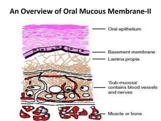
  • 1. An Overview of Oral Mucous Membrane-II
  • 2. • Parts of oral mucous membrane – Covering epithelium – Underlying connective tissue – Submucosa – Periosteum, muscle • Three main types – Masticatory mucosa – Lining mucosa – Specialized mucosa courtesy :www.medscape.com/viewarticle/472915 •Two patterns of maturation a) Keratinized b) Non keratinized Richard B. Presland, Ph.D.; Richard J. Jurevic, D.D.S.. Making Sense of the Epithelial Barrier:What Molecular Biology and Genetics Tell Us About the Functions of Oral Mucosal and Epidermal Tissues. Journal of Dental Education April 2002.
  • 3. Epithelial-Connective Tissue Interface Basement membrane /Basal lamina • Light microscope- 1-2µm thick is seen on the lamina propria side of the junction • Electron microscope- layer appears thinner
  • 4. Basement membrane Richard B. Presland, Ph.D.; Richard J. Jurevic, D.D.S.. Making Sense of the Epithelial Barrier:What Molecular Biology and Genetics Tell Us About the Functions of Oral Mucosal and Epidermal Tissues. Journal of Dental Education April 2002.
  • 5. • Using the electron microscope the following layers of the basement membrane can be distinguished: Lamina rara externa - 10 to 50nm thick Lamina densa -20 to 300nm, mostly 50 nm. Lamina rara interna is only about 10 nm thick Lamina fibroreticularis has a thickness of 200 - 500 nm. Rich in basotubules, 10 nm wide microfibrillike structures. Kimberley Brown. Epithelial Cells: Surface Domains & Junctions. Cell and Tissue Biology 2004
  • 6. Structure of Hemidesmosome Courtesy: xtal.cicancer.org/research.html
  • 7. Protein Alternate terms Site Keratin 5 Basal layer of stratified epithelia Keratin 14 Basal layer of stratified epithelia Plectin/HD1 Intracellular IFAP300 Intracellular P200 Intracellular BPAg1 BP230 or dystonin Intracellular BPAg2 BP180 or TYPE-XVII Transmembrane collagen a6b4 integrin Transmembrane Laminin 5 Epiligrin or nicein or kalinin BMZ Laminin 6 BMZ Ladinin LAD-1 BMZ Uncein BMZ Type-VII collagen BMZ Type-IV collagen BMZ Main hemidesmosome components J Bagan, LL Muzio, C Scully.Mucosal Diseases Series.Number III.Mucous Membrane Pemphigoid.Oral diseases 2005;11:197-218
  • 8. • Basement membrane (basal lamina) is a tough elastic structure – Type IV and VII collagens – heparan sulphate (HS) (perlecan) – chondroitin sulphate (ChS) – proteoglycans (versican), and – glycoproteins laminin nidogen (entactin) thrombospondin tenascin fibronectin David F. Wilson et al. Oral cancer: Role of the basement membrane in invasion. Australian Dental Journal 1999;44:(2):93-97
  • 9. Lamina lucida • Glycoprotein layer • It contains type IV collagen • It contains laminin and entactin. • Laminin and type IV collagen promote epithelial cell growth. Courtesy: www.ncbi.nlm.nih.gov/bookshelf/br.fcgi
  • 10. • Laminins – Laminin is a large, triple chain molecule. – α, β, γ subunits • Integrins – Family of transmembrane proteins consisting of 2 subunits- α and β. • Plectin and BP 230 – Members of the plakin family Courtesy: myworldatlarge.wordpress.com
  • 11. Lamina densa • Layer of finely granular/filamentous material that run parallel to the basal cell membranes of the epithelial cells. • Inserted into the lamina densa are small loops of finely banded fibrils called anchoring fibrils.
  • 12. REGIONAL VARIATIONS OF ORAL MUCOSA
  • 13. Lip • Skin on its outer surface • Labial mucosa on its inner surface • Vermilion zone/ Red zone/ Transitional zone • Striated muscles in their core- muscles of facial expression • Minor mucous salivary glands • Skin – Keratinized layer of epidermis on a bed of connective tissue – Sweat glands, sebaceous glands and bases of hair follicles Section of skin of lip. A=keratinized epidermis, B=shaft of hair, C=sebaceous gland, D=dermis. Masson’s trichrome;x30 Fig.14.31 B.K.B.BERKOVITZ.ORAL ANTOMY, HISTOLOGY AND EMBRYOLOGY. FOURTH EDITION:MOSBY ELSEVIER;2009.CHAP.14;223-252.
  • 14. Vermilion • Lacks the appendages of skin • Angles of the mouth- sebaceous glands • Constant moistening by the tongue- lacks mucous glands • Vermilion zone – Epithelium is keratinized, thin and translucent – Connective tissue papilla are long and narrow • Intermediate zone- lacks a granular layer and has a thick parakeratinized layer Section of red zone of lip. Keratinized epithelium & lamina propria H&E x80 Fig: 14.32 B.K.B.BERKOVITZ.ORAL ANTOMY, HISTOLOGY AND EMBRYOLOGY. FOURTH EDITION:MOSBY ELSEVIER;2009.CHAP.14;223-252.
  • 15. Labial Mucosa • Thick, non keratinized epithelium • Lamina propria is wide with short and irregular papillae • Sub mucosa- Minor salivary glands Section of labial mucosa. A=non-keratinized oral epithellium, B=lamina propria, C=minor salivary gland in submucosa, D=fibres of orbicularis oris H&E; x15 Fig: 14.33 B.K.B.BERKOVITZ.ORAL ANTOMY, HISTOLOGY AND EMBRYOLOGY. FOURTH EDITION:MOSBY ELSEVIER;2009.CHAP.14;223-252.
  • 16. Cheek • Epithelium- non keratinized • Lamina propria- dense with short, irregular papillae • Submucosa- minor mucous salivary glands • Linea alba- keratinized, white line coincident with the occlusal plane • Fordyce’s spots- Sebaceous glands appear as small yellow patches Section of buccal mucosa. B=non keratinized oral epithelium, C=lamina propria, D=minor salivary glands in submucosa, A=fibres of buccinator muscle. H&E;X15 Fig: 14.34 B.K.B.BERKOVITZ.ORAL ANTOMY, HISTOLOGY AND EMBRYOLOGY. FOURTH EDITION:MOSBY ELSEVIER;2009.CHAP.14;223-252.
  • 17. Alveolar Mucosa • Thin, non keratinized epithelium • Lamina propria shows poorly developed dermal papillae • Sub mucosa houses many minor mucous salivary glands Section of lining oral epithelium from the alveolar mucosa. Absence of keratin in the superficial layer H&E;x80 Fig: 14.13 B.K.B.BERKOVITZ.ORAL ANTOMY, HISTOLOGY AND EMBRYOLOGY. FOURTH EDITION:MOSBY ELSEVIER;2009.CHAP.14;223-252.
  • 18. Gingiva • Two recognized regions – Attached gingiva – Free gingiva – Gingival margin – Gingival sulcus • Mucogingival junction Free gingival groove courtesy :www.medscape.com/viewarticle/1893
  • 19. Attached Gingiva • Keratinized • No sub mucosa • Lamina propria-bound directly to the bone Crevicular(sulcular) epithelium • Epithelium on the inner surface of the gingiva • Non keratinized Section of parakeratinized oral epithelium from the gingiva. The superficial layer stain more heavily for keratin but nuclei are retained H&E; x100 Fig: 14.12 B.K.B.BERKOVITZ.ORAL ANTOMY, HISTOLOGY AND EMBRYOLOGY. FOURTH EDITION:MOSBY ELSEVIER;2009.CHAP.14;223-252.
  • 20. Junctional epithelium • Epithelial collar that surrounds the tooth and extends from the region of the cementum-enamel junction to the bottom of the gingival crevice • Two zones: – A single layer of cuboidal cells- stratum germinativum – Several layers of flattened cells- stratum spinosum • Features of the junctional epithelium – Hemidesmosomes and basal lamina- Attachment apparatus/ Epithelial attachment – Internal basal lamina – External basal lamina Demineralized section showing the dentogingival junction .A=region of attached gingiva covered by masticatory epithelium, B=region of free gingiva covered externally by masticatory epithelium,C=non-keratinized crevicular epithelium, D=non-keratinized junctional epithelium H&E; x30 Fig: 14.42 B.K.B.BERKOVITZ.ORAL ANTOMY, HISTOLOGY AND EMBRYOLOGY. FOURTH EDITION:MOSBY ELSEVIER;2009.CHAP.14;223-252.
  • 21. Cont… Internal basal lamina- 2 zones • Electron lucent zone adjacent to the cell • Electron dense layer adjacent to the tooth surface • Lacks type IV collagen and anchoring fibrils • Lamina propria – Rich blood supply • Turnover is rapid
  • 22. Cont… Enamel cuticle • Non-mineralized structure interposed between the junctional epithelium and the underlying hard tissue • Ultrastructurally – Amorphous and biochemically distinct – Proteinaceous and may be derived from serum
  • 23. Gingival crevicular fluid • Tissue fluid and cells passing through the epithelium from the connective tissue- GCF • Can be collected by capillary tubing, gingival washing or absorbent paper strips • Composition of GCF – Immunoglobulins – Complement – Polymorphonucleocytes – Epithelial squames
  • 24. Interdental Gingiva • Part of the gingiva between adjacent teeth • Wedge shaped appearance • Interdental col- curved depression between the buccal and lingual peaks • Epithelium- non keratinized and derived from the reduced enamel epithelium Demineralized section of the interdental papillae cut in the buccolingual plane between two cheek teeth showing the ‘interdental col’. H&E; x4 Fig: 14.53 B.K.B.BERKOVITZ.ORAL ANTOMY, HISTOLOGY AND EMBRYOLOGY. FOURTH EDITION:MOSBY ELSEVIER;2009.CHAP.14;223-252.
  • 25. Collagen fibres • Type I collagen • Functions – Support of the free gingiva – Binding of the attached gingiva to the alveolar bone and tooth – Linkage of one teeth to another
  • 26. Types of Gingival Fibers Diagram showing arrangement of principal collagen fibre groups in the lamina propria of the gingiva. a.buccolingual section, b.mesiodistal section, c.horizontal section, d.buccolingual section along the interdental col. A= DENTOGINGIVAL FIBRES B= LONGITUDINAL FIBRES C= CIRCULAR FIBRES D= ALVEOLO GINGIVAL FIBRES E= DENTOPERIOSTEAL FIBRES F= TRANSSEPTAL FIBRES G= SEMICIRCULAR FIBRES H= TRANSGINGIVAL FIBRES I= INTERDENTAL FIBRES J= VERTICAL FIBRES Fig: 14.55 a c b d B.K.B.BERKOVITZ.ORAL ANTOMY, HISTOLOGY AND EMBRYOLOGY. FOURTH EDITION:MOSBY ELSEVIER;2009.CHAP.14;223-252.
  • 27. Cont… Lamina propria of gingiva • Fibroblasts lack alkaline phosphatase • Less contractile proteins • Releases more prostaglandins • Less ground substance and Type III collagen
  • 28. Palate Hard palate • Keratinized and Para keratinized epithelium • Central region- no sub mucosa • Palate joins the alveolus- sub mucosa contains main neurovascular bundles and minor mucous salivary glands(posteriorly) and adipose tissue(anteriorly) Demineralised section of the hard palate showing the oral surface. A=lined by masticatory epithelium and the nasal epithelium, B=lined by a respiratory epithelium, C=bone of hard palate, D=duct from mucous gland opening onto surface H&E;x110 Fig: 14.61 B.K.B.BERKOVITZ.ORAL ANTOMY, HISTOLOGY AND EMBRYOLOGY. FOURTH EDITION:MOSBY ELSEVIER;2009.CHAP.14;223-252.
  • 29. Cont… • Nasal surface- respiratory epithelium – Ciliated columnar epithelial cells and goblet cells – Vascular sub mucosa- minor glands of both mucous and serous types Section of nasal surface of hard palate. A=cilitated columnar epithelium, B=goblet cell, C=minor gland H&E;x500 Fig: 14.62 B.K.B.BERKOVITZ.ORAL ANTOMY, HISTOLOGY AND EMBRYOLOGY. FOURTH EDITION:MOSBY ELSEVIER;2009.CHAP.14;223-252.
  • 30. Soft Palate • Non keratinized lining mucosa • Lamina propria – Papilla are short and broad – Elastic fibers and collagen bundles are thin • Sub mucosa – Small mucous glands – Attached to the palatal muscles • Nasal surface- respiratory epithelium Section of soft palate showing the oral surface being covered by a non keratinized lining mucosa, numerous minor salivary glands, palatal musculature and nasal epithelium H&E;x20 Fig: 14.63 B.K.B.BERKOVITZ.ORAL ANTOMY, HISTOLOGY AND EMBRYOLOGY. FOURTH EDITION:MOSBY ELSEVIER;2009.CHAP.14;223-252.
  • 31. Tongue and Floor of the Mouth • Epithelium- thin, non keratinized and shows short papillae • Sulcus terminalis – Palatal surface – Pharyngeal surface • Anterior 2/3rd of tongue- numerous papillae – Filiform – Fungiform – Foliate – Circumvallate • Posterior 1/3rd of tongue – Small lymphatic nodules/follicles Section showing the ventral surface of the tongue(A), and floor of mouth(B) being lined by non-keratinized lining epithelium H&E;x20, fig: 14.64 Section of fungiform papillae on dorsal surface of anterior part of tongue showing taste buds H&E;x120, Fig: 14.68 B.K.B.BERKOVITZ.ORAL ANTOMY, HISTOLOGY AND EMBRYOLOGY. FOURTH EDITION:MOSBY ELSEVIER;2009.CHAP.14;223-252.
  • 32. • Anterior tongue – Keratinized/ Para keratinized epithelium – Hair like tufts • Filiform papillae – Central core of lamina propria with smaller, secondary papillae branching from it • Fungiform papillae – Isolated, elevated mushroom-shaped papillae scattered between the filiform papillae – 150-400µm in diameter – Thin epithelium which is non- keratinized – Vascular core of lamina propria – Taste buds may be found on the surface Section showing dorsum of anterior 2/3rd of the tongue H&E;x35 Fig: 14.66 B.K.B.BERKOVITZ.ORAL ANTOMY, HISTOLOGY AND EMBRYOLOGY. FOURTH EDITION:MOSBY ELSEVIER;2009.CHAP.14;223-252.
  • 33. • Foliate papillae – One/ two longitudinal clefts at the side of the tongue – Non keratinized epithelium • Circumvallate papillae – Large and rounded – Surrounded by trench-like feature – Non keratinized epithelium – Taste buds-internal wall of the trench – Small serous glands of von Ebner Section of circumvallate papillae, A=serous glands B= of von Ebner empty via the ducts into the base of the trench C= surrounding the papillae, which is not raised above the surface of the tongue, D= muscle of tongue H&E; x35 Fig: 14.72 B.K.B.BERKOVITZ.ORAL ANTOMY, HISTOLOGY AND EMBRYOLOGY. FOURTH EDITION:MOSBY ELSEVIER;2009.CHAP.14;223-252.
  • 34. Taste Buds • Chemoreceptive organs- Taste • Located within the epithelium – Walls of the circumvallate papillae – Upper surface of fungiform papillae – Lateral walls of foliate papillae – Mucosa of the soft palate and in the epiglottis • 2 types of cells – Supporting cell – Taste cell Section of wall of circumvallate papillae showing two barrel-shaped areas representing two taste buds. Masson’s trichrome; x300 Fig: 14.74 B.K.B.BERKOVITZ.ORAL ANTOMY, HISTOLOGY AND EMBRYOLOGY. FOURTH EDITION:MOSBY ELSEVIER;2009.CHAP.14;223-252.
  • 35. Cont… • Taste cell- 4 distinct cell types – Undifferentiated type IV cells – Type I cells- dark appearance – Type II and III cells- lighter • Type I and III cells – Form synapses with intragemmal nerves
  • 36. Posterior tongue • Collection of lymphoid follicles- Lingual tonsil • This forms a component of Waldeyer’s ring- protects the opening into the pharynx • Follicles are deep crypts lined with epithelium and containing a mass of lymphoid material • Follicles open onto the surface of the tongue • Contains small mucous glands Section of dorsal surface of posterior 1/3rd of tongue containing a lymphoid follicle. This part of the tongue is covered by a non keratinized lining epithelium H&E; x40, Fig: 14.76 Section of foliate papilla showing taste buds. H&E; x200 Fig: 14.70 B.K.B.BERKOVITZ.ORAL ANTOMY, HISTOLOGY AND EMBRYOLOGY. FOURTH EDITION:MOSBY ELSEVIER;2009.CHAP.14;223-252.
  • 37. Functions of the oral mucosa • It is protective mechanically against both compressive and shearing forces • It provides a barrier to microorganisms, toxins and various antigens • It has a role in immunological defense, both humoral and cell mediated • Minor glands within oral mucosa provide lubrication and buffering as well as secretion of some antibodies. The viscoelastic mucous film also acts as a barrier, helping to retain water and electrolytes • The mucosa is richly innervated, providing input for touch, proprioception, pain and taste
  • 38. • Keratinized epithelium – Masticatory mucosa • Gingiva • Hard palate – Vermilion border of the lip • Non keratinized epithelium – Lining mucosa • Lip and cheek • Vestibular fornix and alveolar mucosa • Inferior surface of tongue and floor of oral cavity • Soft palate – Specialized mucosa • Dorsal lingual mucosa • Taste buds B.K.B.BERKOVITZ.ORAL ANTOMY, HISTOLOGY AND EMBRYOLOGY. FOURTH EDITION:MOSBY ELSEVIER;2009.CHAP.14;223-252.
  • 39. References • ANTONIO NANCI.TENCATE’S ORAL HISTOLOGY DEVELOPMENT, STRUCTURE AND FUNCTION.SIXTH EDITION:ELSEVIER;2005.CHAP.12;329-375. • B.K.B.BERKOVITZ.ORAL ANTOMY, HISTOLOGY AND EMBRYOLOGY. FOURTH EDITION:MOSBY ELSEVIER;2009.CHAP.14;223-252. • G S KUMAR.ORBAN’S ORAL HISTOLOGY AND EMBRYOLOGY.TWELFTH EDITION:MOSBY ELSEVIER;2008. CHAP.9;210-257. • David F. Wilson. Oral cancer: Role of the basement membrane in invasion. Australian Dental Journal 1999;44:(2):93-97. • Kimberley Brown. Epithelial Cells: Surface Domains & Junctions. Cell and Tissue Biology 2004. • J Bagan, LL Muzio, C Scully.Mucosal Diseases Series.Number III.Mucous Membrane Pemphigoid.Oral diseases 2005;11:197-218. • Geoffrey M.Cooper, Robert E.Hausman,editors.The Cell.A Molecular Approach.Fourth Edition:ASM Press Washington D.C.;2007.Chap.14;569-575.