2. Medial & larger bone of leg.
It is a long bone
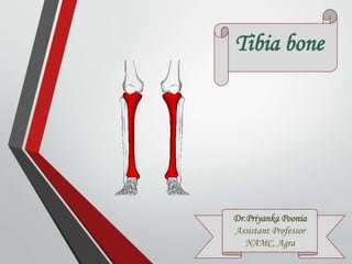
Also known as shin bone
Homologous with radius bone
of forearm.
Upper end is much larger than
lower end.
The medial side of lower end have
a downward projection, medial
malleolus.
The anterior border of shaft is
sharpest border of the bone.
Tibia is held
vertically.
Tibia
3. 1.Upper end
1.Medial condyle
2.Lateral condyle
3.Intercondylar
area
4.Tibial tuberosity
5.Fibular facet
2.Shaft
Borders Surfaces
1.Anterior 1.Medial
2.Medial 2.Lateral
3.Lateral 3. Posterior 3.Lower end
5 surfaces
1. Medial
2. Lateral
3. Anterior
4. Posterior
5. Inferior
Parts of tibia
Upper end
Shaft
Lower end
4. larger than lateral condyle.
Articulate with medial
condyle of femur to form
knee joint.
4 surfaces, anterior, medial,
posterior & superior.
Superior articular, oval shape,
has central & peripheral parts.
Central direct contact with
femoral condyle.
Peripheral is separated by
medial meniscus.
Anterior & Medial -
vascular foramina
Posterior- groove
1.Medial condyle 2. Lateral condyle
Fibular facet oval, lies
posteroinferior aspect.
Forms sup. TBJ with head of
fibula.
Articulate with lateral condyle
of femur to form knee joint.
4 surfaces, anterior, medial,
posterior & superior.
Posterior- groove.
Anterior - Gerdy’s tubercle.
Superior articular, circular shape
has central & peripheral parts.
Central direct contact with
femoral condyle.
Peripheral is separated by lateral
meniscus
5. Elevated part
intercondylar eminence.
Formed by M&L
intercondylar tubercle
B/W superior surface
of 2 condyle.
Narrowest in its
middle.
Prominence located on
anterior aspect of upper
end.
Upper smooth & lower
rough part.
Epiphyseal line of upper
end passes b/w two.
Forms anterior limits
of intercondylar area.
Continuous with
anterior border of shaft.
3. Inter condylar
area
4. Tibial tuberosity
6. Medial border
Round
Extends b/w medial condyle
to posterior border of medial
malleolus.
Anterior border
Sharp & ‘S’ shape
Subcutaneous, forms shin
Extends b/w tibial tuberosity to anterior
border of medial malleolus.
Lateral border
Interosseous
Extends b/w lateral condyle
to anterior border of fibular
notch
Borders
7. Medial surface
b/w medial to anterior border.
Broad & subcutaneous.
Posterior surface
b/w medial to lateral border.
Upper part has rough oblique ridge, soleal
line.
Above soleal line triangular upper area,
below elongated lower area.
Lower area divides into medial & lateral part,
by a vertical ridge.
Nutrient foramen lies in this surface.
Lateral surface
b/w lateral to anterior border.
In lower 1/3 it is directed
forward.
Surfaces
8. Posterior surface
Smaller than anterior.
Anterior surface
Larger, upper smooth &
lower rough grooved
part
Inferior surface
Articular, articulates with
superior trochlear surface of
talus to form ankle joint.
Lower end
Lateral surface
Triangular fibular notch,
articulates with lower end of
fibula to form inferior TFJ.
Medial surface
Subcutaneous, prolonged
downward as medial
malleolus.
9. Medial condyle
Capsular ligament of
knee joint
Medial patellar
ligament- ant. surface
Semimembranosus –
groove on post surface
Intercondylar area
Anterior horn of
medial meniscus
Anterior horn of
lateral meniscus
Anterior cruciate
ligament
Posterior horn of
medial meniscus
Posterior horn of
lateral meniscus
Posterior cruciate
ligament
Lateral condyle
Capsular ligament of
Sup TFJ.
Iliotibial tract-ant.
surface
Tendon of popliteus-
groove on post
surface
Upper
end
Tibial tuberosity
Ligamentum
patellae- upper
smooth part
Infrapatellar bursa-
lower rough part.
Attachments
10. Anterior border
Deep fascia of leg
Superior extensor
retinaculum
Posterior surface
Soleus- soleal line
Popliteus – above the
soleal line
Flexor digitorum longus-
below the soleal line
(medially)
Tibialis posterior-below
the soleal line (laterally)
Medial surface
Sartorious
Gracilis
Semitendinosus
Shaft
Lateral surface-
Tibialis anterior-
upper 2/3
Medial border
Tibial collateral
ligament
F.D.L.
Sartorious
Gracilis
Semitendinosus
Tibialis ant. Tibialis post.
Popliteus Soleous
11. Medial malleolus
Deltiod ligament
of ankle joint
Inferior surface
Capsule of ankle
joint
Fibular notch
Interosseous
tibiofibular ligament
Lower
end
13. Commonest site of
fracture is junction b/w
upper 2/3 & lower 1/3.
Commonest site of
osteomyelitis is upper
end (knee remain safe).
It has 1 primary (appears at 7th
week of IUL)in shaft & 2
secondary (at 16-18th year) in
both ends Ossification Centre.
Applied aspect
Nutrient artery of tibia is largest
& branch of posterior tibial
artery may tears in fracture of
shaft caused necrosis.
Commonest site of
dislocation is ankle joint
in which talus produce
prominence of heel.