SlideShare una empresa de Scribd logo
1 de 38
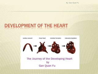
DEVELOPMENT OF THE HEART
By: Gan Quan Fu
CONTENT
1. Introduction
2. Fusion of Heart Tube
3. The Bulbus Cordis
4. The Sinus venosus
5. Formation of Atria
6. Separation of Aorta & Pulmonary
Trunk)
7. The Ventricles
8. The Exterior of Heart
9. Development of Heart Valve
10. Development of the Heart
conducting system
11. Timeline
12. Congenital Anomalies of Heart
By: Gan Quan Fu
By: Gan Quan Fu
INTRODUCTION
Mesodermal origin
Formed from angioblastic tissues that arise from
Splanchnopleuric Mesoderm (Cardiogenic area).
After formation of the head fold, this tube lies dorsal
to pericardial cavity & ventral to foregut.
Tube invaginates pericardial sac from dorsal side,
splanchnopleuric mesoderm lining the dorsal side of
pericardial cavity proliferates to form a thick layer
(myoepicardial mantle).
When invagination complete, myoepicardial mantle
completely surrounds the heart tube and give rise to
myocardium and also the epicardium.
Parietal layer of pericardium is derived from
somatopleuric mesoderm.
By: Gan Quan Fu
By: Gan Quan Fu
HEART TUBE
1st seen in form of right and left
endothelial heart tube.
Fuse and form single tube.
(Cranial to Caudal)
The single tube shows series of
dilatation:
Bulbus Cordis
Primitive Ventricle
Primitive Atrium
Sinus Venosus
Primitive Ventricle and Atrium
connected by a narrow
atrioventricular canal/
atrioventricular sulcus.
By: Gan Quan Fu
BULBUS CORDIS
Lies at arterial end of heart
Divisible into:
Proximal Part = Conus
Distal Part = Truncus
arteriosus
Truncus arteriosus
continuous distally with
aortic sac.
Aortic Sac is continuous
distally with right and left
pharyngeal arch arteries.
Arteries arch backwards to
become continuous with
right and left dorsal aortae.
By: Gan Quan Fu
HEART TUBES IN RELATION TO RIGHT AND LEFT
DORSAL AORTA
By: Gan Quan Fu
SINUS VENOSUS
Lies at venous end of
heart
It has right and left horns
(prolongations of sinus
venosus)
Each Horns are joined by:
One vitelline vein (from the
yolk sac)
One umbilical vein (from
the placenta)
One common cardinal vein
(from the body wall)
By: Gan Quan Fu
By: Gan Quan Fu
FORMATION OF ATRIA (SINU-ATRIAL ORIFICE)
Sinus venosus and
primitive atrial chamber
are 1st connected by wide
opening.
Gradually opening
becomes narrow & shifts
to right.
Finally becomes a narrow
slit. (The slit has right &
left margins known as
right & left venous
valves).
Cranially these 2 valve
fuse to form septum
By: Gan Quan Fu
FORMATION OF ATRIA (ATRIOVENTRICULAR
CANAL)
2 thickenings
(atrioventricular
cushions) appear on
dorsal and ventral
walls.
Grow towards each
other and fuse to form
septum intermedium.
By: Gan Quan Fu
FORMATION OF ATRIA (INTERATRIAL SEPTUM)
Atrial chamber undergoes division into right and
left by formation of 2 septa.
1. Septum Primum
2. Septa Secundum
By: Gan Quan Fu
FORMATION OF ATRIA (SEPTUM PRIMUM)
Arises from roof of atrium
towards atrioventricular canal
and form foramen primum.
Before foramen primum close
(Septum primum fuse with
septum intermedium), a new
gap (foramen secundum) is
form on upper part of foramen
primum by breaking down of
upper part of septum primum.
Septum primum now has a
free upper edge.
This is to ensure
communication between left
and right atria.
By: Gan Quan Fu
FORMATION OF ATRIA (SEPTUM SECUNDUM)
Arises downwards from roof of atrial chamber at
the right of septum primum.
Overlap with free edge of septum primum and
form foramen ovale (valvular aperture that
allows blood to flow from right to left).
After birth, foramen ovale is obliterated by
fusion of septum primum and septum
secundum.
Annulus ovalis  Lower free edge of septum
secundum
Fossa Ovalis  Septum primum
By: Gan Quan Fu
DEVELOPMENT OF RIGHT ATRIUM
Sinus venosus is absorbed into right atrium by
great enlargement of sinu-artrial orifice.
Left horn  Part of coronary sinus
Right common cardinal  Part of superior vena cava
Right vitelline vein  Terminal part of inferior vena
cava
Right margin of original sinu-atrial orifice (right & left
venous valve) expands and divide into:
Crista terminalis
Valve of inferior vena cava
Valve of coronary sinus
Right half of atrioventricular canal is also
absorbed into right atrium.
By: Gan Quan Fu
DEVELOPMENT OF LEFT ATRIUM
During the beginning of
formation of septum
primum, single pulmonary
vein open into left half of
atrium.
This pulmonary vein divide
into left and right branches.
Gradually the parts of
pulmonary veins nearest to
left atrium are absorbed
into the atrium resulting in
4 separate veins (2 each
side).
By: Gan Quan Fu
SEPARATION OF AORTA AND PULMONARY
TRUNK
Spiral Septum appears
within truncus arteriosus
and subdivides it into
ascending aorta and
pulmonary arteries.
They fuse along the axis of
the cylinder extending down
towards the ventricles.
Interventricular foramen
obliterated by mass of
endocardial tissues from
interventricular septum,
endocardial cushions &
By: Gan Quan Fu
DEVELOPMENT OF VENTRICLES
(INTERVENTRICULAR SEPTUM)
Conus of bulbus cordis merge with
cavity of primitive ventricle.
Interventricular septum grows
upwards from the floor of bulbo-
ventricular cavity & divide it into right
& left halves.
2 ridges (right & Left bulbar ridges)
from the wall of the conical part of
bulbar cavity (Spiral septum), grows
towards each other and fuse form
bulbar septum.
The gap between upper edge of
interventricular septum and lower
edge of bulbar septum, is filled by
proliferation of tissues from
atrioventricular cushions.
By: Gan Quan Fu
EXTERIOR OF HEART
Heart tube is suspended from the dorsal wall of the
pericardial cavity by 2 layers of percardium which
constitude dorsal mesocardium.
Mesocardium disappears and heart tube lies within
the pericardial sac.
Caudal part of the heart tube (atrium, sinus venosus)
is embedded within the septum transversum
By: Gan Quan Fu
EXTERIOR OF HEART
Bulbus cordis and ventricle in pericardial cavity grows
rapidly & becomes folded to form U-shape Bulbo-
ventricular loop.
The atrium and sinus venosus freed from septum
transversum. (Lie behind and above the ventricle and
the heart tube is now S shaped)
Arterial and venous ends come closer to each other
and the space between them becomes the transverse
sinus of pericardium.
By: Gan Quan Fu
EXTERIOR OF THE HEART
Till now, bulbus cordis (conus) & ventricle are
separated by deep bulboventricular sulcus.
Sulcus gradually becomes narrow, bulbus
cordis & ventricle come to form 1 chamber
which communicates with truncus arteriosus.
The atrial chamber which lies behind the
upper part of ventricle expands and projects
forwards on either side of the truncus.
Finally heart assumes its definitive shape.
By: Gan Quan Fu
VALVES OF HEART (MITRAL & TRICUSPID)
Formed by proliferation of connective tissue
under endocardium of left and right atrio-
ventricular canals.
By: Gan Quan Fu
VALVES OF HEART (PULMONARY & AORTIC)
Derived from endocardial cushions formed at
junction of truncus arteriosus and the conus
which grows and fuse with each other.
With separation of aortic and pulmonary trunk
each cushions are subdivided into 2 parts (each
in each orifice).
2 more cushions (anterior and posterior) appear.
 3 cushions in each opening which the 3
cusps of the corresponding valve develops.
Pulmonary valve is initially ventral to aortic
valve,. Subsequently rotation happens and
pulmonary valve lie ventral to left aortic valve.
By: Gan Quan Fu
DEVELOPMENT OF CONDUCTING SYSTEM OF
HEART
When there are 2 heart tubes, a pacemaker
(which later forms sinu-atrial node) lies in caudal
part of left tube.
After fusion of 2 tubes it lies in sinus venosus.
Comes to lie near to opening of superior vena
cava when sinus venosus is incorporated into
right ventricle.
Atrio-ventricular node and atrio-ventricular
bundle form in left wall of sinus venosus and in
atrio-ventricular canal.
After sinus venosus is absorbed into right
atrium, atrio-ventricular node comes to lie near
the interatrial septum.
By: Gan Quan Fu
TIMELINE ON FORMATION OF HEART
Early 4th week
The pair of lateral endocardial tubes fuse
5th to 6th week
A pair of septa, Septum Primum & Septum Secundum
separates right & left atria
Development of bicuspid and tricuspid atrioventricular
valve
6th week
Muscular ventricular septum partially separates the
ventricles
7th & 8th week
Spiral septum separates ascending aorta & pulmonary
trunk
Swellings within truncus arteriosus give rise to semilunar
valves of aorta & pulmonary trunk
Complete ventricular septation
By: Gan Quan Fu
CONGENITAL ANOMALIES OF HEART
Anomalies of Position
Atresia or Stenosis
Abnormal Growth
Defective Formation of Septa
Anomalies of Relationship of Chambers to
Great Vessels
By: Gan Quan Fu
ANOMALIES OF POSITION
Dextrocardia
Chambers and blood vessels of heart are reversed
from side to side (i.e. all structures that normally lie
on right side are on left and vice versa)
Maybe a part of the condition called situs inversus
(Organs are transposed)
If not part of situs inversus, usually accompanied by
anomalies of heart chambers and great vessels.
Ectopia Cordis
Heart lies exposed, on front of the chest and can be
seen from outside, due to defective development of
chest wall.
By: Gan Quan Fu
ATRESIA OR STENOSIS
Too narrow opening (stenosis) or no opening
(atresia) of the orifices of heart.
Aortic and pulmonary passages may also show
supravalvular or subvalvular stenosis.
Alternately, the openings may be too large and
valves becomes incompetent.
In pulmonary stenosis  foramen ovale and
ductus arteriosus remain patent.
In aortic stenosis  ductus arteriosus is patent
and blood flows into aorta through it.
By: Gan Quan Fu
ABNORMAL GROWTH
There maybe acessory cusps in the valves
Congenital tumors may be formed
Left atrium may be partially subdivided by a
transverse septum.
Myocardium may be poorly developed
(hypoplasia).
By: Gan Quan Fu
DEFECTIVE FORMATION OF SEPTA
1. INTERATRIAL SEPTAL DEFECTS
Osteum Primum Defect (image A)
Septum primum fail to reach atrio-ventricular
endocardial cushions  Foramen primum persist.
Osteum Secundum Defect (image B)
Septum secundum fail to develop  foramen
secundum remains wide open.
Patent Foramen Ovale (image C)
Septum Primum and Secundum may develop
normally but the Oblique valvular passage between
them may remain patent.
By: Gan Quan Fu
DEFECTIVE FORMATION OF SEPTA
Interventricular Septal Defects (image D)
Maybe seen either in the membranous or muscular
part of the septum
Most common congenital anomalies of the heart.
Patent Truncus Arteriosus
Defects of the spiral septum
Spiral Septum may not be formed at all. Partial
absence of the septum leads to
communications(shunts)between the aorta and
pulmonary trunk.
Atrio-ventricular canal defect / Persistent atrio-
ventricular canal
Defective formation of AV cushions, 4 chambers of
heart may intercommunicate.
By: Gan Quan Fu
COMBINED DEFECTS
Tetralogy of Fallot
Interventricular septal Defect
Aorta overriding the free upper edge of
ventricular septum
Pulmonary Stenosis
Hypertrophy of the Right Ventricle
By: Gan Quan Fu
ANOMALIES OF RELATIONSHIP OF CHAMBERS
TO GREAT VESSELS
Transposition of Great Vessels
The aorta arises from the right ventricle and the
pulmonary trunk from the left ventricle.
Taussig-Bing Syndrome
The aorta arises from the right ventricle and
pulmonary trunk overrides both the right and left
ventricle.
Interventricular defect
The Superior Vena Cava may end in the Left
Atrium
Pulmonary Veins may end in the Right Atrium or
one of it’s tributaries.
By: Gan Quan Fu
SUMMARY
Heart primodium forms in a horseshoe-shape
region of splanchnopleuric mesoderm at cranial
end of germ disc (Cardiogenic Region).
In response to signals from underlying
endoderm, angioblastic cords in this region
coalesce to form a pair of lateral heart tube.
Early of 4th week, they fuse to form primitive
heart tube.
Between weeks 5 and 8, primitive heart tube
undergoes process of looping, remodelling, and
septation that transform it’s single lumen into 4
chambers, thus laying down the basis for
separation of pulmonary and systemic
circulation
By: Gan Quan Fu
REFERENCES
Larsen, W.J. (1997) Human Embryology, 2nd
edn. Hong Kong; Churchill Livingstone.
Singh, I. & Pal, G.P. (1976) Human
Embryology, 7th edn. India; Macmillan.
By: Gan Quan Fu
Q&A SESSION !!!!
By: Gan Quan Fu
By: Gan Quan Fu

Más contenido relacionado

La actualidad más candente

11 development of the heart
11   development of the heart11   development of the heart
11 development of the heartpuneet mahajan
 
Development of Thyroid Gland (Special Embryology)
Development of Thyroid Gland (Special Embryology)Development of Thyroid Gland (Special Embryology)
Development of Thyroid Gland (Special Embryology)Dr. Sherif Fahmy
 
Fibrous skeleton of the heart
Fibrous  skeleton of the heartFibrous  skeleton of the heart
Fibrous skeleton of the heartIdris Siddiqui
 
Embrology of the respiratory system
Embrology of the respiratory systemEmbrology of the respiratory system
Embrology of the respiratory systemOriba Dan Langoya
 
Anatomy of The Diaphragm
 Anatomy of The Diaphragm Anatomy of The Diaphragm
Anatomy of The DiaphragmTrishna Kisiju
 
Development of midgut and pancreas
Development of midgut  and pancreas Development of midgut  and pancreas
Development of midgut and pancreas Sahar Hafeez
 
Development of diaphragm
Development of diaphragmDevelopment of diaphragm
Development of diaphragmNeeta Chhabra
 
Cardiovascular System Development
Cardiovascular System DevelopmentCardiovascular System Development
Cardiovascular System DevelopmentAUC Medical School
 
Internal feature of right and left atria
Internal feature of right and left atriaInternal feature of right and left atria
Internal feature of right and left atriafarranajwa
 
Azygos system of veins
Azygos system of veinsAzygos system of veins
Azygos system of veinsIdris Siddiqui
 
embryology of heart
embryology of heartembryology of heart
embryology of heartSarvesh Raj
 

La actualidad más candente (20)

Development of heart
Development of heartDevelopment of heart
Development of heart
 
Thorax Anatomy
Thorax AnatomyThorax Anatomy
Thorax Anatomy
 
Embryology of heart
Embryology of heartEmbryology of heart
Embryology of heart
 
6. Development of blood vessels
6. Development of blood vessels6. Development of blood vessels
6. Development of blood vessels
 
11 development of the heart
11   development of the heart11   development of the heart
11 development of the heart
 
Development of Thyroid Gland (Special Embryology)
Development of Thyroid Gland (Special Embryology)Development of Thyroid Gland (Special Embryology)
Development of Thyroid Gland (Special Embryology)
 
Internal capsule
Internal capsuleInternal capsule
Internal capsule
 
Fibrous skeleton of the heart
Fibrous  skeleton of the heartFibrous  skeleton of the heart
Fibrous skeleton of the heart
 
Embrology of the respiratory system
Embrology of the respiratory systemEmbrology of the respiratory system
Embrology of the respiratory system
 
Anatomy of The Diaphragm
 Anatomy of The Diaphragm Anatomy of The Diaphragm
Anatomy of The Diaphragm
 
Development of midgut and pancreas
Development of midgut  and pancreas Development of midgut  and pancreas
Development of midgut and pancreas
 
Development of diaphragm
Development of diaphragmDevelopment of diaphragm
Development of diaphragm
 
Coronary circulation
Coronary circulationCoronary circulation
Coronary circulation
 
Development of heart
Development of heartDevelopment of heart
Development of heart
 
Cardiovascular System Development
Cardiovascular System DevelopmentCardiovascular System Development
Cardiovascular System Development
 
Internal feature of right and left atria
Internal feature of right and left atriaInternal feature of right and left atria
Internal feature of right and left atria
 
Azygos system of veins
Azygos system of veinsAzygos system of veins
Azygos system of veins
 
embryology of heart
embryology of heartembryology of heart
embryology of heart
 
mediastinum
mediastinummediastinum
mediastinum
 
Superior vena cava
Superior vena cavaSuperior vena cava
Superior vena cava
 

Destacado

Lecture11 development of the heart and blood vessels
Lecture11   development  of the heart and blood vesselsLecture11   development  of the heart and blood vessels
Lecture11 development of the heart and blood vesselsMUBOSScz
 
Vsd embryology
Vsd embryologyVsd embryology
Vsd embryologySujit Sahu
 
Cardiac development final
Cardiac development finalCardiac development final
Cardiac development finalSandip Gupta
 
Embryology of heart and fetal circulation
Embryology of heart and fetal circulationEmbryology of heart and fetal circulation
Embryology of heart and fetal circulationArchana Koshy
 
Development of heart and respiratory system
Development of heart and respiratory systemDevelopment of heart and respiratory system
Development of heart and respiratory systemAbdul Ansari
 
Fetal circulation
Fetal circulationFetal circulation
Fetal circulationrekha
 
Hypoxia, platypnea orthodeoxia and transient ischemic attacks in a patient
Hypoxia, platypnea orthodeoxia and transient ischemic attacks in a patientHypoxia, platypnea orthodeoxia and transient ischemic attacks in a patient
Hypoxia, platypnea orthodeoxia and transient ischemic attacks in a patientdrucsamal
 
Location & structure of the heart
Location & structure  of the heartLocation & structure  of the heart
Location & structure of the heartVyramuthu Atputhan
 

Destacado (17)

Heart development i
Heart development iHeart development i
Heart development i
 
Lecture11 development of the heart and blood vessels
Lecture11   development  of the heart and blood vesselsLecture11   development  of the heart and blood vessels
Lecture11 development of the heart and blood vessels
 
6 development of the heart akd
6 development of the  heart akd6 development of the  heart akd
6 development of the heart akd
 
Vsd embryology
Vsd embryologyVsd embryology
Vsd embryology
 
Cardiac development final
Cardiac development finalCardiac development final
Cardiac development final
 
Embryology of heart and fetal circulation
Embryology of heart and fetal circulationEmbryology of heart and fetal circulation
Embryology of heart and fetal circulation
 
Development of heart and respiratory system
Development of heart and respiratory systemDevelopment of heart and respiratory system
Development of heart and respiratory system
 
Embryological basis of congenital heart diseases
Embryological basis of congenital heart diseases   Embryological basis of congenital heart diseases
Embryological basis of congenital heart diseases
 
Fetal circulation
Fetal circulationFetal circulation
Fetal circulation
 
Fetalcirculation
FetalcirculationFetalcirculation
Fetalcirculation
 
Heart Anatomy
Heart AnatomyHeart Anatomy
Heart Anatomy
 
Heart
HeartHeart
Heart
 
The boot shaped heart sign
The boot shaped heart signThe boot shaped heart sign
The boot shaped heart sign
 
Hypoxia, platypnea orthodeoxia and transient ischemic attacks in a patient
Hypoxia, platypnea orthodeoxia and transient ischemic attacks in a patientHypoxia, platypnea orthodeoxia and transient ischemic attacks in a patient
Hypoxia, platypnea orthodeoxia and transient ischemic attacks in a patient
 
Ppt18
Ppt18Ppt18
Ppt18
 
Location & structure of the heart
Location & structure  of the heartLocation & structure  of the heart
Location & structure of the heart
 
Cardiac embryology seminar copy
Cardiac embryology seminar   copyCardiac embryology seminar   copy
Cardiac embryology seminar copy
 

Similar a Development of the heart

Cardiac embryology
Cardiac embryologyCardiac embryology
Cardiac embryologyRezaul hayat
 
development of heart and it’s clinical abnormal
development of heart and it’s clinical abnormaldevelopment of heart and it’s clinical abnormal
development of heart and it’s clinical abnormalOmpriyaS
 
Embryology Course VI - Cardiovascular System
Embryology Course VI - Cardiovascular SystemEmbryology Course VI - Cardiovascular System
Embryology Course VI - Cardiovascular SystemRawa Muhsin
 
CVS Embryology (Amoud University)
CVS Embryology (Amoud University)CVS Embryology (Amoud University)
CVS Embryology (Amoud University)moaminnca
 
Development of heart finale copy
Development of heart finale   copyDevelopment of heart finale   copy
Development of heart finale copyRobin Thomas
 
Development of heart cvs anatomy
Development of heart cvs anatomyDevelopment of heart cvs anatomy
Development of heart cvs anatomyRobbinsHobbin
 
Final heart development.pptx
Final heart development.pptxFinal heart development.pptx
Final heart development.pptxSayanRDey
 
Development of Heart (Embryology)
Development of Heart (Embryology)Development of Heart (Embryology)
Development of Heart (Embryology)Yukta Wankhede
 
11 - Development of the Heart.ppt
11 - Development of the Heart.ppt11 - Development of the Heart.ppt
11 - Development of the Heart.pptDr Noorul
 
Aorta, SVC, PULMONARY TRUNK.pptx
Aorta, SVC, PULMONARY TRUNK.pptxAorta, SVC, PULMONARY TRUNK.pptx
Aorta, SVC, PULMONARY TRUNK.pptxMovieDownload83
 
development of heart.pptx
development of heart.pptxdevelopment of heart.pptx
development of heart.pptxKARTHIKD765407
 
Cynotic heart disease plane radiography
Cynotic heart disease plane radiography Cynotic heart disease plane radiography
Cynotic heart disease plane radiography Dr-Girish Gunari
 
Surgery 5th year, 9th, 10th &11th lectures (Dr. Ahmed Al-Azzawi)
Surgery 5th year, 9th, 10th &11th lectures (Dr. Ahmed Al-Azzawi)Surgery 5th year, 9th, 10th &11th lectures (Dr. Ahmed Al-Azzawi)
Surgery 5th year, 9th, 10th &11th lectures (Dr. Ahmed Al-Azzawi)College of Medicine, Sulaymaniyah
 

Similar a Development of the heart (20)

Cardiac embryology
Cardiac embryologyCardiac embryology
Cardiac embryology
 
development of heart and it’s clinical abnormal
development of heart and it’s clinical abnormaldevelopment of heart and it’s clinical abnormal
development of heart and it’s clinical abnormal
 
heart dev.pptx
heart dev.pptxheart dev.pptx
heart dev.pptx
 
Embryology Course VI - Cardiovascular System
Embryology Course VI - Cardiovascular SystemEmbryology Course VI - Cardiovascular System
Embryology Course VI - Cardiovascular System
 
CVS Embryology (Amoud University)
CVS Embryology (Amoud University)CVS Embryology (Amoud University)
CVS Embryology (Amoud University)
 
Evolution of Heart
Evolution of HeartEvolution of Heart
Evolution of Heart
 
Development of heart finale copy
Development of heart finale   copyDevelopment of heart finale   copy
Development of heart finale copy
 
Development of heart cvs anatomy
Development of heart cvs anatomyDevelopment of heart cvs anatomy
Development of heart cvs anatomy
 
Heart development
Heart developmentHeart development
Heart development
 
Heart and lung development
Heart and lung developmentHeart and lung development
Heart and lung development
 
Embryology of the heart
Embryology of the heartEmbryology of the heart
Embryology of the heart
 
Final heart development.pptx
Final heart development.pptxFinal heart development.pptx
Final heart development.pptx
 
Development of Heart (Embryology)
Development of Heart (Embryology)Development of Heart (Embryology)
Development of Heart (Embryology)
 
Manoja_Gullapalli_ug
Manoja_Gullapalli_ugManoja_Gullapalli_ug
Manoja_Gullapalli_ug
 
Final heart development.ppt
Final heart development.pptFinal heart development.ppt
Final heart development.ppt
 
11 - Development of the Heart.ppt
11 - Development of the Heart.ppt11 - Development of the Heart.ppt
11 - Development of the Heart.ppt
 
Aorta, SVC, PULMONARY TRUNK.pptx
Aorta, SVC, PULMONARY TRUNK.pptxAorta, SVC, PULMONARY TRUNK.pptx
Aorta, SVC, PULMONARY TRUNK.pptx
 
development of heart.pptx
development of heart.pptxdevelopment of heart.pptx
development of heart.pptx
 
Cynotic heart disease plane radiography
Cynotic heart disease plane radiography Cynotic heart disease plane radiography
Cynotic heart disease plane radiography
 
Surgery 5th year, 9th, 10th &11th lectures (Dr. Ahmed Al-Azzawi)
Surgery 5th year, 9th, 10th &11th lectures (Dr. Ahmed Al-Azzawi)Surgery 5th year, 9th, 10th &11th lectures (Dr. Ahmed Al-Azzawi)
Surgery 5th year, 9th, 10th &11th lectures (Dr. Ahmed Al-Azzawi)
 

Más de Quan Fu Gan

General sports injuries around the ankle foot complex
General sports injuries around the ankle foot complexGeneral sports injuries around the ankle foot complex
General sports injuries around the ankle foot complexQuan Fu Gan
 
1. risk factors and prevention of sports injuries
1. risk factors and prevention of sports injuries1. risk factors and prevention of sports injuries
1. risk factors and prevention of sports injuriesQuan Fu Gan
 
Chromosomal anomalies
Chromosomal anomaliesChromosomal anomalies
Chromosomal anomaliesQuan Fu Gan
 
Seminar clinical anatomy of upper limb joints and muscles
Seminar clinical anatomy of upper limb joints and musclesSeminar clinical anatomy of upper limb joints and muscles
Seminar clinical anatomy of upper limb joints and musclesQuan Fu Gan
 
Examination of blood
Examination of bloodExamination of blood
Examination of bloodQuan Fu Gan
 
Embryology of kidney
Embryology of kidneyEmbryology of kidney
Embryology of kidneyQuan Fu Gan
 
Histology of gastrointestinal tract
Histology of gastrointestinal tractHistology of gastrointestinal tract
Histology of gastrointestinal tractQuan Fu Gan
 
Exercises (DIY at your own risk)
Exercises (DIY at your own risk)Exercises (DIY at your own risk)
Exercises (DIY at your own risk)Quan Fu Gan
 
Spinal cord injury (sci) Rehab
Spinal cord injury (sci) RehabSpinal cord injury (sci) Rehab
Spinal cord injury (sci) RehabQuan Fu Gan
 
Types of Joint Present in the vertebral column (simplified version)
Types of Joint Present in the vertebral column (simplified version)Types of Joint Present in the vertebral column (simplified version)
Types of Joint Present in the vertebral column (simplified version)Quan Fu Gan
 
Cardiorespiratory Assessment
Cardiorespiratory AssessmentCardiorespiratory Assessment
Cardiorespiratory AssessmentQuan Fu Gan
 
The shoulder joint
The shoulder jointThe shoulder joint
The shoulder jointQuan Fu Gan
 

Más de Quan Fu Gan (12)

General sports injuries around the ankle foot complex
General sports injuries around the ankle foot complexGeneral sports injuries around the ankle foot complex
General sports injuries around the ankle foot complex
 
1. risk factors and prevention of sports injuries
1. risk factors and prevention of sports injuries1. risk factors and prevention of sports injuries
1. risk factors and prevention of sports injuries
 
Chromosomal anomalies
Chromosomal anomaliesChromosomal anomalies
Chromosomal anomalies
 
Seminar clinical anatomy of upper limb joints and muscles
Seminar clinical anatomy of upper limb joints and musclesSeminar clinical anatomy of upper limb joints and muscles
Seminar clinical anatomy of upper limb joints and muscles
 
Examination of blood
Examination of bloodExamination of blood
Examination of blood
 
Embryology of kidney
Embryology of kidneyEmbryology of kidney
Embryology of kidney
 
Histology of gastrointestinal tract
Histology of gastrointestinal tractHistology of gastrointestinal tract
Histology of gastrointestinal tract
 
Exercises (DIY at your own risk)
Exercises (DIY at your own risk)Exercises (DIY at your own risk)
Exercises (DIY at your own risk)
 
Spinal cord injury (sci) Rehab
Spinal cord injury (sci) RehabSpinal cord injury (sci) Rehab
Spinal cord injury (sci) Rehab
 
Types of Joint Present in the vertebral column (simplified version)
Types of Joint Present in the vertebral column (simplified version)Types of Joint Present in the vertebral column (simplified version)
Types of Joint Present in the vertebral column (simplified version)
 
Cardiorespiratory Assessment
Cardiorespiratory AssessmentCardiorespiratory Assessment
Cardiorespiratory Assessment
 
The shoulder joint
The shoulder jointThe shoulder joint
The shoulder joint
 

Último

A Critique of the Proposed National Education Policy Reform
A Critique of the Proposed National Education Policy ReformA Critique of the Proposed National Education Policy Reform
A Critique of the Proposed National Education Policy ReformChameera Dedduwage
 
Kisan Call Centre - To harness potential of ICT in Agriculture by answer farm...
Kisan Call Centre - To harness potential of ICT in Agriculture by answer farm...Kisan Call Centre - To harness potential of ICT in Agriculture by answer farm...
Kisan Call Centre - To harness potential of ICT in Agriculture by answer farm...Krashi Coaching
 
Web & Social Media Analytics Previous Year Question Paper.pdf
Web & Social Media Analytics Previous Year Question Paper.pdfWeb & Social Media Analytics Previous Year Question Paper.pdf
Web & Social Media Analytics Previous Year Question Paper.pdfJayanti Pande
 
Accessible design: Minimum effort, maximum impact
Accessible design: Minimum effort, maximum impactAccessible design: Minimum effort, maximum impact
Accessible design: Minimum effort, maximum impactdawncurless
 
Explore beautiful and ugly buildings. Mathematics helps us create beautiful d...
Explore beautiful and ugly buildings. Mathematics helps us create beautiful d...Explore beautiful and ugly buildings. Mathematics helps us create beautiful d...
Explore beautiful and ugly buildings. Mathematics helps us create beautiful d...christianmathematics
 
fourth grading exam for kindergarten in writing
fourth grading exam for kindergarten in writingfourth grading exam for kindergarten in writing
fourth grading exam for kindergarten in writingTeacherCyreneCayanan
 
APM Welcome, APM North West Network Conference, Synergies Across Sectors
APM Welcome, APM North West Network Conference, Synergies Across SectorsAPM Welcome, APM North West Network Conference, Synergies Across Sectors
APM Welcome, APM North West Network Conference, Synergies Across SectorsAssociation for Project Management
 
Grant Readiness 101 TechSoup and Remy Consulting
Grant Readiness 101 TechSoup and Remy ConsultingGrant Readiness 101 TechSoup and Remy Consulting
Grant Readiness 101 TechSoup and Remy ConsultingTechSoup
 
1029-Danh muc Sach Giao Khoa khoi 6.pdf
1029-Danh muc Sach Giao Khoa khoi  6.pdf1029-Danh muc Sach Giao Khoa khoi  6.pdf
1029-Danh muc Sach Giao Khoa khoi 6.pdfQucHHunhnh
 
Unit-IV- Pharma. Marketing Channels.pptx
Unit-IV- Pharma. Marketing Channels.pptxUnit-IV- Pharma. Marketing Channels.pptx
Unit-IV- Pharma. Marketing Channels.pptxVishalSingh1417
 
Nutritional Needs Presentation - HLTH 104
Nutritional Needs Presentation - HLTH 104Nutritional Needs Presentation - HLTH 104
Nutritional Needs Presentation - HLTH 104misteraugie
 
social pharmacy d-pharm 1st year by Pragati K. Mahajan
social pharmacy d-pharm 1st year by Pragati K. Mahajansocial pharmacy d-pharm 1st year by Pragati K. Mahajan
social pharmacy d-pharm 1st year by Pragati K. Mahajanpragatimahajan3
 
Advanced Views - Calendar View in Odoo 17
Advanced Views - Calendar View in Odoo 17Advanced Views - Calendar View in Odoo 17
Advanced Views - Calendar View in Odoo 17Celine George
 
Z Score,T Score, Percential Rank and Box Plot Graph
Z Score,T Score, Percential Rank and Box Plot GraphZ Score,T Score, Percential Rank and Box Plot Graph
Z Score,T Score, Percential Rank and Box Plot GraphThiyagu K
 
Sports & Fitness Value Added Course FY..
Sports & Fitness Value Added Course FY..Sports & Fitness Value Added Course FY..
Sports & Fitness Value Added Course FY..Disha Kariya
 
Student login on Anyboli platform.helpin
Student login on Anyboli platform.helpinStudent login on Anyboli platform.helpin
Student login on Anyboli platform.helpinRaunakKeshri1
 
Key note speaker Neum_Admir Softic_ENG.pdf
Key note speaker Neum_Admir Softic_ENG.pdfKey note speaker Neum_Admir Softic_ENG.pdf
Key note speaker Neum_Admir Softic_ENG.pdfAdmir Softic
 

Último (20)

A Critique of the Proposed National Education Policy Reform
A Critique of the Proposed National Education Policy ReformA Critique of the Proposed National Education Policy Reform
A Critique of the Proposed National Education Policy Reform
 
Mattingly "AI & Prompt Design: The Basics of Prompt Design"
Mattingly "AI & Prompt Design: The Basics of Prompt Design"Mattingly "AI & Prompt Design: The Basics of Prompt Design"
Mattingly "AI & Prompt Design: The Basics of Prompt Design"
 
Kisan Call Centre - To harness potential of ICT in Agriculture by answer farm...
Kisan Call Centre - To harness potential of ICT in Agriculture by answer farm...Kisan Call Centre - To harness potential of ICT in Agriculture by answer farm...
Kisan Call Centre - To harness potential of ICT in Agriculture by answer farm...
 
Web & Social Media Analytics Previous Year Question Paper.pdf
Web & Social Media Analytics Previous Year Question Paper.pdfWeb & Social Media Analytics Previous Year Question Paper.pdf
Web & Social Media Analytics Previous Year Question Paper.pdf
 
Accessible design: Minimum effort, maximum impact
Accessible design: Minimum effort, maximum impactAccessible design: Minimum effort, maximum impact
Accessible design: Minimum effort, maximum impact
 
Explore beautiful and ugly buildings. Mathematics helps us create beautiful d...
Explore beautiful and ugly buildings. Mathematics helps us create beautiful d...Explore beautiful and ugly buildings. Mathematics helps us create beautiful d...
Explore beautiful and ugly buildings. Mathematics helps us create beautiful d...
 
fourth grading exam for kindergarten in writing
fourth grading exam for kindergarten in writingfourth grading exam for kindergarten in writing
fourth grading exam for kindergarten in writing
 
APM Welcome, APM North West Network Conference, Synergies Across Sectors
APM Welcome, APM North West Network Conference, Synergies Across SectorsAPM Welcome, APM North West Network Conference, Synergies Across Sectors
APM Welcome, APM North West Network Conference, Synergies Across Sectors
 
Grant Readiness 101 TechSoup and Remy Consulting
Grant Readiness 101 TechSoup and Remy ConsultingGrant Readiness 101 TechSoup and Remy Consulting
Grant Readiness 101 TechSoup and Remy Consulting
 
1029-Danh muc Sach Giao Khoa khoi 6.pdf
1029-Danh muc Sach Giao Khoa khoi  6.pdf1029-Danh muc Sach Giao Khoa khoi  6.pdf
1029-Danh muc Sach Giao Khoa khoi 6.pdf
 
Unit-IV- Pharma. Marketing Channels.pptx
Unit-IV- Pharma. Marketing Channels.pptxUnit-IV- Pharma. Marketing Channels.pptx
Unit-IV- Pharma. Marketing Channels.pptx
 
Nutritional Needs Presentation - HLTH 104
Nutritional Needs Presentation - HLTH 104Nutritional Needs Presentation - HLTH 104
Nutritional Needs Presentation - HLTH 104
 
social pharmacy d-pharm 1st year by Pragati K. Mahajan
social pharmacy d-pharm 1st year by Pragati K. Mahajansocial pharmacy d-pharm 1st year by Pragati K. Mahajan
social pharmacy d-pharm 1st year by Pragati K. Mahajan
 
Advanced Views - Calendar View in Odoo 17
Advanced Views - Calendar View in Odoo 17Advanced Views - Calendar View in Odoo 17
Advanced Views - Calendar View in Odoo 17
 
Z Score,T Score, Percential Rank and Box Plot Graph
Z Score,T Score, Percential Rank and Box Plot GraphZ Score,T Score, Percential Rank and Box Plot Graph
Z Score,T Score, Percential Rank and Box Plot Graph
 
Advance Mobile Application Development class 07
Advance Mobile Application Development class 07Advance Mobile Application Development class 07
Advance Mobile Application Development class 07
 
Sports & Fitness Value Added Course FY..
Sports & Fitness Value Added Course FY..Sports & Fitness Value Added Course FY..
Sports & Fitness Value Added Course FY..
 
Student login on Anyboli platform.helpin
Student login on Anyboli platform.helpinStudent login on Anyboli platform.helpin
Student login on Anyboli platform.helpin
 
Key note speaker Neum_Admir Softic_ENG.pdf
Key note speaker Neum_Admir Softic_ENG.pdfKey note speaker Neum_Admir Softic_ENG.pdf
Key note speaker Neum_Admir Softic_ENG.pdf
 
Código Creativo y Arte de Software | Unidad 1
Código Creativo y Arte de Software | Unidad 1Código Creativo y Arte de Software | Unidad 1
Código Creativo y Arte de Software | Unidad 1
 

Development of the heart

  • 1. DEVELOPMENT OF THE HEART By: Gan Quan Fu
  • 2. CONTENT 1. Introduction 2. Fusion of Heart Tube 3. The Bulbus Cordis 4. The Sinus venosus 5. Formation of Atria 6. Separation of Aorta & Pulmonary Trunk) 7. The Ventricles 8. The Exterior of Heart 9. Development of Heart Valve 10. Development of the Heart conducting system 11. Timeline 12. Congenital Anomalies of Heart By: Gan Quan Fu
  • 4. INTRODUCTION Mesodermal origin Formed from angioblastic tissues that arise from Splanchnopleuric Mesoderm (Cardiogenic area). After formation of the head fold, this tube lies dorsal to pericardial cavity & ventral to foregut. Tube invaginates pericardial sac from dorsal side, splanchnopleuric mesoderm lining the dorsal side of pericardial cavity proliferates to form a thick layer (myoepicardial mantle). When invagination complete, myoepicardial mantle completely surrounds the heart tube and give rise to myocardium and also the epicardium. Parietal layer of pericardium is derived from somatopleuric mesoderm. By: Gan Quan Fu
  • 6. HEART TUBE 1st seen in form of right and left endothelial heart tube. Fuse and form single tube. (Cranial to Caudal) The single tube shows series of dilatation: Bulbus Cordis Primitive Ventricle Primitive Atrium Sinus Venosus Primitive Ventricle and Atrium connected by a narrow atrioventricular canal/ atrioventricular sulcus. By: Gan Quan Fu
  • 7. BULBUS CORDIS Lies at arterial end of heart Divisible into: Proximal Part = Conus Distal Part = Truncus arteriosus Truncus arteriosus continuous distally with aortic sac. Aortic Sac is continuous distally with right and left pharyngeal arch arteries. Arteries arch backwards to become continuous with right and left dorsal aortae. By: Gan Quan Fu
  • 8. HEART TUBES IN RELATION TO RIGHT AND LEFT DORSAL AORTA By: Gan Quan Fu
  • 9. SINUS VENOSUS Lies at venous end of heart It has right and left horns (prolongations of sinus venosus) Each Horns are joined by: One vitelline vein (from the yolk sac) One umbilical vein (from the placenta) One common cardinal vein (from the body wall) By: Gan Quan Fu
  • 11. FORMATION OF ATRIA (SINU-ATRIAL ORIFICE) Sinus venosus and primitive atrial chamber are 1st connected by wide opening. Gradually opening becomes narrow & shifts to right. Finally becomes a narrow slit. (The slit has right & left margins known as right & left venous valves). Cranially these 2 valve fuse to form septum By: Gan Quan Fu
  • 12. FORMATION OF ATRIA (ATRIOVENTRICULAR CANAL) 2 thickenings (atrioventricular cushions) appear on dorsal and ventral walls. Grow towards each other and fuse to form septum intermedium. By: Gan Quan Fu
  • 13. FORMATION OF ATRIA (INTERATRIAL SEPTUM) Atrial chamber undergoes division into right and left by formation of 2 septa. 1. Septum Primum 2. Septa Secundum By: Gan Quan Fu
  • 14. FORMATION OF ATRIA (SEPTUM PRIMUM) Arises from roof of atrium towards atrioventricular canal and form foramen primum. Before foramen primum close (Septum primum fuse with septum intermedium), a new gap (foramen secundum) is form on upper part of foramen primum by breaking down of upper part of septum primum. Septum primum now has a free upper edge. This is to ensure communication between left and right atria. By: Gan Quan Fu
  • 15. FORMATION OF ATRIA (SEPTUM SECUNDUM) Arises downwards from roof of atrial chamber at the right of septum primum. Overlap with free edge of septum primum and form foramen ovale (valvular aperture that allows blood to flow from right to left). After birth, foramen ovale is obliterated by fusion of septum primum and septum secundum. Annulus ovalis  Lower free edge of septum secundum Fossa Ovalis  Septum primum By: Gan Quan Fu
  • 16. DEVELOPMENT OF RIGHT ATRIUM Sinus venosus is absorbed into right atrium by great enlargement of sinu-artrial orifice. Left horn  Part of coronary sinus Right common cardinal  Part of superior vena cava Right vitelline vein  Terminal part of inferior vena cava Right margin of original sinu-atrial orifice (right & left venous valve) expands and divide into: Crista terminalis Valve of inferior vena cava Valve of coronary sinus Right half of atrioventricular canal is also absorbed into right atrium. By: Gan Quan Fu
  • 17. DEVELOPMENT OF LEFT ATRIUM During the beginning of formation of septum primum, single pulmonary vein open into left half of atrium. This pulmonary vein divide into left and right branches. Gradually the parts of pulmonary veins nearest to left atrium are absorbed into the atrium resulting in 4 separate veins (2 each side). By: Gan Quan Fu
  • 18. SEPARATION OF AORTA AND PULMONARY TRUNK Spiral Septum appears within truncus arteriosus and subdivides it into ascending aorta and pulmonary arteries. They fuse along the axis of the cylinder extending down towards the ventricles. Interventricular foramen obliterated by mass of endocardial tissues from interventricular septum, endocardial cushions & By: Gan Quan Fu
  • 19. DEVELOPMENT OF VENTRICLES (INTERVENTRICULAR SEPTUM) Conus of bulbus cordis merge with cavity of primitive ventricle. Interventricular septum grows upwards from the floor of bulbo- ventricular cavity & divide it into right & left halves. 2 ridges (right & Left bulbar ridges) from the wall of the conical part of bulbar cavity (Spiral septum), grows towards each other and fuse form bulbar septum. The gap between upper edge of interventricular septum and lower edge of bulbar septum, is filled by proliferation of tissues from atrioventricular cushions. By: Gan Quan Fu
  • 20. EXTERIOR OF HEART Heart tube is suspended from the dorsal wall of the pericardial cavity by 2 layers of percardium which constitude dorsal mesocardium. Mesocardium disappears and heart tube lies within the pericardial sac. Caudal part of the heart tube (atrium, sinus venosus) is embedded within the septum transversum By: Gan Quan Fu
  • 21. EXTERIOR OF HEART Bulbus cordis and ventricle in pericardial cavity grows rapidly & becomes folded to form U-shape Bulbo- ventricular loop. The atrium and sinus venosus freed from septum transversum. (Lie behind and above the ventricle and the heart tube is now S shaped) Arterial and venous ends come closer to each other and the space between them becomes the transverse sinus of pericardium. By: Gan Quan Fu
  • 22. EXTERIOR OF THE HEART Till now, bulbus cordis (conus) & ventricle are separated by deep bulboventricular sulcus. Sulcus gradually becomes narrow, bulbus cordis & ventricle come to form 1 chamber which communicates with truncus arteriosus. The atrial chamber which lies behind the upper part of ventricle expands and projects forwards on either side of the truncus. Finally heart assumes its definitive shape. By: Gan Quan Fu
  • 23. VALVES OF HEART (MITRAL & TRICUSPID) Formed by proliferation of connective tissue under endocardium of left and right atrio- ventricular canals. By: Gan Quan Fu
  • 24. VALVES OF HEART (PULMONARY & AORTIC) Derived from endocardial cushions formed at junction of truncus arteriosus and the conus which grows and fuse with each other. With separation of aortic and pulmonary trunk each cushions are subdivided into 2 parts (each in each orifice). 2 more cushions (anterior and posterior) appear.  3 cushions in each opening which the 3 cusps of the corresponding valve develops. Pulmonary valve is initially ventral to aortic valve,. Subsequently rotation happens and pulmonary valve lie ventral to left aortic valve. By: Gan Quan Fu
  • 25. DEVELOPMENT OF CONDUCTING SYSTEM OF HEART When there are 2 heart tubes, a pacemaker (which later forms sinu-atrial node) lies in caudal part of left tube. After fusion of 2 tubes it lies in sinus venosus. Comes to lie near to opening of superior vena cava when sinus venosus is incorporated into right ventricle. Atrio-ventricular node and atrio-ventricular bundle form in left wall of sinus venosus and in atrio-ventricular canal. After sinus venosus is absorbed into right atrium, atrio-ventricular node comes to lie near the interatrial septum. By: Gan Quan Fu
  • 26. TIMELINE ON FORMATION OF HEART Early 4th week The pair of lateral endocardial tubes fuse 5th to 6th week A pair of septa, Septum Primum & Septum Secundum separates right & left atria Development of bicuspid and tricuspid atrioventricular valve 6th week Muscular ventricular septum partially separates the ventricles 7th & 8th week Spiral septum separates ascending aorta & pulmonary trunk Swellings within truncus arteriosus give rise to semilunar valves of aorta & pulmonary trunk Complete ventricular septation By: Gan Quan Fu
  • 27. CONGENITAL ANOMALIES OF HEART Anomalies of Position Atresia or Stenosis Abnormal Growth Defective Formation of Septa Anomalies of Relationship of Chambers to Great Vessels By: Gan Quan Fu
  • 28. ANOMALIES OF POSITION Dextrocardia Chambers and blood vessels of heart are reversed from side to side (i.e. all structures that normally lie on right side are on left and vice versa) Maybe a part of the condition called situs inversus (Organs are transposed) If not part of situs inversus, usually accompanied by anomalies of heart chambers and great vessels. Ectopia Cordis Heart lies exposed, on front of the chest and can be seen from outside, due to defective development of chest wall. By: Gan Quan Fu
  • 29. ATRESIA OR STENOSIS Too narrow opening (stenosis) or no opening (atresia) of the orifices of heart. Aortic and pulmonary passages may also show supravalvular or subvalvular stenosis. Alternately, the openings may be too large and valves becomes incompetent. In pulmonary stenosis  foramen ovale and ductus arteriosus remain patent. In aortic stenosis  ductus arteriosus is patent and blood flows into aorta through it. By: Gan Quan Fu
  • 30. ABNORMAL GROWTH There maybe acessory cusps in the valves Congenital tumors may be formed Left atrium may be partially subdivided by a transverse septum. Myocardium may be poorly developed (hypoplasia). By: Gan Quan Fu
  • 31. DEFECTIVE FORMATION OF SEPTA 1. INTERATRIAL SEPTAL DEFECTS Osteum Primum Defect (image A) Septum primum fail to reach atrio-ventricular endocardial cushions  Foramen primum persist. Osteum Secundum Defect (image B) Septum secundum fail to develop  foramen secundum remains wide open. Patent Foramen Ovale (image C) Septum Primum and Secundum may develop normally but the Oblique valvular passage between them may remain patent. By: Gan Quan Fu
  • 32. DEFECTIVE FORMATION OF SEPTA Interventricular Septal Defects (image D) Maybe seen either in the membranous or muscular part of the septum Most common congenital anomalies of the heart. Patent Truncus Arteriosus Defects of the spiral septum Spiral Septum may not be formed at all. Partial absence of the septum leads to communications(shunts)between the aorta and pulmonary trunk. Atrio-ventricular canal defect / Persistent atrio- ventricular canal Defective formation of AV cushions, 4 chambers of heart may intercommunicate. By: Gan Quan Fu
  • 33. COMBINED DEFECTS Tetralogy of Fallot Interventricular septal Defect Aorta overriding the free upper edge of ventricular septum Pulmonary Stenosis Hypertrophy of the Right Ventricle By: Gan Quan Fu
  • 34. ANOMALIES OF RELATIONSHIP OF CHAMBERS TO GREAT VESSELS Transposition of Great Vessels The aorta arises from the right ventricle and the pulmonary trunk from the left ventricle. Taussig-Bing Syndrome The aorta arises from the right ventricle and pulmonary trunk overrides both the right and left ventricle. Interventricular defect The Superior Vena Cava may end in the Left Atrium Pulmonary Veins may end in the Right Atrium or one of it’s tributaries. By: Gan Quan Fu
  • 35. SUMMARY Heart primodium forms in a horseshoe-shape region of splanchnopleuric mesoderm at cranial end of germ disc (Cardiogenic Region). In response to signals from underlying endoderm, angioblastic cords in this region coalesce to form a pair of lateral heart tube. Early of 4th week, they fuse to form primitive heart tube. Between weeks 5 and 8, primitive heart tube undergoes process of looping, remodelling, and septation that transform it’s single lumen into 4 chambers, thus laying down the basis for separation of pulmonary and systemic circulation By: Gan Quan Fu
  • 36. REFERENCES Larsen, W.J. (1997) Human Embryology, 2nd edn. Hong Kong; Churchill Livingstone. Singh, I. & Pal, G.P. (1976) Human Embryology, 7th edn. India; Macmillan. By: Gan Quan Fu
  • 37. Q&A SESSION !!!! By: Gan Quan Fu