
Recomendados
Más contenido relacionado
La actualidad más candente
La actualidad más candente (20)
Similar a Programmed Cell Death in Cucumber Plants
Similar a Programmed Cell Death in Cucumber Plants (20)
Último
Último (20)
Programmed Cell Death in Cucumber Plants
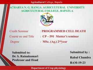
- 1. ACHARYA N. G. RANGA AGRICULTURAL UNIVERSITY AGRICULTURAL COLLEGE, BAPATLA Credit Seminar : PROGRAMMED CELL DEATH Course no and Title : CP – 591 Master’s seminar Degree : MSc. (Ag.) 2ndyear Submitted by : Rahul Chandra BAM-19-23 1 Submitted to: Dr. S. Ratnakumari Professor and Head Agricultural College, Bapatla Department of Crop physiology
- 3. Introduction • Cell death is a regulated process that allows a cell to self-degrade in order for the body to eliminate unwanted or dysfunctional cells. • Cell-cell interactions regulate cell death in two fundamentally different ways. • Cells in multicellular organisms require specific protein hormone signals to stay alive. • In the absence of such survival signals, frequently referred to as trophic factors, cells activate a "suicide" program. 3
- 4. • Second, in some developmental contexts , including the immune system, other specific hormone signals induce a “murder" program that kills cells. • Whether cells commit suicide for lack of survival signals or are murdered by killing signals from other cells, cell death is most often mediated by a common molecular pathway, termed apoptosis. • Term Apoptosis was derived from Greek language meaning ‘shedding’. 4
- 5. • Macrophages and other phagocytic cells recognize apoptotic cells and remove them by phagocytosis without inflammatory phenomena. 5 www.onlinebiology.net Pyknosis, karyorrhexis Pyknosis is the irreversible condensation of chromatin in nucleus of a cell undergoing apoptosis. Karyorrhexis is the fragmentation of nucleus after pyknosis.
- 6. NECROSIS • A different form of cell death, necrosis, occurs when cells are subjected to injury or excessive stresses such as heat, absence of oxygen, or infection by pathogens. • Necrosis creates holes in the plasma membrane, causing leakage of intracellular contents. • The distinction whether necrosis or apoptosis took place is found by detecting Cyt-C in the cytosol, if in addition to Cyt-C other porteins and factors are also present then it is due to necrosis. Eg Fumarse present in cytosol. 6
- 7. Apoptosis compared to necrosis 7 Feature Regulated by organism Apoptosis Yes: involves a series of enzyme-controlled reactions Yes No Necrosis No: can occur as a result of injury Only after cell has lysed Yes Yes No DNA broken down Cell membrane disintegration Nuclear membrane broken down Number of cells affected Energy requirement May be single cells ATP dependent (active process) Fate of dead cells Usually sheets of cells Energy input not required (passive process) Ingested by phagocytes Leakage of cell contents End point Ingested by neighbouring cells or phagocytes No Cell fragments into smaller bodies Yes Lysis of whole cell Caspase activation, chromatin condensation
- 8. 8(Walker et al, 1986., Exp. pathology)
- 9. HISTORY AND IMPORTANT BREAKTHROUGHS • German Scientist Carl Vogt was first to describe the principle of apoptosis in 1842. • In 1972 Kerr first introduced the term apoptosis in publication. Kerr received the Paul Ehlrich and Ludwig Darmstaedter Prize on March 14, 2000 for his description of apoptosis. • The 2002 Nobel Prize in Medicine was awarded to Sydney Brenner, Horvitz and John E. Sulston for their work in identifying genes that control apoptosis. • The genes were identified by studies in the nematode C. elegans and these same genes function in humans for apoptosis. 9
- 10. Time line related to PCD research (Richard et al 2001) 10
- 13. Importance of apoptosis • Apoptosis is needed for proper development. • Apoptosis is needed to destroy cells such as cancer cells , DNA damaged cells etc. • Important in normal physiology –Development: Immune systems maturation, Morphogenesis, Neural development. –Adult: Immune privilege, DNA Damage an wound repair. • Excess apoptosis –Neuro degenerative diseases • Deficient apoptosis –Cancer, Autoimmunity 13
- 14. • Sites of PCD in vascular plants the orange spheres represent the internal dead cells and the the branched 14
- 15. • The functions of PCD in plants : the redd regions represent the cells that have been targeted for PCD and the orange represent cells that have died.15
- 16. – Cancer cells • Radiation and chemicals used in cancer therapy induce apoptosis in some types of cancer cells. SC-1 induced apoptosis in stomach carcinoma cells Left: Before induction Middle: 24h after induction Right: 48h after induction www.biologydiscussions.com 16
- 17. Jean et al , USA (2001) 17
- 18. Caspases • Caspases stands for cysteine aspartate-specific protease. • Caspases have the characteristics of high specificity for substrates containing Asp, and use a Cys for catalyzing peptide bond cleavage. • Synthesized in the cell as precursors named procaspases. 18 NH2-terminal domain Large subunit (~20kD) Small subunit (~10kD) www.wileyonlinelibrary.com
- 19. CaspaseRole in Apoptosis • Cut off contact with surrounding cells • Reorganize cytoskeleton • Shut down DNA replication and repair • Interrupt splicing • Disrupt nuclear structure • Induce cell to display signals marking it for phagocytosis • Disintegrate cells into apoptotic bodies 19
- 20. PATHWAYS OF APOPTOSIS • MITOCHONDRIAL PATHWAY INTRINSIC PATHWAY • THE DEATH RECEPTOR PATHWAY EXTRINSIC PATHWAY 20
- 21. 21
- 22. Intrinsic / Mitochondrial pathway • Intrinsic pathway or mitochondrial pathway occurs due to stimuli from within the cell such as viral infection, DNA damage, growth factor deprivation. • The stimuli is sensed by sensory proteins such as ATM and CHK and activates a protein called P53. • P53 has many roles such as produces proteases , halts cell cycle, activates Bax and Bak proteins and also inhibits Bcl2 protein. 22
- 23. • Bcl2 blocks activity of Bax and hence Bax proteins activated , they form oligomers on the membrane of mitochondria and produces pores and aids in release of Cytochrome C. and IAP (Inhibitor Apoptotic Protein). • Cyt C released into cytosol combines with a protein called Apaf-1(apoptosis activating factor-1) and forms a complex called as Apoptosome. • Apoptosome combines with initiator caspase- procaspase -9 and cleaves at Aspartate site to form caspase-9. • Caspase-9 cleaves procaspase-3 and forms Caspase-3. 23
- 24. • Caspase 3 causes-DNA fragmentation, digestion of nuclear lamins, degrade cytoskeleton( actin, myosin and tubulin), and releases Scramblase enzyme. • The DNA gets fragmented and formation of apoptotic blebs occur, Scramblase translocated Phosphatidyl serine from inner to outer surface of the cell. • Cells having Phosphatidyl serine on the outer surface are recognised by Phagocytic cells and engulfed. 24
- 26. Extrinsic / Death receptor pathway Extrinsic pathway TNF pathway FAS pathway 26 Based on receptor and signalling molecule extrinsic pathway is of two types TNF pathway and FAS pathway. • TNF stands for Tumour necrotic factor in which the signalling molecule is TNF alpha cytokine and receptor on the cell membrane is TNFR-1. • While FAS stands for First Apoptotic Signal pathway, where FAS receptor having death domain accepts FAS ligand signalling molecule.
- 27. • As varied from intrinsic pathway , extrinsic pathway consists of signals for cell death produced externally in other cells to fight against an infection or damage. • The cell which produces the signalling molecule which initiates apoptosis after binding with receptor located on target cell are called Signalling cells commonly – cells associated with immune system such as CytotoxicT- lymphocytes. • The receptor on target cell contains an associated adapter molecules called death domain which undergoes changes to initiate apoptosis. 27
- 28. FAS pathway • FAS ligand produced by signalling cell binds with the FAS receptor on the target cell . • The associated death domain called FADD ( FAS associated Death Domain) gets activated and aggregate to form a DISC ( Death Induced Signalling Cascade). • Procaspase -8 binds to DISC and activated into Caspase- 8, which further cleaves Procaspase-3 to form effector executioner Caspase-3. • Caspase-3 degrade nuclease inhibitor and activate nucleases which enter nucleus and cause DNA fragmentation and finally undergoes phagocytosis. 28
- 29. • Here Procaspase-8 plays a major role in initiating cross talk between two pathways. • Procaspase-8 converted as Caspase-8 , activate a protein BID into TBID which further activates Bax/ Bak proteins which forms pores on mitochondrial membrane to release Cyt-C and trigger Intrinsic pathway. • Thus extrinsic pathway also triggers intrinsic development of Apoptosis. 29
- 31. CASE STUDIES 31
- 32. FEBS Letters 463 (1999), 151-154 Translocation of cytochrome c from the mitochondria to the cytosol occurs during heat-induced programmed cell death in cucumber plants Janneke Balk, Christopher J. Leaver*, Paul F. McCabe University of Oxford, Department of Plant Sciences, South Parks Road, Oxford OX1 3RB, UK • Objectives:To investigate the activation mechanism of plant PCD by induced cell death in cucumber cotyledons to ascertain if mitochondrial components were released during the cell death programme • Materials and methods- PCD was triggered in cucumber cotyledons by subjecting them to a short 55³C heat treatment. And DNA was extracted and southern blot analysis was done to determine inter nucleosomal cleavage in DNA due to PCD. • Immunodetection of cytochrome distribution in cytosol and mitochondria was done by western blot method using antibodies against Cyt-C and fumarase. 32 Case study 1
- 33. Southern blot analysis results on X ray film ( after hybridisation with radioactive prime labelling) showing displacement of Cucumber DNA . Inference: cucumber cotyledons were exposed to 55°C for 10 min and then incubated at 25°C. The cotyledons were frozen in liquid nitrogen at various time points over 12 h after heat treatment prior to extraction of genomic DNA. Clear evidence of DNA laddering was observed at 12 hours after heat treatment.
- 34. 34 Immuno detection of mitochondrial proteins in mitochondrial and cytosolic fractions of cucumber cotyledons following induction of PCD. Western blot was performed and revealed that the mitochondria began to release cytochrome C to the cytosol during the 10-min heat treatment , and that cytochrome C could no longer be detected in mitochondria after 3 hours. ( fumarase not transferred as it is not mere heat damage to mitochondrial walls that cause release of Cyt C).
- 35. Conclusion • Thus the experiment proves that triggering cell death with a known PCD-inducing abiotic stress have shown that the release of cytochrome occurs from the mitochondria into the cytosol, is an early event in plant cell death. 35
- 36. CASE STUDY 2 36
- 37. 37
- 38. 38
- 39. 39
- 40. 40
- 41. Conclusion • It is seen that proteolysis plays a crucial role in apoptosis in plants . • Caspase specific peptide inhibitors were found to be potent inhibitors of chemically induced cell death in tomato cells. • This indicated that as in animal system caspase like proteases are involved in apoptotic cell death pathway in plants. 41
- 42. • Materials and methods Tomato cell suspension culture was grown in liquid MS medium supplemented with 5 micro molar NAA and 1 Micro molar BA and 3% sucrose. Cell death inducers CdSO4 and CdCl2 were added to 5 ml of culture. Inducer effects tested for potency by measured indirectly by H2O2 and ethylene production. Cell death determination using fluorescence microscope. 42 CASE STUDY 3
- 43. 43
- 44. 44
- 45. 45
- 46. 46
- 47. • Chronic exposure to 50–100 μM CdSO4-induced loss of cell viability associated with typical apoptosis-like morphological changes and DNA laddering. • Max. cell death% observed at CdSO4 10mM and CdCl2 100 microM which indicates CdCl2 is more potent chemical apoptosis inducer. 47
- 48. CASE STUDY 5 Journal. Agric. Food Chem. 2008, 56, 10600–10605 Protective Effect of Anthocyanins from Black Soybean Seed Coats on UVB-Induced Apoptotic Cell Death in Vitro and in Vivo Konstantin Tsoyi, Hyung Bin Bark, Young Min Kim,Jong il Chung, Sung Chul Shin. • Objectives: s are not yet known. Thus, the aims of this study were to investigate the protective effect of anthocyanins from black soybean (Glycine max (L.) Merr) on UVB-induced apoptosis. • Materials and methods: Anthocyanins was extracted from black soyabean seed coat. • Uv-B irradiation was done on immortalized human keratinocyte cell line, HaCaT. Anthocyanin application and its effect on UVB induced apoptosis and ROS production was studied. 48
- 49. Effect of anthocyanins on ROS production induced by UVB in HaCaT. Cells were pretreated with anthocyanins (10 or 100 µg/mL) for 24 h and then irradiated with UVB (100 J/m2). After 1 h of incubation with/without anthocyanins, ROS production by confocal microscopy (bright green) analysis was performed.
- 50. 50 Protective effect of anthocyanins on UVB-induced apoptotic cell death in HaCaT by TUNEL assay. UVB irradiation increased TUNEL positive cells (dying cells) about 25% at 100 J/m2, which was reduced to 6% by anthocyanins at 10 µg/mL . Again increasing the amount of Anthocyanin to 100 µg/mL reduces TUNEL positive cells by 25%.
- 51. Conclusion • Pretreatment with anthocyanins reduced UVB-induced reactive oxygen species levels and inhibited UVB-induced apoptotic cell death through the prevention of caspase-3 pathway activation and reduction of proapoptotic Bax protein levels. • It is concluded that anthocyanins from the seed coat of black soy beans may be useful compounds to modulate UVB-induced photoaging. 51
- 52. • Balk. J., Leaver, C.J. and McCabe, P.J. Translocation of cytochrome c from the mitochondria to the cytosol occurs during heat-induced programmed cell death in cucumber plants. FEBS Letters 463 (1999) .University of Oxford, Department of Plant Sciences, UK. 151-154. • De jong. A.J., Hoeberichts. F.A., Yakimova. E.T. and Maximova.E. Chemical induced programmed cell death in tomato cells , involving caspase like proteases. Planta.(2000) 211.656-662 • Prasad. S.H., Amit. J., Anita .S. (2014). Apoptosis and programmed cell death – A review. World Journal of Pharmaceutical Research. 3. 1854-1872. • Tsoyi.K., Hyung. B. P. Young .M. K., Jong I.C and Hye. J. K. Protective Effect of Anthocyanins from Black Soybean Seed Coats on UVB-Induced Apoptotic Cell Death in Vitro and in Vivo. J. Agric. Food Chem. 2008, 56, 10600–10605. • Yakimovaa E.T., Kapchina-Totevab. V.M., Laarhovenc .F.M. and Harrence .E.J. Cadmium-induced programmed cell death in tomato suspension cells. Plant Physiology and Biochemistry 44 (2006) 581–589. 52 References
- 53. 53
Notas del editor
- 444444