SlideShare una empresa de Scribd logo
1 de 2
The four major sutures are the coronal, sagittal, lambdoid, and squamous sutures. They connect
the frontal, parietal, temporal, and occipital bones.

       The coronal suture joins the frontal bone to the parietal bones.
       The sagittal suture joins the two parietal bones to each other.
       The lambdoid suture joins the parietal bones to the occipital bone.
       The squamous suture joins the parietal bones to the temporal bones.


     Although the skull appears to be one large bone, there are actually several major bones that
are connected together. The major bones that compose the skull of a newborn include the
following:

       2 frontal bones
       2 parietal bones
       1 occipital bone

    These bony plates cover the brain and are held together by fibrous material called sutures.

What are sutures?      Some sutures extend to the forehead, while others extend to the sides and
back of the skull. One suture in the middle of the skull extends from the front of the head to the
back. The major sutures of the skull include the following:
metopic suture - extends from the top of the head down the middle of the forehead,
       toward the nose. The two frontal bone plates meet at the metopic suture.
       coronal suture - extends from ear to ear. Each frontal bone plate meets with a parietal
       bone plate at the coronal suture.
       sagittal suture - extends from the front of the head to the back, down the middle of the
       top of the head. The two parietal bone plates meet at the sagittal suture.
       lambdoid suture - extends across the back of the head. Each parietal bone plate meets
       the occipital bone plate at the lambdoid suture.

     Sutures allow the bones to move during the birth process. They act like an expansion joint,
allowing the bone to enlarge evenly as the brain grows and the skull expands, resulting in a
symmetrically shaped head. However, if any of the sutures close too early (fuse prematurely),
there may be no growth in that area. This may force growth to occur in another area or direction,
resulting in an abnormal head shape.
What are fontanelles?       There are two fontanelles (the space between the bones of an infant's
skull where the sutures intersect) that are covered by tough membranes. The fontanelles include:


       anterior fontanelle (Also called soft spot.) - the junction where the two frontal and two
       parietal bones meet. The anterior fontanelle remains soft until about 2 years of age.
       posterior fontanelle - the junction of the two parietal bones and the occipital bone. The
       posterior fontanelle usually closes first, before the anterior fontanelle, during the first
       several months of an infant's life.

Más contenido relacionado

La actualidad más candente

The cranial bones
The cranial bonesThe cranial bones
The cranial bones
Doc Lorie B
 
Nervous tissue (Histology)
Nervous tissue (Histology)Nervous tissue (Histology)
Nervous tissue (Histology)
ozhin araz
 
Nervous System
Nervous SystemNervous System
Nervous System
itutor
 
Skeletal system
Skeletal systemSkeletal system
Skeletal system
coachhuey
 
Axial skeleton parts 1 2
Axial skeleton parts 1  2Axial skeleton parts 1  2
Axial skeleton parts 1 2
Michael Wrock
 

La actualidad más candente (20)

Meninges
MeningesMeninges
Meninges
 
The cranial bones
The cranial bonesThe cranial bones
The cranial bones
 
Nervous tissue (Histology)
Nervous tissue (Histology)Nervous tissue (Histology)
Nervous tissue (Histology)
 
Nervous system ppt
Nervous system pptNervous system ppt
Nervous system ppt
 
The brain
The brainThe brain
The brain
 
Appendicular skeleton
Appendicular skeletonAppendicular skeleton
Appendicular skeleton
 
Nervous System
Nervous SystemNervous System
Nervous System
 
1. Nasal cavity
1. Nasal cavity1. Nasal cavity
1. Nasal cavity
 
Skeletal system
Skeletal systemSkeletal system
Skeletal system
 
Axial skeleton parts 1 2
Axial skeleton parts 1  2Axial skeleton parts 1  2
Axial skeleton parts 1 2
 
Skeletal system. anatomy and physiology of skeletal system. appendicular skel...
Skeletal system. anatomy and physiology of skeletal system. appendicular skel...Skeletal system. anatomy and physiology of skeletal system. appendicular skel...
Skeletal system. anatomy and physiology of skeletal system. appendicular skel...
 
Anatomy of respiratory system
Anatomy of respiratory systemAnatomy of respiratory system
Anatomy of respiratory system
 
Endocrine system 1
Endocrine system 1Endocrine system 1
Endocrine system 1
 
Human anatomy 1
Human anatomy 1Human anatomy 1
Human anatomy 1
 
Introduction to the nervous system
Introduction to the nervous systemIntroduction to the nervous system
Introduction to the nervous system
 
Anatomy and physiology of Lymphatic System
Anatomy and physiology of Lymphatic SystemAnatomy and physiology of Lymphatic System
Anatomy and physiology of Lymphatic System
 
Axial skeleton
Axial skeletonAxial skeleton
Axial skeleton
 
PPT on Lymphatic system
PPT on Lymphatic systemPPT on Lymphatic system
PPT on Lymphatic system
 
Larynx, trachea & bronchi
Larynx, trachea & bronchiLarynx, trachea & bronchi
Larynx, trachea & bronchi
 
Anatomy of Pharynx
Anatomy of PharynxAnatomy of Pharynx
Anatomy of Pharynx
 

Destacado

Positioning and radiographic anatomy of the skull
Positioning and radiographic anatomy of the skullPositioning and radiographic anatomy of the skull
Positioning and radiographic anatomy of the skull
mr_koky
 
Carpal Tunnel Massage
Carpal Tunnel MassageCarpal Tunnel Massage
Carpal Tunnel Massage
guest6c176e
 
Cta 201 module 5 cranium bones and facial bones (1)
Cta 201 module 5 cranium bones and facial bones (1)Cta 201 module 5 cranium bones and facial bones (1)
Cta 201 module 5 cranium bones and facial bones (1)
michgcubed
 
Ch07 a.skeletal
Ch07 a.skeletalCh07 a.skeletal
Ch07 a.skeletal
TheSlaps
 
14 bones of the face
14 bones of the face14 bones of the face
14 bones of the face
garretcj
 
14 bones of the face
14 bones of the face14 bones of the face
14 bones of the face
garretcj
 

Destacado (20)

Sutures of the Skull
Sutures of the SkullSutures of the Skull
Sutures of the Skull
 
Skull bones, features and markings II
Skull bones, features and markings IISkull bones, features and markings II
Skull bones, features and markings II
 
Different Types of Sutures
Different Types of SuturesDifferent Types of Sutures
Different Types of Sutures
 
01 skull
01 skull01 skull
01 skull
 
Anatomy of Skull
Anatomy of SkullAnatomy of Skull
Anatomy of Skull
 
Age estimation by bones
Age estimation by bonesAge estimation by bones
Age estimation by bones
 
Positioning and radiographic anatomy of the skull
Positioning and radiographic anatomy of the skullPositioning and radiographic anatomy of the skull
Positioning and radiographic anatomy of the skull
 
Carpal Tunnel Massage
Carpal Tunnel MassageCarpal Tunnel Massage
Carpal Tunnel Massage
 
Skull
SkullSkull
Skull
 
Cta 201 module 5 cranium bones and facial bones (1)
Cta 201 module 5 cranium bones and facial bones (1)Cta 201 module 5 cranium bones and facial bones (1)
Cta 201 module 5 cranium bones and facial bones (1)
 
Skull bones, features and markings 3
Skull bones, features and markings 3Skull bones, features and markings 3
Skull bones, features and markings 3
 
Ch07 a.skeletal
Ch07 a.skeletalCh07 a.skeletal
Ch07 a.skeletal
 
14 bones of the face
14 bones of the face14 bones of the face
14 bones of the face
 
14 bones of the face
14 bones of the face14 bones of the face
14 bones of the face
 
The nervous system 2014
The nervous system 2014The nervous system 2014
The nervous system 2014
 
Upper limb skeleton 5th october 2013
Upper limb skeleton 5th october 2013Upper limb skeleton 5th october 2013
Upper limb skeleton 5th october 2013
 
Emergency Radiology: Shoulder & Upper Extremities
Emergency Radiology: Shoulder & Upper ExtremitiesEmergency Radiology: Shoulder & Upper Extremities
Emergency Radiology: Shoulder & Upper Extremities
 
Sutures material
Sutures materialSutures material
Sutures material
 
Joints articulations
Joints  articulationsJoints  articulations
Joints articulations
 
Skull bones and features 1
Skull bones and features 1Skull bones and features 1
Skull bones and features 1
 

Similar a The four major sutures are the coronal

Head and neck anat fin
Head and neck anat finHead and neck anat fin
Head and neck anat fin
Emiru Tilahun
 

Similar a The four major sutures are the coronal (20)

fetal skull
 fetal skull fetal skull
fetal skull
 
The skeletal system (slide show)
The skeletal system (slide show)The skeletal system (slide show)
The skeletal system (slide show)
 
SWERNY SKELETAL 2.pdf
SWERNY SKELETAL 2.pdfSWERNY SKELETAL 2.pdf
SWERNY SKELETAL 2.pdf
 
Axial skeletal
Axial skeletalAxial skeletal
Axial skeletal
 
Head and neck anat fin
Head and neck anat finHead and neck anat fin
Head and neck anat fin
 
Fetal skull
Fetal skullFetal skull
Fetal skull
 
Axial skeleton ANATOMY AND PHYSIOLOGY
Axial skeleton ANATOMY AND PHYSIOLOGYAxial skeleton ANATOMY AND PHYSIOLOGY
Axial skeleton ANATOMY AND PHYSIOLOGY
 
Fetal head diameters
Fetal head diametersFetal head diameters
Fetal head diameters
 
a_4_skull.ppt
a_4_skull.ppta_4_skull.ppt
a_4_skull.ppt
 
Lec 5 skull
Lec 5 skullLec 5 skull
Lec 5 skull
 
Osseous System Part I.pptx
Osseous System Part I.pptxOsseous System Part I.pptx
Osseous System Part I.pptx
 
SKELETAL system- axial.ppt
SKELETAL system- axial.pptSKELETAL system- axial.ppt
SKELETAL system- axial.ppt
 
anatomy of human skull
anatomy of human skullanatomy of human skull
anatomy of human skull
 
Osteology of facial skeleton
Osteology of facial skeletonOsteology of facial skeleton
Osteology of facial skeleton
 
Skull of rabbit
Skull of rabbit Skull of rabbit
Skull of rabbit
 
Topic 5 bone of skull neck
Topic 5 bone of skull neckTopic 5 bone of skull neck
Topic 5 bone of skull neck
 
The Skeletal Sytem
The Skeletal SytemThe Skeletal Sytem
The Skeletal Sytem
 
skeletal system
skeletal systemskeletal system
skeletal system
 
Axial skeleton 2.pptx
Axial skeleton 2.pptxAxial skeleton 2.pptx
Axial skeleton 2.pptx
 
Osteology of head and neck i
Osteology of head and neck iOsteology of head and neck i
Osteology of head and neck i
 

The four major sutures are the coronal

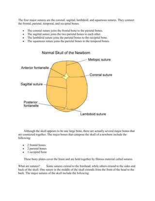
  • 1. The four major sutures are the coronal, sagittal, lambdoid, and squamous sutures. They connect the frontal, parietal, temporal, and occipital bones. The coronal suture joins the frontal bone to the parietal bones. The sagittal suture joins the two parietal bones to each other. The lambdoid suture joins the parietal bones to the occipital bone. The squamous suture joins the parietal bones to the temporal bones. Although the skull appears to be one large bone, there are actually several major bones that are connected together. The major bones that compose the skull of a newborn include the following: 2 frontal bones 2 parietal bones 1 occipital bone These bony plates cover the brain and are held together by fibrous material called sutures. What are sutures? Some sutures extend to the forehead, while others extend to the sides and back of the skull. One suture in the middle of the skull extends from the front of the head to the back. The major sutures of the skull include the following:
  • 2. metopic suture - extends from the top of the head down the middle of the forehead, toward the nose. The two frontal bone plates meet at the metopic suture. coronal suture - extends from ear to ear. Each frontal bone plate meets with a parietal bone plate at the coronal suture. sagittal suture - extends from the front of the head to the back, down the middle of the top of the head. The two parietal bone plates meet at the sagittal suture. lambdoid suture - extends across the back of the head. Each parietal bone plate meets the occipital bone plate at the lambdoid suture. Sutures allow the bones to move during the birth process. They act like an expansion joint, allowing the bone to enlarge evenly as the brain grows and the skull expands, resulting in a symmetrically shaped head. However, if any of the sutures close too early (fuse prematurely), there may be no growth in that area. This may force growth to occur in another area or direction, resulting in an abnormal head shape. What are fontanelles? There are two fontanelles (the space between the bones of an infant's skull where the sutures intersect) that are covered by tough membranes. The fontanelles include: anterior fontanelle (Also called soft spot.) - the junction where the two frontal and two parietal bones meet. The anterior fontanelle remains soft until about 2 years of age. posterior fontanelle - the junction of the two parietal bones and the occipital bone. The posterior fontanelle usually closes first, before the anterior fontanelle, during the first several months of an infant's life.