Chemical Tests; flame test, positive and negative ions test Edexcel Internati...
Electromagnetic spectrum, interaction with matter
1. Electromagnetic spectrum
I would doubt if I use the term ‘ light’ , a listener usually interprets it as “visible light.” Instead
of the entire electromagnetic spectrum.
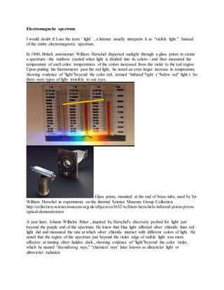
In 1800, British astronomer William Herschel dispersed sunlight through a glass prism to create
a spectrum—the rainbow created when light is divided into its colors—and then measured the
temperature of each color. temperatures of the colors increased from the violet to the red region.
Upon putting the thermometer past the red light, he noted an even larger increase in temperature
showing evidence of “light”beyond the color red, termed “infrared”light ( “below red” light.) So
there were types of light invisible to our eyes.
Glass prism, mounted at the end of brass tube, used by Sir
William Herschel in experiments on the thermal Science Museum Group Collection
http://collection.sciencemuseum.org.uk/objects/co3632/william-herschels-infrared-prism-prism-
optical-demonstration
A year later, Johann Wilhelm Ritter , inspired by Herschel's discovery probed for light just
beyond the purple end of the spectrum. He knew that blue light affected silver chloride than red
light did and measured the rate at which silver chloride interact with different colors of light. He
noted that the region of the spectrum just beyond the violet edge of visible light was more
effective at turning silver halides dark., showing evidence of “light”beyond the color violet,
which he named “deoxidizing rays,” “chemical rays’ later known as ultraviolet light or
ultraviolet radiation
2. https://de-film.com/v-johann-ritter-ultraviolet-light-discovery-4M5vooCpai0.html
In 1820, Danish scientist Hans Ørsted studied the deflection of a needle on a nearby compass
when the current was turned on and off using a voltaic cell showing a connection between
electricity and magnetism.
In 1821, Michael Faraday verified Ørsted experments by placing a small magnet around a
current-carrying wire proving the magnetic field of the current. In 1831 he produced electrical
current from a changing magnetic field, through electromagnetic induction.
In 1845, Faraday discovered that a magnetic field influenced polarized light in the magneto-
optical effect or Faraday effect or Faraday rotation, He observed rotation of plane of
polarisation of a beam of linearly polarized light incident on a piece of glass , when a magnetic
field was applied in the direction of propagation.he indicated that electromagnetism and light
were related
this is applied in polarimeters and chiroptical
spectroscopy.
https://www.bbvaopenmind.com/en/faraday-electromagnetic-theory-light/
‘Experimental Researches in Electricity,” Philosophical Transactions (vol 136, pp. 1-20) he
prophesized that light could be a vibration of the electric and magnetic lines of force in his article
Thoughts on Ray Vibrations "Experimental Researches in Electricity", Vol III, M. Faraday,
p447-452 also Philosophical Magazine, S.3, Vol XXVIII, N188, May 1846.
In 1945 Faraday introduced the concepts of field and field lines, metaphors of Michael Faraday
to represent the influence of sources of magnetism and electricity. This concept , according to
Einstein brought the great change in physics in providing electricity, magnetism and optics with
a common framework of physical theories.
3. In the 1855, James Clerk Maxwell took effort developing mathematical sense of Faraday’s
observations and theories. In paper called “On Faraday’s Lines of Force”. Maxwell Combined
the works of Oersted, Coulomb, Gauss, and Faraday, and developed the all-embracing theory of
electromagnetism
In 1865, Maxwell presented his theory uniting electricity and magnetism before the Royal
Society of London. Phil. Trans. R. Soc. Lond. 1865 155, 459-512,
Eventhough Maxwell’s formulation took the form of 20 simultaneous equations, with
20 variables, Maxwell’s theory can be demonstrated by four equations.
Gauss law for electricity represents the relation between electric charge , Q, and electric field
intensity, E
Gauss’ equation for electric flux through a closed surface of area A defines that electric field
lines start on positive charges and end on negative charges. The electric field is defined as the
force per unit charge on a test charge, and the strength of the force is related to the electric
constant , є0 . Fields diverge on point sources.
.∫ 𝐸. 𝑑𝐴 =
𝑄 𝑒𝑛𝑐𝑙𝑜𝑠𝑒𝑑
𝜀0𝐴
Q enclosed is the charge enclosed by the surface
є0 is the permittivity of free space = 8.85 x 10 -12C2/N.m2.
Application of Gauss’ law for magnetism gives the law of conservation of magnetic flux
∫ 𝐵. 𝑑𝐴 = 0
𝐴
B is magnetic field, and the total magnetic flux passing through any closed surface is 0 showing
absence of magnetic monopoles. Hence no magnetic flow sources occurs, and the magnetic flux
lines always close upon themselves.
A time varying magnetic field through a closed loop of length l induces an electromotive force
(emf) in-turn creating an electric field. The direction of the emf opposes the ∫ 𝐸. 𝑑𝑙 =𝐶
−
𝑑𝜙𝐵
𝑑𝑡
change. The voltage accumulated around a closed circuit is proportional to the
time rate of change of the magnetic flux it encloses. This comprises Faraday's Law of Induction.
ΦB - magnetic flux
Mathematical relation for the dependence of produced magnetic field on electric flux change is
given by Ampere’s law. It shows that electric current I or a time varying electric flux through a
surface creates a circulating magnetic field around any path that bounds that surface. Electric
currents and changes in electric fields are proportional to the magnetic fields circulating about
the areas where they accumulate.
4. ∫ 𝐵. 𝑑𝑙 = 𝜇0 𝐼𝑒𝑛𝑐𝑙𝑜𝑠𝑒𝑑 + 𝜇0𝜀0
𝑑𝜙𝐸
𝑑𝑡
𝐶
I enclosed - change in current induced,
μ0is the permeability of free space which is equal to 4πx 10-7T.m/A
ΦE is the electric flux through a closed path.
Maxwell’s Equations show that time varying electric field generates magnetic field with time
varying magnitude . Similarly changing magnetic fields promote generation of electric fields
with changing magnitude. Direction of corresponding electric and magnetic fields are
perpendicular to each other.
Electrical charges accelerated in an alternating electric field generate an alternating magnetic
field. Such electric fields with sinusoidal alternations create electric field and magnetic field
couples propagating through space in form of “harmonic electromagnetic waves” . So EM wave
propagation requires energy and is regarded as photonic energy where the energy of a photon E
= hν = hc/λ
electromagnetic waves was calculated to propagate at a speed given by the equation 𝑐 =
1
√ 𝜇0𝜀0
= 3 ∗ 10 ^8 = speed of light. So light is an electromagnetic wave with detectable
wavelength. Nothing in Maxwell’s equations limits the possible values
of the wavelength/frequency of electromagnetic radiation; they only confine its velocity. Infrared
light, visiblelight, and ultraviolet light are all the same phenomenon, but with different values of
wavelength and frequency(and the same velocity).
In 1867, Maxwell predicted existence of light with even longer wavelengths than infrared light,
but proved nothing.
In 1887, Heinrich Hertz had proved Maxwell's theory by discovering radiowaves using a
transmitter, and detected them by using a loop of having a small gap between the ends. . In 1895,
Wilhelm Conrad Röntgen detected X-rays on a fluorescent
screen of barium platinocyanide by accident using a cathode ray tube,
but it took scientists until 1912 to determine they were another type of light. He he took the first
X-ray of the human body using his wife’s hands.
An X-ray of Anna Röntgen’s hand
wilhelmconradroentgen.de
Paul Villard found gamma-rays in 1900 while studying properties of radioactivity. They were
first thought to be particles emitted during the course of radioactivity, so was named after alpha
5. particles and beta particles . Ernest Rutherford discovered it to be another type of light by
measuring their wavelengths by diffracting them with crystals.
6. Real nature of light
https://www.sps.ch/en/articles/progresses/wave-particle-duality-of-light-for-the-classroom-13/
http://photonterrace.net/en/photon/duality/
https://www.nature.com/articles/ncomms7407
https://phys.org/news/2015-03-particle.html
What is the nature of light?
In order to study the refraction, René Descartes assumed that the propagation of light is similar
to the movement of a projectile, like a tennis ball. By analysing the motion of a light ray into a
parallel and a normal components (on boundary between the two media, ) he assumed that light
was a wavelike disturbance of plenum , a continuous substance filling the universe.
drawing from Descartes’ La dioptrique illustrating the light reflection and refraction.
When light goes through lenses or bounces off mirrors , Isaac Newton stated in 1650 that light
was composed of corpuscles or particles emitted in all directions from a source , and that light
should be treated as a particle . He disregarded light to be waves, because waves have a
tendency
towards a spherical propagation, while light rays propagate through space in straight lines
proving corpuscular theory.
his treatise New Theory of Light and Colours in 1672 revealed colours and the method for
extracting them from a sunbeam with the use of a prism.
Thomas Young and Augustin Fresnel developed , the wave theory of light.
7. What is the true nature of photon?
Photons , when considered as an elementary particle, are the carriers of light or “quantum" of the
electromagnetic field. The photon has no rest mass and displays properties of both
waves and particles. This duality is still a mystery.
According to classical standard Maxwell's model, photons are solitary wave-packets . how can
we relate a photon and electromagnetic waves (waves emanating from an antenna).
Photons reside at diverse dimensional scales, like in constituents of nuclei, in atomic and
molecular interactions and even are part of biological processes.
Bats has the ability to recognize objects, from sound waves of certain frequencies, on striking the
objects to vibrate them. Similarly scientists can recognize molecules by exposing them to
vibrations in the electromagnetic spectrum and observes the responses of its atoms and
molecules. Similarly the atoms and molecules may be induced to emit radiation, and from the
region of emission in the electromagnetic spectrum we can establish chemical structure.
A moving electric charge creates a disturbance (perturbation) in which there is both an electric
component and a magnetic component forming a wavelike electromagnetic vibration.
Range of such vibrations, arranged in the increasing order of their oscillation frequency,
constitutes the electromagnetic spectrum.
The distinction between X and gamma rays is based on sources: gamma rays are generated from
nuclear decay or other nuclear and sub nuclear/particle process, while X-rays are generated by
electronic transitions involving highly energetic inner atomic electrons. Nuclear transitions being
more energetic than electronic transitions, shows that gamma-rays are more energetic than X-
rays.
Radio Waves are generated by charges accelerating through conducting wires and found in such
electronic devices as LC oscillators and are used in communication systems.
Microwaves are generated in klystron and magnetron tubes, and with solid state diodes such as
Gunn and IMPATT devices. Microwaves are absorbed by molecules having permanent dipole
moment and is applied to heat food.
Infrared Waves can be divided into three parts:
Far-infrared is absorbed by rotational modes in gases , by molecular motions in liquids, and by
phonons in solids. The water in the Earth's atmosphere absorbs this region making atmosphere
opaque.
Mid-infrared is generated from hot objects and is absorbed by molecular vibrations,
Near-infrared
Visible light
8. Ultraviolet light UV can break chemical bonds, ionizing molecules
X-rays generated bombarding a metal target using high-energy electrons .
Why does atoms and molecules respond to electromagnetic spectrum ?
Matter (charged ones unlike neutrons ) is electromagnetic in nature. Matter receives or absorbs
passing radiation whose frequency matches with the distinctive frequencies of its atoms and
atom groups it is composed of , and can be induced to emit the same characteristic radiations if
delivered with the required energy. Matter is composed of atoms with a nucleus surrounded by
electrons.
When two atoms form molecules inter-penetration and redistribution of the electron cloud about
the two atoms take place. electromagnetic responses arises from redistributed
electron cloud in a chemical.bond A chemical bond can be considered as a model of 1 D
Harmonic oscillator, comprising a spring coil connecting weighed balls. This is same in case of
H atom connected to C atom, by single spring. C being tetrahedral will be connected to 4 such
springs. This spring like a violin string can absorb and emit radiation of a characteristic
frequency. Energy to stretch inter-atomic bonds, when translated into electromagnetic radiation
reveals in the infrared portion of the spectrum. This comprises infrared spectroscopy.
When gaseous atoms are electronically disturbed by placing them between the electrodes of an
arc or spark, as in Townsend discharge, and hit by electrons, interesting phenomenon happened.
The incident electrons then impact on the electron cloud around the atomic nuclei and provide
additional energy to them. Then the excited molecules produced in inelastic electron- molecule
collision, releases in higher frequency range of visible and ultraviolet region. This visible and
ultraviolet region was earlier used for electronic excitation. Each atomic species will register its
own characteristic spectrum on the photographic plate. the unique wave length and frequency of
spectral lines enables to recognize corresponding chemical element. This is used in atomic
absorption and emission spectroscopy. The relative intensity of its lines informs amount of the
element present.
This is applied in UV absorption spectra to study unsaturated organic compounds. X-rays are
generated by electronic transitions involving highly energetic inner atomic electrons.
The techniques we have seen so far enables us to probe into molecular structure, as in
spectroscopy, using tool of electromagnetic radiation.
How does atoms and molecules respond to electromagnetic spectrum ?
General description of emission, absorption, refraction, scattering, and dispersion of light implies
that the atoms contain charged particles performing harmonic oscillations round positions of
stable equilibrium, and exchange energy and momentum with the radiation fields according to
the classical Maxwell laws of electrodynamics.
9. But many contradictions to the consequences of the classical electrodynamical theory were
revealed.
Case 1 . black body radiation or temperature radiation showing spectrum of light emitted by hot
objects.
A black body is an object entirely absorbing all light or radiation that falls on it, hence making it
a perfect source of thermal radiation too. It then emits thermal radiation in a continuous spectrum
according to its temperature.
black body resembles an oven with a small hole, with very
small probability of all radiation that enters through the opening to leave through it again.
Radiation emitted is thermal radiation. which is function of frequency and the oven temperature .
Wein tried to explain that according to electromagnetic theory , oscillating electromagnetic
charges produce electromagnetic waves, and hence the radiation emitted by a hot object could
be due to the oscillations of electric charges of the molecules in object
𝜌( 𝜆, 𝑇) =
𝑊(𝜆𝑇)
𝜆5 Wien's formula was accurate at short wavelengths but deviated at longer
wavelengths.
This was explained by classical Rayleigh-Jeans law which expresses
𝜌( 𝜆, 𝑇) =
8𝜋𝑘𝑇
𝜆4 , lying between wavelengths λ and λ+dλ
ρ =density of states =
𝑑𝐸
𝑑𝜆
dE = total energy per unit volume of all oscillators
Boltzmann's constant, k = 1; 38 * 10-23 JK-1 , kT - the dimension of energy,
The total energy in range dλ = E(T) = ∫ 𝜌( 𝜆, 𝑇) 𝑑𝜆
∞
𝜆
𝜌( 𝜆, 𝑇) =
8𝜋𝑘𝑇
𝜆4 tends to infinity at λ tends to 0.
This curve approaches the experimental curve at longer wavelengths, but it deviates badly at
short wavelengths. causing the ultraviolet catastrophe
Rayleigh-Jeans formula was accurate at long wavelengths but deviated at shorter wavelength.
10. Planck assumed that the radiation in the cavity was emitted and absorbed by “oscillators.”
Plank proposed that only certain distinguished states of the oscillating particles have to be
considered. with the energy equal to a multiple of the quantum hν, where ν is the natural
frequency of the oscillator and h is a universal constant.
𝜌( 𝜆, 𝑇) =
8𝜋ℎ𝜈
𝜆4 ( 𝑒 −1
ℎ𝜈
𝑘𝑇
)
The oscillators (of electromagnetic origin) have certain discrete energies, En= nhν,
n is an integer,
ν is the frequency,
h = Planck’s constant = 6.6261 ×10⁻³⁴J·s.
The oscillators can absorb or emit energy in discrete multiples of the fundamental quantum of
energy E = hν
Examples of blackbody radiation
Thermal Imaging: We All Glow in
the Dark
Red stars are cooler, emitting in the red wavelengths.
A hotter star like our sun emits the most radiation in the yellow/green part of the spectrum.
stars with peak wavelengths in the green also emit lots of radiation in the red and blue part of the
spectrumwhich are combined to white colour
11. hotter stars and other objects emit the most radiation in the blue, ultraviolet or even x-ray and
gamma ray part of the spectrum
cooler objects like planets and humans emit the most radiation in the infrared.
The child emits plenty of midwave IR light, while the TV emits almost none because the picture
tube does not heat up in operation significantly and the color phosphors do not emit significant
infrared light
Can we illuminate a dark room by lighting a cigarette?
The glowing coal on the end of a cigarette is at a temperature of about 800 C (1472 F), hence
emit light in 900–1700 nm band.
12. Shortwave IR image of a lit cigarette illuminating a room. (Courtesy of FLIR)
Even cooler objects emit microwaves and radio waves.
In 1855, Anders Angstrom published the results of his spectroscopic investigations on the line
spectrum of hydrogen. He identified a red-line at 6562.852 Å, a blue-green-line at 4861.33 Å
and a violet line, formed by two closer distinct lines ,at 4340.47 Å.
table of Angstrom on the study of light spectrum
in 1885 Johann Jakob Balmer predicted the wavelength in atomic hydrogen spectra for the lines
between 656 and 377 nm
in 1888 Janne Robert Rydberg generalized the formula
1
𝜆
= (
1
𝑛1
2 −
1
𝑛2
2 )𝑅 𝐻,
wave number (reciprocalof wavelength, in units of cm−1) is related to a difference of terms.
13. RH = 109677,58 cm−1 = Rydberg constant for the hydrogen atom.
In the Balmer series, n1 > 2 and n2 = 2.
This was agrreable to discoveries in the series
for the ultraviolet and infrared spectral ranges
in 1906 Lyman Series n1 > 1 and n2 = 1
in 1908 Paschen series n1 > and n2 = 3
in 1922 Brackett series n1 > 4 and n2 = 4.
in 1924 Pfund series n1 > 5 and n2 = 5.
In 1953 hydrogen spectrum was completed by physicist C.J. Humphreys in the microwave
region n1 > 6 and n2 = 6
the structure of hydrogen emission spectra with the placement of the line series
14. Energy level diagram and spectral series of the H atom.
Hα (656nm) emission (red glow) in HII regions in Horsehead nebula and cats eye nebula
In 1913, theoretical explanation of the hydrogen spectrum was obtained by N. Bohr using
using the Plank’s concept of quanta.
15. In beginning of 19th century planetary model was applied to the hydrogen atom where the
electron of mass me and charge qe rotates on a circular orbit about the atomic core of mass mn
and charge qn
According to electromagnetism, the rotation of the electron around the charged core leads to the
emission of electromagnetic radiation (energy) till electron collapses to the atomic core
(nucleus). hydrogen atom being physically stable , the electron never fall on the nucleus, and this
idea was quite illogical.
positions of the electron re and core rn respect to the center of mass is given by
re=
𝑚𝑒
𝑚𝑒+𝑚𝑛
r
rn=
𝑚𝑒
𝑚𝑒+𝑚𝑛
r
r=re -rn = radius of the circular orbit
K.E =
1 𝑚 𝑒
2
𝑟𝑒
2 + 1 𝑚 𝑛
2
𝑟𝑛
2 = 1
2
𝑚 𝑒 𝑚 𝑛
𝑚 𝑒 + 𝑚 𝑛
r2 = 1
2
𝜇r2 =T
μ - reduced mass of the atom
Hamiltonian of the atom = H = K.E + P.E
P.E = V
Coulomb force between nucleus and electron = F Coulomb =
−1
4𝜋𝜀0
𝑞 𝑒 𝑞 𝑛
𝑟
𝑟2 |𝑟|
=
−1
4𝜋 𝜀0
𝑞 𝑒2
𝑟
𝑟2 |𝑟|
the interaction force between electron and nucleus is balanced by the centrifugal force due to the
circular motion, creating stable system
F circular =
−1
𝑚𝜔2
𝑟
𝑟
|𝑟|
Equalizing the two forces
−1
𝑚𝜔2
𝑟
𝑟
| 𝑟|
=
−1
4𝜋𝜀0
𝑞 𝑒2
𝑟
𝑟2 | 𝑟|
𝑚𝜔2
𝑟
𝑟
| 𝑟|
=
1
4𝜋𝜀0
𝑞 𝑒2
𝑟
𝑟2 | 𝑟|
gives
1
4𝜋𝜀0
𝑞 𝑒2
1
𝑟2 = 𝑚 𝑒 𝑟𝜔2
= 𝑚 𝑒
1
𝑚 𝑒
2
𝑟4 𝐼2
r
This gives position r =
4𝜋𝜀0
𝑚 𝑒 𝑞 𝑒
2 𝐼2
Bohr proposed angular momentum is conserved, so I = r× 𝑝 = 𝑚 𝑒 v× 𝑟 = 𝑚 𝑒 𝜔𝑟2
𝑛̂
Gives angular velocity ω = I/𝑚 𝑒 𝑟2
n - angular momentum vector
Bohr used quantisation of anglar momentum I = nh/2π = 𝑚 𝑒 v× 𝑟
So 2πr = nh/𝑚 𝑒 v = nh/p
16. orbit of the electron was associated to a standing wave with wavelength λ = h/p, showing
electron as material wave.
Total energy of the planetary model of the atom =
1
2
𝜇𝜔2
𝑟2
+
−1
4𝜋 𝜀0
𝑞 𝑒 𝑞 𝑒
1
𝑟
= E
Given angular velocity ω = I/𝑚 𝑒 𝑟2
Hence E = =
1
2
𝜇𝐼2 1
𝑚 𝑒
2
𝑟2 +
−1
4𝜋𝜀0
𝑞 𝑒 𝑞 𝑒
1
𝑟
Given r =
4𝜋𝜀0
𝑚 𝑒 𝑞 𝑒
2 𝐼2
So
E =
1
2
𝜇𝐼2 1
𝑚 𝑒
2
𝑚 𝑒
2
𝑞 𝑒
4
𝐼4 (4𝜋𝜀0)2 -
1
4𝜋𝜀0
1
4𝜋𝜀0𝐼2 𝑞 𝑒
2
𝑚 𝑒 𝑞𝑒
2
=
1
2
𝜇
𝑞 𝑒
4
𝐼2 (4𝜋𝜀0)2 -
1
𝐼2 (4𝜋𝜀0)2 𝑞 𝑒
4
𝑚 𝑒
mn >>me , so μ = (mn me)/( mn +me) ≅(mn me)/mn = me
center of mass of the atom coincide with the nucleus
so E =
1
2
𝑚 𝑒
𝑞 𝑒
4
𝐼2(4𝜋𝜀0)2 -
1
𝐼2 (4𝜋𝜀0)2 𝑞 𝑒
4
𝑚 𝑒 = -
1
2 𝐼2 (4𝜋𝜀0)2 𝑞 𝑒
4
𝑚 𝑒
given I = nh/2π
so E = −
1
2 𝑛2 (4𝜋𝜀0)2 𝑞 𝑒
4
𝑚 𝑒
4
ℎ2
𝜋2
electron can assume only quantized energy values depending on the integer number n called
principal quantum number . Increasing , n, lessens the spacing between the energy levels.
The equations obtained by Bohr describes the hydrogen spectrum: each spectral line is due to an
electronic transition from a given orbit to another one.
The Bohr model, was unable to explain both the fine structure of the hydrogen spectrum ,
Zeeman effect and the spectrum of atoms with more than one electron.
Bohr, Heisenberg, Schrödinger and Dirac tested their theories on the spectrum of the
hydrogen atom
each spectral line arises by electronic transition from a given orbit to another one
E = En1 –En2 =−
1
2 𝑛12 (4𝜋𝜀0)2 𝑞 𝑒
4
𝑚 𝑒
4
ℎ2
𝜋2
+
1
2 𝑛22 (4𝜋𝜀0)2 𝑞 𝑒
4
𝑚 𝑒
4
ℎ2
𝜋2
= hc/λ
Hence 1/λ ∝ (
1
𝑛1
2 −
1
𝑛2
2 )
17. http://cronodon.com/Atomic/AtomTech3.html
Einstein introduced light quanta, proposing radiation should not be propagated through space as
continuous wavelets( classical theory of light) , but as entities, each having the energy hv,
concentrated in a minute volume, where h is Planck’s constant and v is the number of waves
passing in unit time. This is verified by photoelectric effect.
The emission and absorption spectra of the elements speculated that an atom possesses a number
of distinguished, ‘stationary states’, having remarkable stability,
18. Emission and absorption involves any change of the state of the atom involving transition from
one of these stationary states to another.
hv = E1 — E2
E1 and E2 are the values of the energy of the atom in the initial and in the final state of the
process. The
reversed process of this transition is obtained by illumination with light of same frequency
Einstein used Planks law of thermal radiation and proposed that an atom in a given stationary
state may possess a certain probability of a ‘spontaneous’ transition in unit time to a stationary
state of smaller energy content,
spontaneous emission
induced emission
induced absorption
Illuminating an atom with external radiation of suitable frequency, provides it a certain
probability of an ‘induced’ transition to another stationary state with higher or smaller energy
content.
Energy exchange in the transition process is accompanied by an exchange of momentum hv/c,
Spontaneous transitions are generated by a virtual field of radiation among other atoms which is
connected with the virtual harmonic oscillators conjugated with the motion of the atom itself,
this is called collisions.
According to the quantum theory , common optical phenomena depends on discontinuous
transition processes.
19. Reflection, refraction, and dispersion are attributed to a scattering of light by the atoms.
Electromagnetic forces of the radiation field induce forced vibrations in individual charged
particles in atoms, causing an absorption and emission of virtual atomic oscillators.
Compton studied reflection of X-rays from crystals and recorded that scattering was
accompanied by a change of frequency differing in different directions. He proposed that the
electron absorbs a quantum of the incident light and simultaneously re-emit a light- quantum in
some other direction. He determined the velocity of the electron in a certain direction and the
frequency of the re-emitted light, using laws of conservation of energy and momentum.
Each light quantum has an energy of hv and a momentum hv/c .
Due to the large mass of the atomic nucleus , the velocity change associated with these
transitions is so small, hence causing little effect on the energy of the atom and the frequency of
the scattered radiation.
The Schrödinger representation enables us to view a state as a complex wave in space. In QM,
we find the eigenvalues(spectrum) of the Hamiltonian(energy) operator. Observing absorption
and emission spectrum when atoms transit between different energy eigenstates are important
than studying how the single particles (e.g. atomic electrons) move.
Due to discrete or specific energies of atomic orbitals, transitions among them have discrete or
specific energies. So atomic absorption spectra consist of a series of “lines” at the frequency of
radiation corresponding to each allowable electronic transition.
Refer
A. Einstein, Phys. Zs. 18 (1917) 121.
20. Time dependent describes the semiclassical interaction between a charged particle and
the electromagnetic field.
A molecule has following degrees of freedom
Translational
Rotational
Vibrational
Electronic energy
Nuclear energy
Energy level
diagram for a molecule showing electronic, vibrational and rotational states. Arrows represent
possible absorption transitions.
Translational behavior
Translation is the displacement of center of mass. 3 components of translation are the 3
components of center of mass. Energy must be quantized for molecule. Permitted energy for
translation are obtained from solving Schrodinger wave equation EΨ=HΨ
2 simple approaches or methods used in quantum mechanics
Variable separable method- all the variables are separated and analyzed
21. Taking simple methods- the molecule is confined in XY plane, so Z component is 0 and
2nd order differential is solved for 2 variables instead of 3. The molecule is confined to X
axis, 2nd order differential in 1 variable ( particle in 1D box) . this gives En = n2h2/(8ml2).
Taking average molar mass 100gl-1, translational energy spacing = 10-44J( VERY SMALL
QUANTITY COMPARED TO THERMAL ENERGY = 10-22J). reference in case of energy is
thermal energy. It is based on Boltzmann exponential law Nc œ exp(-Ec/kT). So translational
energy is assumed as continuous, so classifies classical mechanics. So it donot obey QM and
give no spectroscopic technique, being insignificantly quantized.
Translation of electron can be quantized, so obey QM . free electron has l = infinity, E = 0.
Quantisation arises from confined restrictions. Very small particles like electron, confined to a
region has confined translation (but not for molecules and atoms)
Rotational behavior
Rotation is the change in orientation keeping center of mass same. Rotational energy Er = Iω2/2
I – moment of inertia
ω angular velocity
to solve Schrodinger wave equation for rotating system, simplest model used is diatomic system,
confined to plane (planar rotation or particle in a ring). In space, 3D rotator gives spherical
rotator. It has spherical symmetry, so spherical coordinates are used.
The coordinate systems are selected based on symmetry of the system.so schrodinger equation is
transformed for spherical coordinates. If rotational energy increases , centrifugal forces tend to
keep the 2 masses away, increasing the bond length. So exact solution of schrodinger equation
for rigid rotator is not possible. It is wrongly assumed that diatomic molecule rotates with
constant bond length { hypothetical rotation with rigid rotator [ stiff bond length]}. Legendre
equation is solved by power series method, for rigid rotator. This gives exact solution. Spherical
polar coordinate Schrodinger equation is transferred to mathematical treatment { Legendre
equation}.
EJ = h2J(J+1)/8π2I ; J = 0,1,2,…
L – angular momentum quantum number in QM
J – is used here.
In translational and rotational, energy levels diverge.
EJ = h2J(J+1)/8π2I =10-24 for rotational, if molar mass = 100g mol-1 with intermolecular
distance= 1 A
22. {I = μr2}
Rotations are quantized, so used as spectroscopic technique.
Vibrational behavior
Simple harmonic oscillator is the model used. Restoring force F = -kr; k- hookes law constant;
r – displacement; V=kr2/2 , so resembles parabola and is continuous. QM says energy must be
quantised. So schrodinger equation is solved. Simple harmonic oscillator is transformed to
Hermite equation. It is solved by power series method.
EV = (V+1/2)hω
ω- fundamental vibration frequency = 1/2π (k/μ)1/2
vibration – change in bond length and bond angle keeping the center on mass constant
k depends on bond strength; k deermines strength of spring
Spacing of vibrational energy level = hω = constant, unlike rotational and translational energy
level
Thermal energy level = 10-24J
Vibrational energy level = 10-21J , vibration is quantized
Ideal harmonic oscillator never breaks down, when extended to any length. Real harmonic
oscillator extends only till 10% of bond length then dissociation occurs. Real molecule are
unharmonic oscillators. So energy levels diverge, spacing decreases as vibrational level
increases.
water molecules have a strong absorption band at a 1450-nm wavelength corresponding to
stretching vibration.
23. a glass of water viewed in both visible light and IR light in the 900–1680-nm
Electronic behavior is quantized
Nuclear behavior – shell model is used. Like electronic energy levels nuclear energy levels are
also quantized.
Tools for spectroscopy
Let E1 and E2 be permitted energy levels of molecule. Molecule is comfortable in ground state.
E is given.
Simplest form of energy is heat, other sources are electrical energy. In spectroscopy is by EM
radiation (photon) E= hν. When EM radiation matches energy level spacing, excitation
(absorption ) occurs. Transmitted radiation has less intensity than incident. Intensity difference
gives ν of absorbed radiation. This gives energy level spacing.
Rotational energy level spacing gives moment of inertia , hence shape of molecule.
Vibrational energy level spacing gives hω; ω=1/2π(k/μ)1/2 . each functional group has a
characteristic bonds strength k. so functional group can be identified .
Electronic energy level spacing gives type of bonding ( electron reside in MO. MO are formed
from suitable LCAO. Suitable LC is by suitable bonding hybridization} energy level spacing
gives molecular parameters. So molecular structure is found.
Source of radiation
Sun is the best sunlight. But it has no sufficient intensity for detection and measurement. (Raman
used sunlight for spectroscopy, but had to wait for weeks)
Artificial sources are used. But these aren’t universal sources like sun. so source is based on type
of excitation energy provided.
24. Detector analyses transmitted rays. Sources and detectors depend on type of energy of radiation.
High and low energy detectors.
Eradiation = hν = hc/m = hcṽ
RF 3000cm – 1cm (NMR)
MW 1 -102cm-1 (ESR/EPR)
IR 102-104cm-1 (vibrational spectroscopy)
UV-Vis 104-106 cm-1 (electronic spectroscopy)
X ray hundreds of eV
γ- million of eV (Mossbauer spectroscopy)( nuclear excitation)
all regions of EM radiation, except Xray, are used in spectroscopy. But X rays are developed for
spectroscopy.
Electronic spectroscopy is the 1st spectroscopic technique ( so was early called absorption
spectroscopy). Electron is promoted from 1 electron state to another electron state (1 MO to
another MO).
If energy of radiation is greater than energy spacing , electron ionization occurs (eject electron
from atom) – photo ionization. X rays ( high energy radiation) can aid photo ionization, so
overcome binding energy.
hν0 = B.E ( binding energy ) + K.E (kinetic energy of electron)
K.E of electron of determined, hence B.E of electron in atom or molecule can be found. This is
called electron spectroscopy. This is different form electronic spectroscopy. ( absorption
spectroscopy). Electron spectroscopy (photo electron spectroscopy) uses X-rays , so is called
XPS (X-ray photo electron spectroscopy).
If B.E is very small, ultra high energy UV can photoionise(blue region)
Potential is adjusted (stopping potential), so electron is neither attracted or repelled , so K.E of
electron is analysed. OS of species being absorbed can be found from XPS. XPS is used for
chemical analysis (quantitative, qualitative)
ESCA- electron spectroscopy for chemical analysis
IR and UV region
Sample is kept in sample holder ( glass tube). Radiation is passed through sample. Lens and
mirror focus rays to sample. Assumption- holder and optics are transparent to radiation.
25. For coloured radiation, silica glass is transparent and donot absorb visible rays. Quartz in UV
transparent holder and optics; alkali halides (NaCl, CsCl, CsBr) in IR transparent.
If NaCl is used, only 650-4000cm-1 (mid IR region MIR)can be used- cheapest IR material. { for
most organic functional groups absorb in this region.
Metal organic frameworks absorb below 650cm-1 so use costly CsCl, CsI materials.
100-650 cm-1 far IR FIR
4000-10000 near IR NIR
With respect to visible light.
UV-Vis region
Electron is in σ, π,n(NBO) orbitals.
σ-σ*, π-π*, n-σ* are possible transition states. Any
molecule in air give σ-σ* transition, so can affect measurement of system. so 10-185 nm vaccum
Uv is used. 185-385 nm (Uv region) for π-π* and n-π*.
385-850 visible region, highly conjugated n-π*, π-π* lead to visible absorption.
Particle in 1D gives shift in λ, for conjugated system. Consider C8 system
C=C-C=C-C=C-C=C HERE ELECTRON ARE FREE TO MOVE.
n=4-n=5 is the lowest energy transition in octatetraene.
26. E = n2h2/(8πml2) = h2(52-42)/(8πml2) gives λ = 262nm.if the number of double bonds increases
from 4 to 8, chain length l increases, so E decreases. As extend of conjugation increases,
electron transition move from UV to visible eg. Carotenoids.
Visible (left) and near-UV (right) images of Black-Eyed Susans. (Courtesy of Prof. Tom Eisner)
dark color is due to the presence of UV-absorbing compounds called flavanols
Absorption spectroscopy
Beer’s and Lambert’s Law
As the number of molecules that absorb light of a given wavelength increases , degree of light
absorption and peak intensity in absorption spectrum too increases.
If there are only a few molecules that absorb radiation, the total absorption of energy is less and
subsequently lower intensity peak is observed. This introduces Beer-Lambert Law stating that
the fraction of incident radiation absorbed is proportional to the number of absorbing molecules
in its path.
the amount of light absorbed or transmitted is an exponential function of product of molecular
concentration of the solute and of length of the path of radiation through the sample.
log Io
/ I = ε c l
Io
= Intensity of the incident light
c = concentration of the solute in mol l
-1
l = path length of the sample in cm
I = Intensity of transmitted light through the sample solution
ε = characteristic molar absorptivity or the molar extinction coefficient of the absorbing
substance
ratio I / Io
= transmittance T
-log T = absorbance A.
In absorption spectroscopy electron is promoted to EM radiation. When phototn is absorbed,
intensity decreases. Molecules in excited state tends to return to ground state. It has 2
mechanisms for de-excitation.
27. Thermal or non radiative de-excitation- molecule collide with other molecule,
exchanging heat energy
Radiative de-excitation-molecule emits radiaton. If energy between ground and excited
levels are great, radiative de-excitation is preferred (in electronic spectra. If energy is
less, thermal de-excitation occurs (in rotational energy levels) {used in MW oven}
Photon released in radiative de-exciation is analysed in emission spectroscopy. Detector is
unable to detect all photons released in all directions.
Emission spectroscopy
Excitation energy is provided from
Heat energy (flame photometry { analogues to flame test}
Electrical energy {conventionally called emission spectroscopy} Inductively coupled
plasma spectroscopy. Here plasma is prepared {atoms in excited state in high
temperature}, when they de-excite, emission spectra is analysed {simultaneously upto 80
elemnts can be analysed. This is a new version of emission spectrosocopy.
Photons (fluorimetry) – more specific in absorption
Only 1%of radiation is scattered by molecule.
Elastic scattering – no energy exchange between molecule and photon { Rayleigh
scattering}. It gives no information.
Inelastic scattering – energy exchange between molecule and photon {Raman scattering}.
Here molecule can gain or lose energy from photon.
In stokes lines, molecule gain energy, causing low energy photons.
In anti-stokes , molecule lose energy, giving high energy for scattered phototn
In raman interaction, molecules’ vibrational or rotational levels excite giving
Vibrational raman spectroscopy
Rotational raman spectroscopy
Rayleigh
h
Anti stokes
stokes
28. NC œ exp(-EC/kT), lowest possible energy = 0
Boltzmann factor is a negative exponential.{ these are the simplest mathematical functions, with
value between 0 and1.
e-
Nc = 0, EC =
1, EC = 0
Maximum population is on ground state. As EC increases, population decreases on ground state.
In stokes region ,transition ground – excited state.
Due to large energy level spacing, vibrational Raman spectra, very small population on excited
state. So weak anti stokes, intense stokes. In rotational Raman spectra, lines are on stokes, due to
low energy spacing.
In vibrational Raman, visible souce can be used. Raman lines are very weak. Only 1% of
photons are scattered by molecule , in all directions. Detector must be perpendicular to incident
radiation.
Laser can provide coherent radiation, so LASER Raman spectroscopy is used.
If exciting radiation of visible light match one of the electronic energy spacing, intensity of
Raman line is increased by 106 fold, it is called Resonance Raman spectra.
Rotational spectra
H-Cl can interact with E of EM , but H-H cannot interact with E. –Cl gives MW spectra.
Various orientations of H-Cl
It generates oscillating E, which interact with E of EM waves.
Electronic interaction – rotational, vibrational, electronic spectra
Various orientations of H-H is silent in MW region.
Selection rules in MW spectra
Gross selection rule – general selection rule for molecule to interact with rotational MW
spectra. Molecule must have permanent dipole moment. Heteronuclear diatomic
molecule –MW active, Homonuclear diatomic molecule – MW inactive
29. Triatomic system (linear or non linear)
Non linear system has dipole moment
If linear system has centre of inversion, it is MW inactive eg. CO2. But H-CN is MW
active. Consider BCl3 , NH3. NH3 is Mw active, being bent structure.
To predict permanent dipole moment , vector addition method is the standard method. If many
atoms are present in molecule, group theory is used ( based on Schloenflies symbol). CS, Cn, Cnv
has permanent dipole moment.
Specific selection rule – about permissible change in quantum number, during spectroscopic
excitation. J =+-1
Electronic spectroscopy has no gross selection rule ( all molecules give excitation), but has
specific selection rule.