SlideShare una empresa de Scribd logo
1 de 16
DENTAL ANATOMY AND HISTOLOGY 2018
SHAHID EQUABAL Page 1
Part -1
1. Classify oral mucous membrane. Describe the clinical feature and
histology of gingival. 10
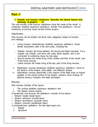
The oral mucosa is the mucous membrane lining the inside of the mouth. It
comprises stratified squamous epithelium, termed "oral epithelium", and an
underlying connective tissue termed lamina propria
Classification
Oral mucosa can be divided into three main categories based on function
and histology:
i. Lining mucosa nonkeratinized stratified squamous epithelium, found
almost everywhere else in the oral cavity, including the:
o Alveolar mucosa the lining between the buccal and labial mucosae. It is a
brighter red, smooth, and shiny with many blood vessels, and is not
connected to underlying tissue by rete pegs
o Buccal mucosa the inside lining of the cheeks and floor of the mouth; part
of the lining mucosa.
o Labial mucosa the inside lining of the lips; part of the lining mucosa.
ii. Masticatory mucosa keratinized stratified squamous epithelium, found on
the dorsum of the tongue, hard palate, and attached gingiva.
iii. Specialized mucosa specifically in the regions of the taste buds on lingual
papillae on the dorsal surface of the tongue; contains nerve endings for
general sensory reception and taste perception
Structure
Oral mucosa consists of two layers,
i. The surface stratified squamous epithelium and
ii. The deeper lamina propria.
In keratinized oral mucosa, the epithelium consists of four layers:
 Stratum basale (basal layer)
 Stratum spinosum (prickle layer)
 Stratum granulosum (granular layer)
 Stratum corneum (keratinized layer)
In nonkeratinized epithelium, the two deep layers are
i. Basale
ii. Spinosum
DENTAL ANATOMY AND HISTOLOGY 2018
SHAHID EQUABAL Page 2
Depending on the region of the mouth, the epithelium may be
i. Nonkeratinized or
ii. Keratinized.
 Nonkeratinized squamous epithelium covers the soft palate, inner lips,
inner cheeks, and the floor of the mouth, and ventral surface of the tongue.
 Keratinized squamous epithelium is present in the gingiva and hard
palate as well as areas of the dorsal surface of the tongue.
 Keratinization is the differentiation of keratinocytes in the stratum
granulosum.
 The lamina propria is a fibrous connective tissue layer that consists of a
network of type I and III collagen and elastin fibers in some regions.
 The main cells of the lamina propria are the fibroblasts, which are
responsible for the production of the fibers as well as the extracellular
matrix
Function
Protection - One of the main functions of the oral mucosa is to physically protect
the underlying tissues from the mechanical forces, microbes and toxins in the
mouth.
Keratinized masticatory mucosa is tightly bound to the hard palate and
gingivae.
It accounts for 25% of all oral mucosa.
Lining mucosa in the cheeks, lips and floor of mouth is mobile to create space
when chewing and talking.
During mastication, it allows food to move freely around the mouth and
physically protects the underlying tissues from trauma.
It accounts for 60% of oral mucosa
Secretion - Saliva is the primary secretion of the oral mucosa.
It has many functions including lubrication, pH buffering and immunity.
Sensation - The oral mucosa is richly innervated, meaning it is a very good at
sensing pain, touch, temperature and taste.
A number of cranial nerves are involved in sensations in the mouth including
trigeminal (V), Facial (VII), glossopharyngeal (IX) and Vagus (X) nerves.
Thermal regulation.
DENTAL ANATOMY AND HISTOLOGY 2018
SHAHID EQUABAL Page 3
Histology of Gingival
The gingival epithelium encompasses the external surface of the gingiva including
the mobile and fixed areas as well as the gingival sulcus and the junctional
epithelium.
It is divided up into three major sections known as the:
i. Oral epithelium: The oral epithelium is comprised of stratified squamous
keratinizing epithelium and covers the oral and vestibular gingival surfaces.
ii. The sulcular epithelium: The sulcular epithelium is continuous with the oral
epithelium and lines the gingival sulcus.
iii. The junctional epithelium
 Clinical feature of gingival:
 Knife-edged marginal gingiva.
 Pink in color.
 Pointed papillae filling interproximal space.
 Moderately scalloped contours.
 Firm, stippled attached gingiva.
 Well demarcated mucogingival junction.
2. Discuss in detail the stages of development of the tooth. Add a short
note on root formation of multirooted tooth. 10
Based on the shape of enamel organ during the development of tooth,
developmental stages of the tooth are divided into the three morphological
stages.
1) Bud stage.
2) Cap stage.
3) Bell stage.
 Early.
 Late or advanced.
Bud stage:
 Enamel organ is bud shaped with peripheral cuboidal cell and central
polyhedral cell.
 All cell is attached to each other by desmosomal junction .
 Dental papilla adjacent to enamel organ.
 Dental follicle surrounding the dental papilla and enamel organ.
DENTAL ANATOMY AND HISTOLOGY 2018
SHAHID EQUABAL Page 4
Cap stage:
 The first signs of an arrangement of cells in the tooth bud occur in
the cap stage.
 Three layers in enamel organ.
i. Inner enamel epithelium.
ii. Stellate reticulum.
iii. Outer enamel epithelium.
 Dental papilla with condensation of ectomesenchyme and budding
capillaries.
 Dense fibrous dental follicle.
DENTAL ANATOMY AND HISTOLOGY 2018
SHAHID EQUABAL Page 5
Early bell stage:
 Enamel organ having bell shaped.
 Four layer in enamel organ:
i. Inner enamel epithelium.
ii. Stratum intermedium.
iii. Stellate reticulum.
iv. Outer enamel epithelium.
 Dental papilla with peripheral cells differentiating to odontoblast.
 Distinct dental follicle.
Advanced bell stage:
 Dentin and enamel formation.
 Four distinct layer of enamel organ collapse of Stellate reticulum.
 Distinct layer of odontoblast.
 Distinct dental follicle.
DENTAL ANATOMY AND HISTOLOGY 2018
SHAHID EQUABAL Page 6
Formation of multirooted tooth:
 The root start to develop after the crown is completed through the
formation of a cervical loop.
 The first formed part of the rot sheath bends to form a disc like structure.
 The root of the tooth is composed by dentin and cementum.
 Dentin formed when the outer cells of the dental papilla are induced to
differentiation into ODCs.
 The development of the multirooted tooth teeth takes place in a same
manner until the furcation area.
 When the furcation area is reached the epithelial diaphragm develops
tongue like extensions that grow until they contact each other
3. Write a short note on: 5×3=15
a) Maxillary central incisor.
b) Type of cement enamel junction.
c) Stages of deglutition.
 Maxillary central incisor:
 Centered in the maxilla, one on either side of median line, with
mesial surface of each in contact with mesial surface of other
 Two in number
 Larger than the lateral incisor
 Major function is to punch and cut food material during the process
of mastication
DENTAL ANATOMY AND HISTOLOGY 2018
SHAHID EQUABAL Page 7
CHRONOLOGY:
 First evidence of calcification 3-4 months
 Crown completion 4-5 years
 Eruption 7-8 years
 Root completion 10-11 years
Labial Aspect:
 Labial surface of crown is usually convex, especially toward cervical 3rd,
some central incisors are flat at middle & incisal portions.
 Mesial outline of crown is only slightly convex, with crest of curvature.
 Distal outline of the crown is more convex than mesial outline, with the
crest of curvature higher toward the cervical line
Lingual Aspect:
 The lingual outline is the reverse of that found on labial aspect
 Lingual aspect has convexities and a concavity
 Outline of cervical line is similar, but immediately below cervical line a
smooth convexity is found – CINGULUM
 Between marginal ridges, below cingulum, a shallow concavity is present -
lingual fossa
DENTAL ANATOMY AND HISTOLOGY 2018
SHAHID EQUABAL Page 8
Lingual fossa is bordered by:
 Mesially: Mesial marginal ridge,
 Incisally: Lingual portion of the Incisal ridge,
 Distally: Distal marginal ridge,
 Cervically: Cingulum.
 Developmental grooves extending from the cingulum into the lingual fossa
Mesial Aspect:
 Lingual outline
 Convex: crest of curvature at the cingulum
 Concave: at Middle portion
 Slightly convex: at linguo-incisal ridge & incisal edge
 Cervical line
 Mesially on maxillary central incisor curves incisally to a noticeable
degree.
DENTAL ANATOMY AND HISTOLOGY 2018
SHAHID EQUABAL Page 9
Distal Aspect:
 The distal surface closely resembles mesial surface, with following
exceptions:
 Distal surface is generally smaller than mesial surface, because inciso-
cervical dimension is shorter.
 Distal surface is more convex inciso-gingivally.
 The cervical margin does not curve as far incisally.
 Curvature of cervical line outlining the CEJ is less in extent on the distal
than on the mesial surfaces
Incisal Aspect:
 Outline of lingual portion tapers lingually toward cingulum.
 Cingulum of crown makes up cervical portion of lingual surface.
 Crown of this tooth shows more bulk from incisal aspect than from
mesial or distal aspect.
Root:
 The root is single, conical, relatively straight, and tapers to a rounded apex
 Horizontal cross section of root near cervical line shows a rounded
triangular outline
 Types of cement enamel junction:
 Cemento-enamel junction is the junction between enamel and
cementum that occurs at cervical region of the tooth.
 There are three types of CEJ
I. Sharp junction:
 Cementum and enamel meet at sharp point.
 Seen in 30% of teeth.
DENTAL ANATOMY AND HISTOLOGY 2018
SHAHID EQUABAL Page 10
II. Gap junction:
 A gap is present between enamel and cementum.
 Occur due to lack of degeneration of HERS.
 Seen in 15% of teeth.
III. Overlap junction:
 Cementum overlap cervical portion of enamel.
 Acellular afibrillar cementum is seen in the region of overlap.
 Seen in 60% of teeth.
DENTAL ANATOMY AND HISTOLOGY 2018
SHAHID EQUABAL Page 11
 Deglutition:
 The process by which swallowing of food is helped.
 It also includes reflex action.
 Helped by movement of pharynx.
 Closure of epiglottis to close larynx
 Upward movement of palate
 Stages of deglutition:
 Oral stage
 Pharyngeal stage
 Oesophageal stage
 Oral stage:
 Oral stage is voluntary stage of deglutition.
 When food is ready for swallowing, bolus formed is put over the
dorsum of the tongue.
 Tongue pressed against the palate and is moved backward, moving
the bolus from mouth to pharynx.
 Pharyngeal stage:
 When bolus moves from mouth to pharynx receptors around opening
of pharynx are stimulated impulses from these areas pass to the
brain stem deglutition centre to initiate series of muscular
contraction.
 Soft palate moves upwards and close posterior nasal openings to
prevent food into the nose.
 Upward movement of larynx enlarges opening of esophagus.
 Pharyngo-oesophageal sphincter relaxes.
 Oesophageal stage:
 Oesophageal stage conducts food from esophagus to stomach by
movements as follows.
 Primary Peristalsis:
o It is simply continuation of peristaltic wave initiated in
pharynx.
o It takes 8-10 seconds to carry food to stomach.
 Secondary Peristalsis:
o If primary peristaltic wave fails to carry all the food to the
stomach, secondary peristaltic wave is initiated in
esophagus due to distension of esophagus with food.
DENTAL ANATOMY AND HISTOLOGY 2018
SHAHID EQUABAL Page 12
Part- 2
4. Describe the functions, components and histological structure of
periodontal ligament: 10
 Periodontal ligament is a connective tissue structure that attaches the
tooth to the alveolar bone.
 It is a part of attachment apparatus of the tooth.
 Function:
1. Physical function.
2. Formative function.
3. Nutritional function.
4. Homeostatic function.
5. Sensory function.
 Physical function:
i. Transmission of Occlusal forces to the bone.
ii. Attachment of the bone to the teeth.
iii. Maintenance of the gingival tissue in their proper
relationship to the teeth.
 Formative function:
Cells of the PDL participate in the formation and Resorption of
cementum and bone, which occur in,
o Physiological tooth movement.
o Accommodation of the periodontium to Occlusal force.
o In the repair of injuries.
 Nutritional function:
o PDL supplies nutrient to the cementum, bone and
gingiva by way of blood vessels and provide lymphatic
drainage.
 Homeostatic function:
o It is evident that the cell of PDL has the ability to
reabsorb and synthesize the extracellular substance of
the connective tissue of the ligament, alveolar bone and
cementum.
Components of PDL:
DENTAL ANATOMY AND HISTOLOGY 2018
SHAHID EQUABAL Page 13
Structure of PDL:
5. Describe the development of occlusion in primary, mixed and
permanent dentition: 10
 Term occlusion is derived from the Latin word, “occlusio”.
 “Defined as the relationship between all the components of the
masticatory system in normal function, dysfunction and
parafunction.”
 Occlusion in primary dentition:
DENTAL ANATOMY AND HISTOLOGY 2018
SHAHID EQUABAL Page 14
 Occlusion in mixed dentition:
 The period during which both primary and permanent teeth are in oral
cavity together is known as mixed dentition period.
 Extend from 6-12 year of age.
 Successional teeth: those permanent teeth that follow into the arch
once held by primary tooth;
Example: incisor, canine, premolar
 Accessional teeth: those permanent teeth that erupt posteriorly to the
primary teeth.
Example: molars
 Occlusion in permanent dentition:
6. Write a short note on: 5×3=15
a) Structure and function of alveolar bone.
b) Histology of salivary gland.
c) Shedding of primary teeth.
 Structure and function of alveolar bone.
 Alveolar process is that part of maxilla and mandible that forms
and supports the sockets of teeth.
 Development: it starts in the second month of fetal life.
DENTAL ANATOMY AND HISTOLOGY 2018
SHAHID EQUABAL Page 15
 Structure of Alveolar bone:
 Alveolar bone proper:
 It surrounds the root of the tooth and gives attachment to the
periodontal ligament fibers.
 It consists of Lamellated bone and Bundle bone
 Lamellated bone consists of osteons.
 Part of the alveolar bone where periodontal ligament fibers are
inserted is known as Bundle bone.
 Supporting alveolar bone:
It consists of two parts –
 Cortical plates: these are made up of compact bone & form the outer and
inner plates of alveolar bone.
 Spongy bone: it fills the area between the cortical plates and the alveolar
bone proper.
 Function of alveolar bone:
 Alveolar bone holds the teeth firmly in position to masticate.
 Supplies vessels to periodontal ligaments and cementum.
 Houses and protects developing permanent teeth while supporting primary
teeth
Alveolar bone
proper
Lamallated
bone
Bundle
bone
Supporting
alveolar bone
Cortical
palte
Spongy
bone
DENTAL ANATOMY AND HISTOLOGY 2018
SHAHID EQUABAL Page 16
 Histology of salivary gland:
 Saliva is produced by three pairs of major salivary glands,
Parotid, Sublingual and Submandibular as well as minor accessory
glands found throughout the mucosa.
 Salivary glands are made up of secretory acini and ducts.
 There are two types of secretions - serous and mucous.

Más contenido relacionado

La actualidad más candente

La actualidad más candente (20)

Gingiva in health and disease
Gingiva in health and diseaseGingiva in health and disease
Gingiva in health and disease
 
Gingiva
GingivaGingiva
Gingiva
 
oral mucous membranes-6 /certified fixed orthodontic courses by Indian dental...
oral mucous membranes-6 /certified fixed orthodontic courses by Indian dental...oral mucous membranes-6 /certified fixed orthodontic courses by Indian dental...
oral mucous membranes-6 /certified fixed orthodontic courses by Indian dental...
 
486 junctional epithelium
486 junctional epithelium486 junctional epithelium
486 junctional epithelium
 
Gingiva
GingivaGingiva
Gingiva
 
Gingiva
GingivaGingiva
Gingiva
 
Gingiva (Macroscopic features)
Gingiva (Macroscopic features)Gingiva (Macroscopic features)
Gingiva (Macroscopic features)
 
Gingiva
GingivaGingiva
Gingiva
 
Gingiva
GingivaGingiva
Gingiva
 
Dentinogingival junction
Dentinogingival junctionDentinogingival junction
Dentinogingival junction
 
gingiva
 gingiva gingiva
gingiva
 
Gingiva.
Gingiva.Gingiva.
Gingiva.
 
Anatomical landmarks-7620033
Anatomical landmarks-7620033Anatomical landmarks-7620033
Anatomical landmarks-7620033
 
Oral landmarks
Oral landmarksOral landmarks
Oral landmarks
 
Gingiva (2)
Gingiva (2)Gingiva (2)
Gingiva (2)
 
Gingiva......
Gingiva......Gingiva......
Gingiva......
 
Aberrant frenum and its treatment
Aberrant frenum and its treatmentAberrant frenum and its treatment
Aberrant frenum and its treatment
 
landmarks of face and oral cavity
landmarks of face and oral cavitylandmarks of face and oral cavity
landmarks of face and oral cavity
 
Gingiva
GingivaGingiva
Gingiva
 
Maxillary and mandbular anatomical landmarks
Maxillary and mandbular anatomical landmarksMaxillary and mandbular anatomical landmarks
Maxillary and mandbular anatomical landmarks
 

Similar a Dental anatomy and histology 2018

Ultrastructure of gingiva
Ultrastructure of gingiva Ultrastructure of gingiva
Ultrastructure of gingiva naseemashraf2
 
Himanshu meghwal (1).pptxygbugugvugvggg
Himanshu meghwal  (1).pptxygbugugvugvgggHimanshu meghwal  (1).pptxygbugugvugvggg
Himanshu meghwal (1).pptxygbugugvugvgggmoditirth170904
 
GINGIVA SEMINAR .pptx
GINGIVA SEMINAR .pptxGINGIVA SEMINAR .pptx
GINGIVA SEMINAR .pptxANIL KUMAR
 
Oral cavity anatomy and histology
Oral cavity anatomy and histologyOral cavity anatomy and histology
Oral cavity anatomy and histologyAliLaith5230
 
Clinical Periodontology - Introduction & Anatomy
Clinical Periodontology - Introduction & AnatomyClinical Periodontology - Introduction & Anatomy
Clinical Periodontology - Introduction & AnatomySarang Suresh Hotchandani
 
Gingiva Periodontology
Gingiva PeriodontologyGingiva Periodontology
Gingiva PeriodontologySaiLakshmi128
 
Periodontium seminar
Periodontium seminar Periodontium seminar
Periodontium seminar PrajaktaGir
 
Clinical anatomy and histology of periodontiumby mohamed assaadwy
Clinical anatomy and histology of periodontiumby mohamed assaadwyClinical anatomy and histology of periodontiumby mohamed assaadwy
Clinical anatomy and histology of periodontiumby mohamed assaadwyDrMohamed Assadawy
 
Development of dentition and occlsion
Development of dentition and occlsionDevelopment of dentition and occlsion
Development of dentition and occlsionAmritha James
 
GINGIVA Macroscopic features
GINGIVA Macroscopic featuresGINGIVA Macroscopic features
GINGIVA Macroscopic featuresSaiLakshmi128
 
2000 biologia celular y molecular de la encia
2000 biologia celular y molecular de la encia2000 biologia celular y molecular de la encia
2000 biologia celular y molecular de la enciakenigal
 

Similar a Dental anatomy and histology 2018 (20)

Ultrastructure of gingiva
Ultrastructure of gingiva Ultrastructure of gingiva
Ultrastructure of gingiva
 
Oral mucosa part 2
Oral mucosa part 2Oral mucosa part 2
Oral mucosa part 2
 
Oral Cavity
Oral CavityOral Cavity
Oral Cavity
 
Gingiva
Gingiva Gingiva
Gingiva
 
Himanshu meghwal (1).pptxygbugugvugvggg
Himanshu meghwal  (1).pptxygbugugvugvgggHimanshu meghwal  (1).pptxygbugugvugvggg
Himanshu meghwal (1).pptxygbugugvugvggg
 
Gingiva
Gingiva Gingiva
Gingiva
 
GINGIVA SEMINAR .pptx
GINGIVA SEMINAR .pptxGINGIVA SEMINAR .pptx
GINGIVA SEMINAR .pptx
 
Oral cavity anatomy and histology
Oral cavity anatomy and histologyOral cavity anatomy and histology
Oral cavity anatomy and histology
 
Clinical Periodontology - Introduction & Anatomy
Clinical Periodontology - Introduction & AnatomyClinical Periodontology - Introduction & Anatomy
Clinical Periodontology - Introduction & Anatomy
 
Gingiva
GingivaGingiva
Gingiva
 
Gingiva Periodontology
Gingiva PeriodontologyGingiva Periodontology
Gingiva Periodontology
 
Gingiva
GingivaGingiva
Gingiva
 
Anatomy of periodontium
Anatomy of periodontiumAnatomy of periodontium
Anatomy of periodontium
 
Gingiva
Gingiva Gingiva
Gingiva
 
Periodontium seminar
Periodontium seminar Periodontium seminar
Periodontium seminar
 
Clinical anatomy and histology of periodontiumby mohamed assaadwy
Clinical anatomy and histology of periodontiumby mohamed assaadwyClinical anatomy and histology of periodontiumby mohamed assaadwy
Clinical anatomy and histology of periodontiumby mohamed assaadwy
 
Development of dentition and occlsion
Development of dentition and occlsionDevelopment of dentition and occlsion
Development of dentition and occlsion
 
Gingiva
GingivaGingiva
Gingiva
 
GINGIVA Macroscopic features
GINGIVA Macroscopic featuresGINGIVA Macroscopic features
GINGIVA Macroscopic features
 
2000 biologia celular y molecular de la encia
2000 biologia celular y molecular de la encia2000 biologia celular y molecular de la encia
2000 biologia celular y molecular de la encia
 

Último

Call Girls Jayanagar Just Call 7001305949 Top Class Call Girl Service Available
Call Girls Jayanagar Just Call 7001305949 Top Class Call Girl Service AvailableCall Girls Jayanagar Just Call 7001305949 Top Class Call Girl Service Available
Call Girls Jayanagar Just Call 7001305949 Top Class Call Girl Service Availablenarwatsonia7
 
Hemostasis Physiology and Clinical correlations by Dr Faiza.pdf
Hemostasis Physiology and Clinical correlations by Dr Faiza.pdfHemostasis Physiology and Clinical correlations by Dr Faiza.pdf
Hemostasis Physiology and Clinical correlations by Dr Faiza.pdfMedicoseAcademics
 
Book Call Girls in Yelahanka - For 7001305949 Cheap & Best with original Photos
Book Call Girls in Yelahanka - For 7001305949 Cheap & Best with original PhotosBook Call Girls in Yelahanka - For 7001305949 Cheap & Best with original Photos
Book Call Girls in Yelahanka - For 7001305949 Cheap & Best with original Photosnarwatsonia7
 
Call Girls Hebbal Just Call 7001305949 Top Class Call Girl Service Available
Call Girls Hebbal Just Call 7001305949 Top Class Call Girl Service AvailableCall Girls Hebbal Just Call 7001305949 Top Class Call Girl Service Available
Call Girls Hebbal Just Call 7001305949 Top Class Call Girl Service Availablenarwatsonia7
 
call girls in Connaught Place DELHI 🔝 >༒9540349809 🔝 genuine Escort Service ...
call girls in Connaught Place  DELHI 🔝 >༒9540349809 🔝 genuine Escort Service ...call girls in Connaught Place  DELHI 🔝 >༒9540349809 🔝 genuine Escort Service ...
call girls in Connaught Place DELHI 🔝 >༒9540349809 🔝 genuine Escort Service ...saminamagar
 
Call Girls Service Chennai Jiya 7001305949 Independent Escort Service Chennai
Call Girls Service Chennai Jiya 7001305949 Independent Escort Service ChennaiCall Girls Service Chennai Jiya 7001305949 Independent Escort Service Chennai
Call Girls Service Chennai Jiya 7001305949 Independent Escort Service ChennaiNehru place Escorts
 
Call Girls Electronic City Just Call 7001305949 Top Class Call Girl Service A...
Call Girls Electronic City Just Call 7001305949 Top Class Call Girl Service A...Call Girls Electronic City Just Call 7001305949 Top Class Call Girl Service A...
Call Girls Electronic City Just Call 7001305949 Top Class Call Girl Service A...narwatsonia7
 
Call Girls Frazer Town Just Call 7001305949 Top Class Call Girl Service Avail...
Call Girls Frazer Town Just Call 7001305949 Top Class Call Girl Service Avail...Call Girls Frazer Town Just Call 7001305949 Top Class Call Girl Service Avail...
Call Girls Frazer Town Just Call 7001305949 Top Class Call Girl Service Avail...narwatsonia7
 
Housewife Call Girls Hsr Layout - Call 7001305949 Rs-3500 with A/C Room Cash ...
Housewife Call Girls Hsr Layout - Call 7001305949 Rs-3500 with A/C Room Cash ...Housewife Call Girls Hsr Layout - Call 7001305949 Rs-3500 with A/C Room Cash ...
Housewife Call Girls Hsr Layout - Call 7001305949 Rs-3500 with A/C Room Cash ...narwatsonia7
 
Call Girls Service Noida Maya 9711199012 Independent Escort Service Noida
Call Girls Service Noida Maya 9711199012 Independent Escort Service NoidaCall Girls Service Noida Maya 9711199012 Independent Escort Service Noida
Call Girls Service Noida Maya 9711199012 Independent Escort Service NoidaPooja Gupta
 
Call Girl Bangalore Nandini 7001305949 Independent Escort Service Bangalore
Call Girl Bangalore Nandini 7001305949 Independent Escort Service BangaloreCall Girl Bangalore Nandini 7001305949 Independent Escort Service Bangalore
Call Girl Bangalore Nandini 7001305949 Independent Escort Service Bangalorenarwatsonia7
 
Call Girls Thane Just Call 9910780858 Get High Class Call Girls Service
Call Girls Thane Just Call 9910780858 Get High Class Call Girls ServiceCall Girls Thane Just Call 9910780858 Get High Class Call Girls Service
Call Girls Thane Just Call 9910780858 Get High Class Call Girls Servicesonalikaur4
 
call girls in green park DELHI 🔝 >༒9540349809 🔝 genuine Escort Service 🔝✔️✔️
call girls in green park  DELHI 🔝 >༒9540349809 🔝 genuine Escort Service 🔝✔️✔️call girls in green park  DELHI 🔝 >༒9540349809 🔝 genuine Escort Service 🔝✔️✔️
call girls in green park DELHI 🔝 >༒9540349809 🔝 genuine Escort Service 🔝✔️✔️saminamagar
 
Russian Call Girls Chickpet - 7001305949 Booking and charges genuine rate for...
Russian Call Girls Chickpet - 7001305949 Booking and charges genuine rate for...Russian Call Girls Chickpet - 7001305949 Booking and charges genuine rate for...
Russian Call Girls Chickpet - 7001305949 Booking and charges genuine rate for...narwatsonia7
 
Call Girls Whitefield Just Call 7001305949 Top Class Call Girl Service Available
Call Girls Whitefield Just Call 7001305949 Top Class Call Girl Service AvailableCall Girls Whitefield Just Call 7001305949 Top Class Call Girl Service Available
Call Girls Whitefield Just Call 7001305949 Top Class Call Girl Service Availablenarwatsonia7
 
Dwarka Sector 6 Call Girls ( 9873940964 ) Book Hot And Sexy Girls In A Few Cl...
Dwarka Sector 6 Call Girls ( 9873940964 ) Book Hot And Sexy Girls In A Few Cl...Dwarka Sector 6 Call Girls ( 9873940964 ) Book Hot And Sexy Girls In A Few Cl...
Dwarka Sector 6 Call Girls ( 9873940964 ) Book Hot And Sexy Girls In A Few Cl...rajnisinghkjn
 
Call Girls ITPL Just Call 7001305949 Top Class Call Girl Service Available
Call Girls ITPL Just Call 7001305949 Top Class Call Girl Service AvailableCall Girls ITPL Just Call 7001305949 Top Class Call Girl Service Available
Call Girls ITPL Just Call 7001305949 Top Class Call Girl Service Availablenarwatsonia7
 
Call Girl Koramangala | 7001305949 At Low Cost Cash Payment Booking
Call Girl Koramangala | 7001305949 At Low Cost Cash Payment BookingCall Girl Koramangala | 7001305949 At Low Cost Cash Payment Booking
Call Girl Koramangala | 7001305949 At Low Cost Cash Payment Bookingnarwatsonia7
 
Low Rate Call Girls Mumbai Suman 9910780858 Independent Escort Service Mumbai
Low Rate Call Girls Mumbai Suman 9910780858 Independent Escort Service MumbaiLow Rate Call Girls Mumbai Suman 9910780858 Independent Escort Service Mumbai
Low Rate Call Girls Mumbai Suman 9910780858 Independent Escort Service Mumbaisonalikaur4
 
Asthma Review - GINA guidelines summary 2024
Asthma Review - GINA guidelines summary 2024Asthma Review - GINA guidelines summary 2024
Asthma Review - GINA guidelines summary 2024Gabriel Guevara MD
 

Último (20)

Call Girls Jayanagar Just Call 7001305949 Top Class Call Girl Service Available
Call Girls Jayanagar Just Call 7001305949 Top Class Call Girl Service AvailableCall Girls Jayanagar Just Call 7001305949 Top Class Call Girl Service Available
Call Girls Jayanagar Just Call 7001305949 Top Class Call Girl Service Available
 
Hemostasis Physiology and Clinical correlations by Dr Faiza.pdf
Hemostasis Physiology and Clinical correlations by Dr Faiza.pdfHemostasis Physiology and Clinical correlations by Dr Faiza.pdf
Hemostasis Physiology and Clinical correlations by Dr Faiza.pdf
 
Book Call Girls in Yelahanka - For 7001305949 Cheap & Best with original Photos
Book Call Girls in Yelahanka - For 7001305949 Cheap & Best with original PhotosBook Call Girls in Yelahanka - For 7001305949 Cheap & Best with original Photos
Book Call Girls in Yelahanka - For 7001305949 Cheap & Best with original Photos
 
Call Girls Hebbal Just Call 7001305949 Top Class Call Girl Service Available
Call Girls Hebbal Just Call 7001305949 Top Class Call Girl Service AvailableCall Girls Hebbal Just Call 7001305949 Top Class Call Girl Service Available
Call Girls Hebbal Just Call 7001305949 Top Class Call Girl Service Available
 
call girls in Connaught Place DELHI 🔝 >༒9540349809 🔝 genuine Escort Service ...
call girls in Connaught Place  DELHI 🔝 >༒9540349809 🔝 genuine Escort Service ...call girls in Connaught Place  DELHI 🔝 >༒9540349809 🔝 genuine Escort Service ...
call girls in Connaught Place DELHI 🔝 >༒9540349809 🔝 genuine Escort Service ...
 
Call Girls Service Chennai Jiya 7001305949 Independent Escort Service Chennai
Call Girls Service Chennai Jiya 7001305949 Independent Escort Service ChennaiCall Girls Service Chennai Jiya 7001305949 Independent Escort Service Chennai
Call Girls Service Chennai Jiya 7001305949 Independent Escort Service Chennai
 
Call Girls Electronic City Just Call 7001305949 Top Class Call Girl Service A...
Call Girls Electronic City Just Call 7001305949 Top Class Call Girl Service A...Call Girls Electronic City Just Call 7001305949 Top Class Call Girl Service A...
Call Girls Electronic City Just Call 7001305949 Top Class Call Girl Service A...
 
Call Girls Frazer Town Just Call 7001305949 Top Class Call Girl Service Avail...
Call Girls Frazer Town Just Call 7001305949 Top Class Call Girl Service Avail...Call Girls Frazer Town Just Call 7001305949 Top Class Call Girl Service Avail...
Call Girls Frazer Town Just Call 7001305949 Top Class Call Girl Service Avail...
 
Housewife Call Girls Hsr Layout - Call 7001305949 Rs-3500 with A/C Room Cash ...
Housewife Call Girls Hsr Layout - Call 7001305949 Rs-3500 with A/C Room Cash ...Housewife Call Girls Hsr Layout - Call 7001305949 Rs-3500 with A/C Room Cash ...
Housewife Call Girls Hsr Layout - Call 7001305949 Rs-3500 with A/C Room Cash ...
 
Call Girls Service Noida Maya 9711199012 Independent Escort Service Noida
Call Girls Service Noida Maya 9711199012 Independent Escort Service NoidaCall Girls Service Noida Maya 9711199012 Independent Escort Service Noida
Call Girls Service Noida Maya 9711199012 Independent Escort Service Noida
 
Call Girl Bangalore Nandini 7001305949 Independent Escort Service Bangalore
Call Girl Bangalore Nandini 7001305949 Independent Escort Service BangaloreCall Girl Bangalore Nandini 7001305949 Independent Escort Service Bangalore
Call Girl Bangalore Nandini 7001305949 Independent Escort Service Bangalore
 
Call Girls Thane Just Call 9910780858 Get High Class Call Girls Service
Call Girls Thane Just Call 9910780858 Get High Class Call Girls ServiceCall Girls Thane Just Call 9910780858 Get High Class Call Girls Service
Call Girls Thane Just Call 9910780858 Get High Class Call Girls Service
 
call girls in green park DELHI 🔝 >༒9540349809 🔝 genuine Escort Service 🔝✔️✔️
call girls in green park  DELHI 🔝 >༒9540349809 🔝 genuine Escort Service 🔝✔️✔️call girls in green park  DELHI 🔝 >༒9540349809 🔝 genuine Escort Service 🔝✔️✔️
call girls in green park DELHI 🔝 >༒9540349809 🔝 genuine Escort Service 🔝✔️✔️
 
Russian Call Girls Chickpet - 7001305949 Booking and charges genuine rate for...
Russian Call Girls Chickpet - 7001305949 Booking and charges genuine rate for...Russian Call Girls Chickpet - 7001305949 Booking and charges genuine rate for...
Russian Call Girls Chickpet - 7001305949 Booking and charges genuine rate for...
 
Call Girls Whitefield Just Call 7001305949 Top Class Call Girl Service Available
Call Girls Whitefield Just Call 7001305949 Top Class Call Girl Service AvailableCall Girls Whitefield Just Call 7001305949 Top Class Call Girl Service Available
Call Girls Whitefield Just Call 7001305949 Top Class Call Girl Service Available
 
Dwarka Sector 6 Call Girls ( 9873940964 ) Book Hot And Sexy Girls In A Few Cl...
Dwarka Sector 6 Call Girls ( 9873940964 ) Book Hot And Sexy Girls In A Few Cl...Dwarka Sector 6 Call Girls ( 9873940964 ) Book Hot And Sexy Girls In A Few Cl...
Dwarka Sector 6 Call Girls ( 9873940964 ) Book Hot And Sexy Girls In A Few Cl...
 
Call Girls ITPL Just Call 7001305949 Top Class Call Girl Service Available
Call Girls ITPL Just Call 7001305949 Top Class Call Girl Service AvailableCall Girls ITPL Just Call 7001305949 Top Class Call Girl Service Available
Call Girls ITPL Just Call 7001305949 Top Class Call Girl Service Available
 
Call Girl Koramangala | 7001305949 At Low Cost Cash Payment Booking
Call Girl Koramangala | 7001305949 At Low Cost Cash Payment BookingCall Girl Koramangala | 7001305949 At Low Cost Cash Payment Booking
Call Girl Koramangala | 7001305949 At Low Cost Cash Payment Booking
 
Low Rate Call Girls Mumbai Suman 9910780858 Independent Escort Service Mumbai
Low Rate Call Girls Mumbai Suman 9910780858 Independent Escort Service MumbaiLow Rate Call Girls Mumbai Suman 9910780858 Independent Escort Service Mumbai
Low Rate Call Girls Mumbai Suman 9910780858 Independent Escort Service Mumbai
 
Asthma Review - GINA guidelines summary 2024
Asthma Review - GINA guidelines summary 2024Asthma Review - GINA guidelines summary 2024
Asthma Review - GINA guidelines summary 2024
 

Dental anatomy and histology 2018

  • 1. DENTAL ANATOMY AND HISTOLOGY 2018 SHAHID EQUABAL Page 1 Part -1 1. Classify oral mucous membrane. Describe the clinical feature and histology of gingival. 10 The oral mucosa is the mucous membrane lining the inside of the mouth. It comprises stratified squamous epithelium, termed "oral epithelium", and an underlying connective tissue termed lamina propria Classification Oral mucosa can be divided into three main categories based on function and histology: i. Lining mucosa nonkeratinized stratified squamous epithelium, found almost everywhere else in the oral cavity, including the: o Alveolar mucosa the lining between the buccal and labial mucosae. It is a brighter red, smooth, and shiny with many blood vessels, and is not connected to underlying tissue by rete pegs o Buccal mucosa the inside lining of the cheeks and floor of the mouth; part of the lining mucosa. o Labial mucosa the inside lining of the lips; part of the lining mucosa. ii. Masticatory mucosa keratinized stratified squamous epithelium, found on the dorsum of the tongue, hard palate, and attached gingiva. iii. Specialized mucosa specifically in the regions of the taste buds on lingual papillae on the dorsal surface of the tongue; contains nerve endings for general sensory reception and taste perception Structure Oral mucosa consists of two layers, i. The surface stratified squamous epithelium and ii. The deeper lamina propria. In keratinized oral mucosa, the epithelium consists of four layers:  Stratum basale (basal layer)  Stratum spinosum (prickle layer)  Stratum granulosum (granular layer)  Stratum corneum (keratinized layer) In nonkeratinized epithelium, the two deep layers are i. Basale ii. Spinosum
  • 2. DENTAL ANATOMY AND HISTOLOGY 2018 SHAHID EQUABAL Page 2 Depending on the region of the mouth, the epithelium may be i. Nonkeratinized or ii. Keratinized.  Nonkeratinized squamous epithelium covers the soft palate, inner lips, inner cheeks, and the floor of the mouth, and ventral surface of the tongue.  Keratinized squamous epithelium is present in the gingiva and hard palate as well as areas of the dorsal surface of the tongue.  Keratinization is the differentiation of keratinocytes in the stratum granulosum.  The lamina propria is a fibrous connective tissue layer that consists of a network of type I and III collagen and elastin fibers in some regions.  The main cells of the lamina propria are the fibroblasts, which are responsible for the production of the fibers as well as the extracellular matrix Function Protection - One of the main functions of the oral mucosa is to physically protect the underlying tissues from the mechanical forces, microbes and toxins in the mouth. Keratinized masticatory mucosa is tightly bound to the hard palate and gingivae. It accounts for 25% of all oral mucosa. Lining mucosa in the cheeks, lips and floor of mouth is mobile to create space when chewing and talking. During mastication, it allows food to move freely around the mouth and physically protects the underlying tissues from trauma. It accounts for 60% of oral mucosa Secretion - Saliva is the primary secretion of the oral mucosa. It has many functions including lubrication, pH buffering and immunity. Sensation - The oral mucosa is richly innervated, meaning it is a very good at sensing pain, touch, temperature and taste. A number of cranial nerves are involved in sensations in the mouth including trigeminal (V), Facial (VII), glossopharyngeal (IX) and Vagus (X) nerves. Thermal regulation.
  • 3. DENTAL ANATOMY AND HISTOLOGY 2018 SHAHID EQUABAL Page 3 Histology of Gingival The gingival epithelium encompasses the external surface of the gingiva including the mobile and fixed areas as well as the gingival sulcus and the junctional epithelium. It is divided up into three major sections known as the: i. Oral epithelium: The oral epithelium is comprised of stratified squamous keratinizing epithelium and covers the oral and vestibular gingival surfaces. ii. The sulcular epithelium: The sulcular epithelium is continuous with the oral epithelium and lines the gingival sulcus. iii. The junctional epithelium  Clinical feature of gingival:  Knife-edged marginal gingiva.  Pink in color.  Pointed papillae filling interproximal space.  Moderately scalloped contours.  Firm, stippled attached gingiva.  Well demarcated mucogingival junction. 2. Discuss in detail the stages of development of the tooth. Add a short note on root formation of multirooted tooth. 10 Based on the shape of enamel organ during the development of tooth, developmental stages of the tooth are divided into the three morphological stages. 1) Bud stage. 2) Cap stage. 3) Bell stage.  Early.  Late or advanced. Bud stage:  Enamel organ is bud shaped with peripheral cuboidal cell and central polyhedral cell.  All cell is attached to each other by desmosomal junction .  Dental papilla adjacent to enamel organ.  Dental follicle surrounding the dental papilla and enamel organ.
  • 4. DENTAL ANATOMY AND HISTOLOGY 2018 SHAHID EQUABAL Page 4 Cap stage:  The first signs of an arrangement of cells in the tooth bud occur in the cap stage.  Three layers in enamel organ. i. Inner enamel epithelium. ii. Stellate reticulum. iii. Outer enamel epithelium.  Dental papilla with condensation of ectomesenchyme and budding capillaries.  Dense fibrous dental follicle.
  • 5. DENTAL ANATOMY AND HISTOLOGY 2018 SHAHID EQUABAL Page 5 Early bell stage:  Enamel organ having bell shaped.  Four layer in enamel organ: i. Inner enamel epithelium. ii. Stratum intermedium. iii. Stellate reticulum. iv. Outer enamel epithelium.  Dental papilla with peripheral cells differentiating to odontoblast.  Distinct dental follicle. Advanced bell stage:  Dentin and enamel formation.  Four distinct layer of enamel organ collapse of Stellate reticulum.  Distinct layer of odontoblast.  Distinct dental follicle.
  • 6. DENTAL ANATOMY AND HISTOLOGY 2018 SHAHID EQUABAL Page 6 Formation of multirooted tooth:  The root start to develop after the crown is completed through the formation of a cervical loop.  The first formed part of the rot sheath bends to form a disc like structure.  The root of the tooth is composed by dentin and cementum.  Dentin formed when the outer cells of the dental papilla are induced to differentiation into ODCs.  The development of the multirooted tooth teeth takes place in a same manner until the furcation area.  When the furcation area is reached the epithelial diaphragm develops tongue like extensions that grow until they contact each other 3. Write a short note on: 5×3=15 a) Maxillary central incisor. b) Type of cement enamel junction. c) Stages of deglutition.  Maxillary central incisor:  Centered in the maxilla, one on either side of median line, with mesial surface of each in contact with mesial surface of other  Two in number  Larger than the lateral incisor  Major function is to punch and cut food material during the process of mastication
  • 7. DENTAL ANATOMY AND HISTOLOGY 2018 SHAHID EQUABAL Page 7 CHRONOLOGY:  First evidence of calcification 3-4 months  Crown completion 4-5 years  Eruption 7-8 years  Root completion 10-11 years Labial Aspect:  Labial surface of crown is usually convex, especially toward cervical 3rd, some central incisors are flat at middle & incisal portions.  Mesial outline of crown is only slightly convex, with crest of curvature.  Distal outline of the crown is more convex than mesial outline, with the crest of curvature higher toward the cervical line Lingual Aspect:  The lingual outline is the reverse of that found on labial aspect  Lingual aspect has convexities and a concavity  Outline of cervical line is similar, but immediately below cervical line a smooth convexity is found – CINGULUM  Between marginal ridges, below cingulum, a shallow concavity is present - lingual fossa
  • 8. DENTAL ANATOMY AND HISTOLOGY 2018 SHAHID EQUABAL Page 8 Lingual fossa is bordered by:  Mesially: Mesial marginal ridge,  Incisally: Lingual portion of the Incisal ridge,  Distally: Distal marginal ridge,  Cervically: Cingulum.  Developmental grooves extending from the cingulum into the lingual fossa Mesial Aspect:  Lingual outline  Convex: crest of curvature at the cingulum  Concave: at Middle portion  Slightly convex: at linguo-incisal ridge & incisal edge  Cervical line  Mesially on maxillary central incisor curves incisally to a noticeable degree.
  • 9. DENTAL ANATOMY AND HISTOLOGY 2018 SHAHID EQUABAL Page 9 Distal Aspect:  The distal surface closely resembles mesial surface, with following exceptions:  Distal surface is generally smaller than mesial surface, because inciso- cervical dimension is shorter.  Distal surface is more convex inciso-gingivally.  The cervical margin does not curve as far incisally.  Curvature of cervical line outlining the CEJ is less in extent on the distal than on the mesial surfaces Incisal Aspect:  Outline of lingual portion tapers lingually toward cingulum.  Cingulum of crown makes up cervical portion of lingual surface.  Crown of this tooth shows more bulk from incisal aspect than from mesial or distal aspect. Root:  The root is single, conical, relatively straight, and tapers to a rounded apex  Horizontal cross section of root near cervical line shows a rounded triangular outline  Types of cement enamel junction:  Cemento-enamel junction is the junction between enamel and cementum that occurs at cervical region of the tooth.  There are three types of CEJ I. Sharp junction:  Cementum and enamel meet at sharp point.  Seen in 30% of teeth.
  • 10. DENTAL ANATOMY AND HISTOLOGY 2018 SHAHID EQUABAL Page 10 II. Gap junction:  A gap is present between enamel and cementum.  Occur due to lack of degeneration of HERS.  Seen in 15% of teeth. III. Overlap junction:  Cementum overlap cervical portion of enamel.  Acellular afibrillar cementum is seen in the region of overlap.  Seen in 60% of teeth.
  • 11. DENTAL ANATOMY AND HISTOLOGY 2018 SHAHID EQUABAL Page 11  Deglutition:  The process by which swallowing of food is helped.  It also includes reflex action.  Helped by movement of pharynx.  Closure of epiglottis to close larynx  Upward movement of palate  Stages of deglutition:  Oral stage  Pharyngeal stage  Oesophageal stage  Oral stage:  Oral stage is voluntary stage of deglutition.  When food is ready for swallowing, bolus formed is put over the dorsum of the tongue.  Tongue pressed against the palate and is moved backward, moving the bolus from mouth to pharynx.  Pharyngeal stage:  When bolus moves from mouth to pharynx receptors around opening of pharynx are stimulated impulses from these areas pass to the brain stem deglutition centre to initiate series of muscular contraction.  Soft palate moves upwards and close posterior nasal openings to prevent food into the nose.  Upward movement of larynx enlarges opening of esophagus.  Pharyngo-oesophageal sphincter relaxes.  Oesophageal stage:  Oesophageal stage conducts food from esophagus to stomach by movements as follows.  Primary Peristalsis: o It is simply continuation of peristaltic wave initiated in pharynx. o It takes 8-10 seconds to carry food to stomach.  Secondary Peristalsis: o If primary peristaltic wave fails to carry all the food to the stomach, secondary peristaltic wave is initiated in esophagus due to distension of esophagus with food.
  • 12. DENTAL ANATOMY AND HISTOLOGY 2018 SHAHID EQUABAL Page 12 Part- 2 4. Describe the functions, components and histological structure of periodontal ligament: 10  Periodontal ligament is a connective tissue structure that attaches the tooth to the alveolar bone.  It is a part of attachment apparatus of the tooth.  Function: 1. Physical function. 2. Formative function. 3. Nutritional function. 4. Homeostatic function. 5. Sensory function.  Physical function: i. Transmission of Occlusal forces to the bone. ii. Attachment of the bone to the teeth. iii. Maintenance of the gingival tissue in their proper relationship to the teeth.  Formative function: Cells of the PDL participate in the formation and Resorption of cementum and bone, which occur in, o Physiological tooth movement. o Accommodation of the periodontium to Occlusal force. o In the repair of injuries.  Nutritional function: o PDL supplies nutrient to the cementum, bone and gingiva by way of blood vessels and provide lymphatic drainage.  Homeostatic function: o It is evident that the cell of PDL has the ability to reabsorb and synthesize the extracellular substance of the connective tissue of the ligament, alveolar bone and cementum. Components of PDL:
  • 13. DENTAL ANATOMY AND HISTOLOGY 2018 SHAHID EQUABAL Page 13 Structure of PDL: 5. Describe the development of occlusion in primary, mixed and permanent dentition: 10  Term occlusion is derived from the Latin word, “occlusio”.  “Defined as the relationship between all the components of the masticatory system in normal function, dysfunction and parafunction.”  Occlusion in primary dentition:
  • 14. DENTAL ANATOMY AND HISTOLOGY 2018 SHAHID EQUABAL Page 14  Occlusion in mixed dentition:  The period during which both primary and permanent teeth are in oral cavity together is known as mixed dentition period.  Extend from 6-12 year of age.  Successional teeth: those permanent teeth that follow into the arch once held by primary tooth; Example: incisor, canine, premolar  Accessional teeth: those permanent teeth that erupt posteriorly to the primary teeth. Example: molars  Occlusion in permanent dentition: 6. Write a short note on: 5×3=15 a) Structure and function of alveolar bone. b) Histology of salivary gland. c) Shedding of primary teeth.  Structure and function of alveolar bone.  Alveolar process is that part of maxilla and mandible that forms and supports the sockets of teeth.  Development: it starts in the second month of fetal life.
  • 15. DENTAL ANATOMY AND HISTOLOGY 2018 SHAHID EQUABAL Page 15  Structure of Alveolar bone:  Alveolar bone proper:  It surrounds the root of the tooth and gives attachment to the periodontal ligament fibers.  It consists of Lamellated bone and Bundle bone  Lamellated bone consists of osteons.  Part of the alveolar bone where periodontal ligament fibers are inserted is known as Bundle bone.  Supporting alveolar bone: It consists of two parts –  Cortical plates: these are made up of compact bone & form the outer and inner plates of alveolar bone.  Spongy bone: it fills the area between the cortical plates and the alveolar bone proper.  Function of alveolar bone:  Alveolar bone holds the teeth firmly in position to masticate.  Supplies vessels to periodontal ligaments and cementum.  Houses and protects developing permanent teeth while supporting primary teeth Alveolar bone proper Lamallated bone Bundle bone Supporting alveolar bone Cortical palte Spongy bone
  • 16. DENTAL ANATOMY AND HISTOLOGY 2018 SHAHID EQUABAL Page 16  Histology of salivary gland:  Saliva is produced by three pairs of major salivary glands, Parotid, Sublingual and Submandibular as well as minor accessory glands found throughout the mucosa.  Salivary glands are made up of secretory acini and ducts.  There are two types of secretions - serous and mucous.