Spio mri studies by dr naghavi - amersham oct 2003 - part2
•Descargar como PPT, PDF•
0 recomendaciones•396 vistas

SHAPE Society

Recomendados
Más contenido relacionado
Destacado
Destacado (17)
Similar a Spio mri studies by dr naghavi - amersham oct 2003 - part2
Similar a Spio mri studies by dr naghavi - amersham oct 2003 - part2 (20)
Más de Society for Heart Attack Prevention and Eradication
Más de Society for Heart Attack Prevention and Eradication (20)
Último
Último (20)
Spio mri studies by dr naghavi - amersham oct 2003 - part2
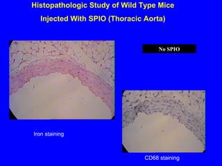
- 1. Histopathologic Study of Wild Type Mice Injected With SPIO (Thoracic Aorta) CD68 staining Iron staining No SPIO
- 2. Comparison of the Number of the Iron Particles (per HPF) in ApoE KO Mice Plaque vs. Normal Wall 0 5 10 15 Atherosclerotic Aorta Average number of iron particles per sample P <0.001
- 3. MR Image of Abdominal Aorta After SPIO Injection in ApoE and Control Mice ApoE deficien t mouse C57B1 (control) mouse Before Injection After Injection (5 Days ) Dark (negatively enhanced) aortic wall, full of iron particles Bright aortic lumen and wall without negative enhancement and no significant number of iron particles
- 4. Injection of Cytokine Increases SPIO-Loaded Macrophage Density in Aortic Plaques (Apo E Deficient Mice) Naghavi et al Circulation 2003
- 5. Naghavi et al Circulation 2003 Injection of Cytokine Increases SPIO-Loaded Macrophage Density in a Coronary Plaque (Apo E Deficient Mice)
- 6. Histopathologic studies of Thoracic aorta in Watanabe Hereditary Hypercholesterolemic rabbit after SPIO injection H&E staining Iron staining Iron staining
- 7. Histopathologic studies of Thoracic aorta in Watanabe Hereditary Hypercholesterolemic rabbit after SPIO injection H&E staining Iron staining Iron staining Iron particles
- 8. Plaque Cell Density vs SPIO 0 10 20 30 40 50 60 0 10 20 30 40 50 60 70 Cell Denity in H&E staining SPIOpositivecell-Iron staining Series1 R=0.956 Correlation between Iron positive cells in Iron staining and cell density in H&E staining in rabbit atherosclerotic aorta.
- 9. MR Angiography 3D with Gadolinium-DTPA in Watanabe Rabbit Before SPIO injection After SPIO injection
- 10. MRI Identifies Plaque Inflammation by SPIO Nanoparticles Watanabe rabbit Post-SPIO Watanabe rabbit control NZW rabbit control NZW rabbit Post-SPIO Watanabe Rabbit Post-SPIO Watanabe Rabbit Control NZW Rabbit Post-SPIO NZW Rabbit Control
- 11. Ex-vivo MR study of the thoracic aorta in Watanabe and Wild type rabbit after SPIO injection compared to control. (Gradient echo) Watanabe rabbit Post-SPIO Watanabe rabbit control NZW rabbit Post-SPIO NZW rabbit control
- 12. Schmitz et al
- 13. Schmitz et al
- 14. Schmitz et al
- 15. Ruhem et al
- 16. Ruhem et al
- 17. Ruhem et al
- 18. Ruhem et al
- 19. Ruhem et al
- 20. Kooi et al
- 21. Kooi et al
- 22. Kooi et al
- 23. Kooi et al
- 24. Ho et al
- 25. Ho et al
- 26. MR signal changes after CsA treatment. A group with allotransplants was treated with CsA for either 7 days (n=5) or 4 days (n=5) after initial MRI experiments showed decrease of MR signal intensity according to degree of graft rejection. On POD 14, transplanted rats were reinjected with USPIO particles, and then MRI experiments were performed. Animals treated 7 days showed minimal changes in MR signal intensity 24 hours after reinjection (A and B), and animals treated 4 days showed MR signal intensity that was significantly decreased after USPIO injection (C and D). MR images of short-axis view of graft are shown. MR images before USPIO infusion are at A and C, and images taken 24 hours after infusion are at B and D. Ho et al
- 27. Ho et al
- 28. • Imaging Macrophage Activity in the Brain Using Ultrasmall Particles of Iron Oxide Jeff W.M. Bultea and Joseph A. Franka Laboratory of Diagnostic Radiology Research National Institutes of Health Bethesda, MD
- 30. • SPIO MR imagine of macrophage will is feasible for clinical applications. • It will be of great interest to investigate the cost-effectiveness of this technique in comparison with alternative diagnostic approaches. Conclusion
- 31. • Poor spatial and temporal resolution for coronary applications • Cost • Cumbersome (injection and delayed imaging) Limitations