
Recomendados
Más contenido relacionado
Similar a CHAPTER 5 NUCLEIC ACIDS_EA.pdf
Similar a CHAPTER 5 NUCLEIC ACIDS_EA.pdf (20)
Más de SOLOMONKIPSEREK
Más de SOLOMONKIPSEREK (12)
Último
Último (20)
CHAPTER 5 NUCLEIC ACIDS_EA.pdf
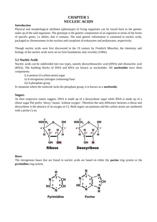
- 1. CHAPTER 5 NUCLEIC ACIDS Introduction Physical and morphological attributes (phenotype) of living organisms can be traced back to the genetic make up of the said organisms. The genotype is the genetic composition of an organism in terms of the forms of specific genes, i.e alleles, that it contains. The total genetic information is contained in nucleic acids, packaged as chromosomes in the nucleus and cytoplasm of eukaryotes and prokaryotes, respectively. Though nucleic acids were first discovered in the 19 century by Friedrich Miescher, the chemistry and biology of the nucleic acids were set on firm foundations only recently (1940s). 5.1 Nucleic Acids Nucleic acids can be subdivided into two types, namely deoxyribonucleic acid (DNA) and ribonucleic acid (RNA). The building blocks of DNA and RNA are known as nucleotides. All nucleotides have three components: i) A pentose (5-carbon-atom) sugar ii) A nitrogenous (nitrogen containing) base iii) A phosphate group In situations where the molecule lacks the phosphate group, it is known as a nucleoside. Sugars As their respective names suggest, DNA is made up of a deoxyribose sugar while RNA is made up of a ribose sugar.The prefix ‘deoxy’ means ‘without oxygen’. Therefore the only difference between a ribose and deoxyribose is the absence of an oxygen at C2. Both sugars are pentoses and the carbon atoms are numbered with a prime (') on. Bases The nitrogenous bases that are found in nucleic acids are based on either the purine ring system or the pyrimidine ring system.
- 2. Both DNA and RNA contain two major purine bases, adenine (A) and guanine (G). Both nucleic acids also contain the pyrimidine, cytosine (C), and a second pyrimidine that is thymine (T) in DNA and uracil (U) in RNA. Only occasionally does thymine occur in RNA or uracil in DNA. In some cases the names of the bases reflect the sources from which they originally were isolated. Guanine, for example, was first isolated from guano (bird manure) and thymine was first isolated from thymus tissue. Chemical structure of the five bases In addition to these major bases, there is a wide range of the so called minor bases which occur less frequently e.g methyl cytosine and pseudouracil. It is important to note that purine and pyrimidine bases can exist in two or more tautomeric forms (isomers of a molecule in a solution arising from rearrangement of chemical bonds) depending on the pH. Certain tautomeric forms predominate at neutral pH, and these are the structures shown for the five common purines and pyrimidines. Uracil, for example, occurs in lactam, lactim, and double lactim forms.
- 3. Inorganic phosphate There are phosphate residues in the nucleic acids and they are of the type derived from phosphoric acid. Nomenclature of Nucleotides and Nucleosides: The names of the nucleosides and nucleotides containing the five common bases are listed in the table below. Deoxyribonucleotides of DNA The structures and names of the four major deoxyribonucleotides (deoxyribonucleoside 5’-monophosphates) of DNA are shown below. All nucleotides are shown in their free form at pH 7.0. The deoxyribonucleotide units of DNA are usually symbolized as A, G, T, and C, and sometimes as dA, dG, dT, and dC. In their free forms, the deoxyribonucleotides are commonly abbreviated dAMP, dGMP, cTMP, and dCMP. For each nucleotide in the figure, the more common name is followed by the complete name in parentheses. All abbreviations assume that the phosphate group is at the 5’ position. The nucleoside portion of each molecule is shaded in light orange.
- 4. Ribonucleotides of RNA The structures and names of the four major ribonucleotides (ribonucleoside 5’-monophosphates) of RNA are shown below. All nucleotides are shown in their free form at pH 7.0. The ribonucleotide units of RNA are usually symbolized as A, G, U, and C. In their free forms, the ribonucleotides are commonly abbreviated AMP, GMP, UMP, and CMP. For each nucleotide in the figure, the more common name is followed by the complete name in parentheses. All abbreviations assume that the phosphate group is at the 5’ position. The nucleoside portion of each molecule is shaded in light orange. 5.2 Nucleoside di- and triphosphates One or two additional phosphates can be added to the first phosphate group of a nucleoside monophosphate (NMP) by means of a pyrophosphate linkage. Molecules formed in this manner are known as nucleoside diphosphates (NDPs) and nucleoside triphosphates (NTPs). The most important base involved in these compounds is adenine, forming the adenosine mono- di- and triphosphates (AMP, ADP and ATP), which fulfil very important cellular processes.
- 5. 5.3 Phosphodiester Linkages in the covalent backbone of DNA and RNA Nucleic acids are polymeric, formed by the combination of nucleotide molecules through sugar-phosphate bonds known as phosphodiester linkages. The successive nucleotides in DNA and RNA are covalently linked through phosphate-group bridges in which the 5’-phosphate of one nucleotide unit is joined to the 3’- hydroxyl group of the next, creating a phosphodiester linkage. Thus, the covalent backbones of nucleic acids consist of alternating phosphate and pentose residues, and the nitrogenous bases may be regarded as side groups joined to the backbone at regular intervals. The backbones of both DNA and RNA are hydrophilic. The hydroxyl groups of the sugar residues form hydrogen bonds with water. The phosphate groups, with a pKa near 0, are completely ionized and negatively charged at pH 7.
- 6. The negative charges are generally neutralized by ionic interactions with positive charges on proteins, metal ions, and polyamines. All the phosphodiester linkages in DNA and RNA have the same orientation along the chain giving each linear nucleic acid strand a specific polarity and distinct 5’ and 3’ ends. By definition, the 5’ end lacks a nucleotide at the 5’ position and the 3’ end lacks a nucleotide at the 3’ position. 5.3.1 Alkaline hydrolysis of RNA Under extreme alkaline conditions, DNA is relatively stable save for the separation of the double stranded DNA molecule into single stranded DNA (ssDNA) molecules. On the other hand, the covalent backbone of RNA is susceptible to slow non enzymatic hydrolysis of its phosphodiester bonds, resulting in free nucleotides. RNA is easily hydrolysed because the 2’-hydroxyl group in the ribose moieties of RNA is directly involved in the cleavage process. This OH-group allows the formation of an intermediate 2'-3' cyclic phosphate. Subsequently 2’,3’-cyclic monophosphate nucleotides are released as the first products of the action of alkali on RNA which are hydrolyzed further to yield a mixture of 2’- and 3’-nucleoside monophosphates. 5.3.2 Representations of Nucleotides The nucleotide sequences of nucleic acids can be represented as illustrated below for a segment of DNA with five nucleotide units. The phosphate groups are symbolized by circled Ps, and each deoxyribose is symbolized by a vertical line, from C-1’ at the top to C-5’ at the bottom. The connecting lines between nucleotides (which pass through the P symbols) are drawn diagonally from the middle (C-3’) of the deoxyribose of one nucleotide to the bottom (C-5’) of the next. Some simpler representations of this pentadeoxyribonucleotide are pA-C-G-T-AOH, pApCpGpTpA, and pACGTA. Note that the sequence of a single strand of nucleic acid is always written with the 5’ end at the left and the 3’ end at the right, that is in the 5’ 3’ direction. A short nucleic acid such as shown in the figure is
- 7. referred to as an oligonucleotide. This term is generally applied to nucleotides of 50 or fewer residues. Longer nucleic acids are referred to as polynucleotides. 5.4 DNA structure; the double helix The functional groups of pyrimidines and purines are ring nitrogens, carbonyl groups, and exocyclic amino groups. Hydrogen bonds involving the amino and carbonyl groups are the most important mode of interactions between two complementary strands of nucleic acid. The most common hydrogen-bonding patterns are those defined by Watson and Crick in 1953, in which A bonds specifically to T (or U) and G bonds to C. These two types of base pairs predominate in double- stranded DNA and RNA, and the tautomers shown below are responsible for these types of base pairs. It is this specific pairing of bases that permits the duplication of genetic information in DNA. Bases fit in the double helical model if pyrimidine on one strand is always paired with purine on the other. From Chargaff's rules, the two strands will pair A with T and G with C. This pairs a keto base with an amino base, a purine with a pyrimidine. Two H-bonds can form between A and T, and three can form between G and C. This third H-bond in the G:C base pair is between the additional exocyclic amino group on G and the C2 keto group on C. The pyrimidine C2 keto group is not involved in hydrogen bonding in the A:T base pair. The complementary strand which are in opposite direction (antiparallel) coil around a common axis giving a helical organization in which the sugar-phosphate backbone is on the outside and the bases are inside. Due
- 8. the helical turns, there are major and minor grooves. These are exposed regions that are important in protein- nucleic acid interaction. Base stacking accounts for the 3.4 Å repeat along the length of the helix. The secondary repeat of about 34 Å was accounted for by the presence of 10 base pairs in each complete turn of the double helix. This was later modified to 10.5 base pairs per turn for DNA in aqueous solution. In the Watson-Crick model, A/T and G/C base pairing was proposed based on the fact that these combinations of bases fit well inside the double helix. Finally, the offset pairing of the two strands creates a major and a minor groove on the surface of the duplex. It should be noted that the double helix not only is stabilized by Watson-Crick base pairing between residues in the helix, but is also stabilized by base-stacking interactions that remove the bases from contact with water. The features of the double-helical model of DNA structure are supported by much chemical and biochemical evidence. Worked example on DNA base pairing: Q In samples of DNA isolated from two unidentified species of bacteria, X and Y, adenine makes up 32% and 17%, respectively of the total bases. What relative proportions of adenine, guanine, thymine and cytosine would you expect to find in the two DNA samples? What assumptions have you made? One of these species was isolated from a hot spring (64 °C), which species is most likely the thermophilic bacterium and why? Solution: For any double-helical DNA, A=T and G=C. The DNA from species X has 32% A and therefore must contain 32% T. This accounts for 64% of the bases and leaves 36% as G=C pairs i.e 18% G and 18% C. The sample from species Y, with 17% A, must contain 17% T, accounting for 34% of the base pairs. The remaining 66% of these are thus equally distributed as 33% G and 33% C. This calculation is based on the assumption that both DNA molecules are double stranded. The higher the G + C content of a DNA molecule, the higher the melting temperature. Species Y, having the DNA with the higher G + C content (66%), most likely is the thermophilic bacterium: its DNA has a higher melting temperature and thus is more stable at the temperature of the hotspring.
- 9. 5.4.1 The A, B, and Z Forms of DNA The Watson-Crick structure of DNA is also referred to as B-form DNA, or B-DNA. B-DNA is the most stable structure for a random-sequence DNA molecule under physiological conditions and is therefore the standard structural reference in any study of the properties of DNA. A-form DNA is a thicker right-handed duplex with a shorter distance between the base pairs. This is a dehydrated form of nucleic acids that may not occur naturally in cells, but has been described in RNA-DNA duplexes and RNA-RNA duplexes. A third form of duplex DNA has a strikingly different, left-handed helical structure. This Z-form DNA is formed by stretches of alternating purines and pyrimidines, e.g. GCGCGC, especially in negatively supercoiled DNA. A small amount of the DNA in a cell exists in the Z form. It has been tantalizing to propose that this different structure is involved in some way in regulation of some cellular function, such as transcription or regulation, but conclusive evidence for or against this proposal is not available yet. Comparisons of B-form, A-form and Z-DNA A-Form B-Form Z-Form helix sense Right Handed Right Handed Left Handed base pairs per turn 11 10 12 vertical rise per bp 2.56 Å 3.4 Å 7.4 Å helical diameter 26 Å 20 Å 18 Å height of helical turns 28.6 Å 34 Å 44 Å 5.5 DNA replication Living systems have the inherent ability to replicate. Here, the content of the parent cells duplicate and divide into two daughter cells. In the case of DNA, replication is the process by which DNA makes a copy of itself during cell division. Each parent strand is the template from which a new complementary strand is formed resulting into formation of replicons composed of parent strand and a complementary daughter strand (Figure 4.5). The first step in DNA replication is known as initiation, which involves ‘unzipping’ the double helix structure of the DNA molecule. This is carried out by an enzyme known as a helicase which breaks the hydrogen bonds holding the complementary bases of the DNA together. The separation of the two single strands of DNA creates a ‘Y’ shape called a replication ‘fork’. Unzipping of DNA strands in its entire length is unfeasible due to high energy input.
- 10. The second step is elongation; As the strands are separated, the polymerase enzymes start synthesizing the complementary sequence in each of the strands. The parental strands will act as a template for newly synthesizing daughter strands. It is to be noted that elongation is unidirectional i.e. DNA is always polymerized only in the 5′ to 3′ direction. Therefore, in one strand (the template 3‘→5‘) it is continuous, hence called continuous replication while on the other strand (the template 5‘→3‘) it is discontinuous replication. They occur as fragments called Okazaki fragments. The enzyme called DNA ligase joins them later. Termination is the last step. Termination of replication occurs in different ways in different organisms. The process of expanding or elongating the new strand continues until there is no more DNA strand left to replicate or when two replication forks meet and 'terminate'. The newly synthesized strands are then bound and stabilized. For the lagging strand, an enzyme known as DNA ligase joins the Okazaki fragments together to create one complete strand. 5.6 Types of Ribonucleic acids (RNA) Ribonucleic acids include messenger RNA (mRNAs), transfer RNA (tRNAs) and ribosomal RNA (rRNAs). They are involved in storage and transmission of genetic information as well as catalysis (ribozymes). mRNA