
Recomendados
Más contenido relacionado
La actualidad más candente
La actualidad más candente (20)
Destacado
Destacado (20)
Similar a Mscaroline
Similar a Mscaroline (20)
Mscaroline
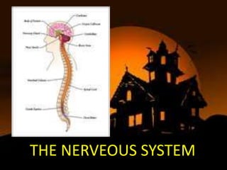
- 3. THE BRAIN
- 4. 1.Constitutes about one-fifth of body weight 2.Lies within the cranial cavity. 3.Parts are: -cereberum -midbrain -pons -medulla oblongata -cerebellum
- 5. CEREBRUM 1.Occupies the anterior and middle cranial fossae. 2.Divided by a deep cleft,the longitudinal cerebral fissure into right and left cerebral hemisphere,each containing one of the lateral ventricles. 3.The hemispheres connected by a mass of white matter called corpus callosum. 4.Divided into lobes which take the names of the bones of the cranium: -frontal -parietal -temporal -occipital
- 6. INFERIOR OF THE CEREBRUM 1.Surface of the cerebral cortex composed of gray matter. 2.the lobes connected by masses of nerve fibres or tract. Association(arcuate)-connect different parts of a cerebral hemisphere by extending from one gyrus to another. Commissural tract-connect corresponding areas of the two cerebral hemisphere. Projection tract-connect the cerebral cortex with grey matter of lower part of the brain and with the spinal cord.
- 7. Function of the cerebral cortex 1.Mental activities ex:memory,thinking,intelligence,moral sense,learning,reasoning 2.Sensory perception Ex:the perception of the pain,temperature,touch,hearing,taste,smell, sight 3.Initiation and control of skeletal(voluntary) muscle contraction.
- 8. Functional areas of cerebral Functional areas of cerebral cortex cortex 1.Somatic sensory area – receives impulses from the body’s sensory receptors 2.Primary motor area – sends impulses to skeletal muscles 3.Association-intergration nad processing of complex mental function
- 9. Motor area of the of the cerebral cortex a)Primary motor area-control voluntary contraction of specific muscle on the opposite side of the body.e.g:finger maneuver (b)broca’s (motor speech) area-involve in the translation of thoughts into speech Sensory areas of the cerebral cortex (a)Somatosensory area-sensation of pain,temperature,pressure and touch,awareness of muscular movement and the position of joints are perceived. (b)Auditory area-nerve cells receive and interpet impulses transmitted from inner ear by the cochlear.
- 10. c)Olfactory area-impulses from the nose,transmitted via the olfactory nerves. (d)Taste area-impulses from sensory receptors in taste buds are received and perceived as taste (e)Visual area-optic nerve pass from the eye to this area,which receive and interprets the impulses as visual impression.
- 12. Association area (a)Premotor area-neurones coordinate movement initiated by the primary motor cortex. (b)Prefrontal area-intellectual function controlled such as perception and comprehension of passage of time. (c)wernicke’s (sensory speech)area-the spoken word is perceived and comprehension and intelligence are based. (d)parieto-occipitotemporal area- spatial awareness,interpreting written language and ability to name object.
- 13. Others areas of the cerebrum BASAL GANGLIA 1.Lying deep within the cerebral hemisphere. 2.Involved in initiation and fine control of complex movement and learned coordinated activities THALAMUS 1.Consist of two masses of nerve sense and fibres situated within cerebral hemisphere. 2.Sensory input from the skin,viscera and special sense organ is relayed to thalamus
- 14. HYPOTHALAMUS 1.Situated below and in front of the thalamus,above the pituitary gland. 2.Funtion are: -the autonomic nervous system -appetite and satiety -thirst and water balance -body temperature -emotional reaction -sexual behaviour -biological clocks
- 15. BRAIN STEM MIDBRAIN 1.Situated around the cerebral aqueduct between the cerebrum above and the pons below. 2.Consist of nucleic and nerve fibres(tract). 3.Nucleic act as relay station for the ascending and descending nerve fibres.
- 16. PONS 1.Situated in front of the cerebellum,below the midbrain and above the medulla oblongata. 2.Nucleic within the pons that act as relay station and associated with the cranial nerves. MEDULLA OBLONGATA 1.Extend from pons above and is continous with spinal cord below. 2.Has several special features: -decussation(crossing) of the pyramid.motor nervedescending from the motor area in the cerebrum to the spinal cord in the pyramidal tracts.
- 17. -sensory decussation.sensory nerve ascending to the cerebrum from the spinal cord cross from one side to other in the medulla. -the cardiovascular centre(CVC).control the rate and force cardiac contraction and blood pressure. -the respiratory centre.control the rate depth of respiration -reflex centres.irritants present in the stomach or respiratory tract stimulate the medulla oblongata,activating the reflex centres.
- 18. RETICULAR FORMATION 1.Is a collection of neurones in the core of the brain stem,surrounded by neural pathway. 2.function: -coordination of skeletal muscle activity associated with voluntary motor movement and the maintenance of balance. -coordination of activity controlled by the autonomic nervous system. -selective awareness that function through the reticular activating system(RAS).
- 19. CEREBELLUM 1.Situated behind the pons and immediately below the posterior portion of the cerebrum occupying the posterior cranial fossae. 2.function: -coordination of voluntary muscular movement,posture and balance . -role in learning and language processing.
- 20. SPINAL CORD
- 21. 1.elongated,almost cylindrical part of the CNS. 2.Continuos above with the medulla oblongata and extend from the upper border of the atlas to the lower border of the 1st lumbar. 3.Is the nervous tissue link between the brain and the rest of the body. 4.Incompletely divided into two equal parts,anteriorly by a short,shallow median fissure and posteriorly by a deep narrow septum.
- 22. GREY MATTER 1.Have 2 posterior,2 anterior and 2 lateral columns. 2.Is the transverse commisure and it is pierced by the central canal an extension from the fourth ventricle. 3.The nerve cell bodies may be: -sensory neurones,receive impulses from the periphery of the body -lower motor neurones,transmit impulses to the skeletal muscle. -connector neurones,form spinal reflex arcs.
- 23. POSTERIOR COLUMNS OF GREY MATTER 1.Composed of cell bodies that stimulate by sensory impulses from the periphery. 2.Contribute to the formation of white matter of the cord and transmit the sensory impulses upwards to the brain. ANTERIOR COLUMNS OF GREY MATTER 1.Composedof cell bodies of the lower motor neurones that stimulate the upper motor neurnes.
- 24. WHITE MATTER 1.Arranged in 3 column:anterior,posterior and lateral. 2.function: -ascending tract:sensory toward brain -descending tract:motor from brain
- 26. 1.Consist of: -31 pairs of spinal nerves -12 pairs of cranial nerves -the autonomic nervous system 2.Each nerve consist of numerous nerve fibres collected into bundle and 3.Has covering of protective connective tissue: ENDONEURIUM-delicate tissue,surrounding each individual fibre. PERINEURIUM-smooth connective tissue,surrounding each bundle of fibres. EPINEURIUM-fibrous tissue which surround and encloses a number of bundles of nerve fibres
- 27. 1. 31 pairs of spinal nerves that leave the vertebral canal. -8 cervical -12 thoracic -5 lumbar -5 sacral -1 coccygeal 2. Nerve is formed by the union: Mixed nerves – both sensory and motor fibers Afferent (sensory) nerves – carry impulses toward the CNS Efferent (motor) nerves – carry impulses away from the CNS 3.The Anterior Nerve root -consists of motor nerve fibres. The Posterior nerve root: -consists of sensory nerve fibres
- 30. CERVICAL PLEXUS 1.Cervical plexus (C1-C4) innervates the muscles and skin of the neck and shoulder. 2.The superficial branches supply the structures at the back and side of the head and skin of the front of neck. 3.The deep branches supply muscle of the neck.e.g:the sternoleidomastoid
- 31. BRACHIAL PLEXUS Main nerves (be able to label): •Musculocutaneous C8,T1 –passes downwards to the lateral aspect of the forearm. •Median C5,6,7,8,T1 – passes down the midline of arm in close association with the brachial artery. •Ulnar C7,8,T1 – passes behind the medial epicondyle of humerus. •Axillary C5,6– winds round the humerus at the level of the surgical neck. •Radial C5,6,7,8,T1– to posterior part of limb 31
- 32. LUMBAR PLEXUS 1.Formed by anterior rami of the L1-L4. 2.Lies within the psoas major muscle. 3.Main branches: -iliohypogastric nerve L1 -ilioinguinal nerve L1 -genitofemoral L1,2 -lateral cutaneous nerve of thigh L2,3 (supplies the skin of the lateral aspect o thigh) -femoral nerve L2,3,4 (passes behind the inguinal ligament to enter thigh) -obturator nerve L2,3,4 (supplies the adductor muscle of thigh and skin of the medial aspect of thigh) -lumbosacral trunk L4,5 (trunk dsecends into the pelvis)
- 34. SACRAL PLEXUS 1. formed L4-S4 2.Supplies muscles and skin of posterior thigh and almost all of the leg 3.Main branch is the large sciatic nerve, which consists of: Tibial nerve – to most of hamstrings, calf and sole Common fibular nerve – to muscles of anterior and lateral leg and skin 4.Other branches supply pelvic girdle (gluteus muscles) and perineum (pudental nerve)
- 35. COCCYGEAL PLEXUS 1.Formed by part of the 4th and 5th sacral and the coccygeal nerves. 2.Supply the skin around the coccyx and anal area
- 36. 1.Do not intermingle to form plexus. 2.There are 12 pairs and 11 are the intercoastal nerves and 12th pair comprise the subcostal nerves. 3.The 7th to 12th nerves supply the muscle and skin of the posterior and anterior abdominal wall
- 37. 1.There are 12 pairs of cranial nerves originating from nucleic in the inferior surface of the brain,some sensory,some motor and some mixed. 2.Names and numbers: I. olfactory:sensory II. Optic:sensory III. Oculomotor:motor IV. Trochlear:motor V. Trigeminal:mixed VI. Abducent:motor VII.Facial:mixed VIII.Vestibulocochlear(auditory):sensory IX. Glossopharyngeal:mixed X. Vagus:mixed XI. Accessory:motor XII.Hypoglossal:motor
- 38. CN Name Attached Foramen Function # to I Olfactory Forebrain Cribriform Sense of smell plate II Optic Forebrain Optic canal Sense of vision (sight) from retina III Oculomotor Midbrain Superior Motor to 4 of the 6 (brainste orbital muscles of eye m) fissure movement (up & in); eyelid; constriction of pupil IV Trochlear Midbrain Superior Motor to superior (brainste orbital oblique muscle of eye m) fissure (down & out) V Trigeminal Pons V1: superior All three divisions: facial V1 (brainste orbital sensation ophthalmic m) fissure V2 maxillary V2: foramen V3 rotundum V3 (mandibular mandibular V3: foramen division): chewing also ovale
- 39. VI Abducens Pons Superior orbital Motor to lateral rectus muscle (brainstem) fissure of eye (abducts outwards) VII Facial Pons Internal Facial expression (motor) (brainstem) auditory canal Taste anterior 2/3 tongue Salivary & lacrimal glands (saliva and tears) VIII Vestibulocochlear Pons Internal Equilibrium (vestibular) (brainstem) auditory Hearing (cochlear) canal IX Glossopharyngeal Medulla Jugular Taste & touch from posterior (brainstem) foramen 1/3 tongue (sour, bitter); pharynx (throat) muscles of swallowing; parotid gland (saliva); senses carotid BP X Vagus Medulla Jugular Senses aortic BP, slows heart (brainstem) foramen rate, stimulates digestive organs; larynx (vocal cords), taste, swallowing XI Accessory Medulla Jugular Sternocleidomastoid, trapezius, (brainstem) foramen swallowing; part joins Vagus XII Hypoglossal Medulla Hypoglossal Innervation of tongue muscles (brainstem) canal
- 40. TH@NK YOU