SlideShare una empresa de Scribd logo
1 de 66
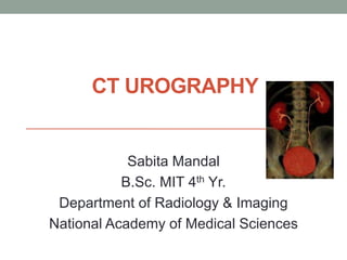
CT UROGRAPHY
Sabita Mandal
B.Sc. MIT 4th Yr.
Department of Radiology & Imaging
National Academy of Medical Sciences
CONTENTS
• Anatomy
• Introduction
• Procedure (Indications, Contraindications,
Preparation, Protocols, Aftercare)
• Dose reduction techniques
• Summary
• References
Anatomy
• Urinary system consists of
1. Kidneys (2)
2. Ureter (2)
3. Urinary bladder (1)
4. Urethra (1)
• Function: filters blood and
create urine as a waste by-
product.
Kidneys
• Bean shaped
retroperitoneal structure
• Located: T12-L3
• Right kidney is 2 cm lower
than left kidney
• Long axis of kidney is
directed downwards,
outward and parallel to long
axis of psoas muscle.
InternalAnatomy of kidney
Radiological Anatomy of Urinary System
Ureter
• Ureter are narrow tubes that carry
urine from the kidneys to the
bladder.
• The ureters are constricted at 5
places
Ureteropelvic junction
At the crossing of iliac artery
At juxta position of vas deferens or
broad ligament
As the ureter pass through the wall
of the bladder
At ureteric orifice
Urinary Bladder
• Triangle-shaped, hollow muscular
organ
• Lies in pelvic cavity as a reservoir
of urine.
• Its size and position vary,
depending on the amount of urine
it contains.
• Bladder capacity: 300-600 ml
• First urge to void is felt at a
bladder volume of 150ml
Vascular supply of Urinary System
• Kidneys have an
extensive blood
supply via the renal
arteries which leave
the kidneys via the
renal vein.
How contrast media reaches to Kidney ?
Median antecubital vein- Internal jugular vein
– SVC – RA- RV- PA – LA – LV – Arch of aorta
– Thoracic aorta – Abdominal Aorta – Renal
Arteries – micro vasculature of kidney – Renal
Vein – Iliac vein - IVC
Imaging Modalities
• Conventional radiography (KUB)
• Special radiography (IVU-Intravenous Urography)
• Ultrasound
• CT (KUB and Urography)
• Magnetic Resonance Urography
• Nuclear medicine ( DTPA , DMSA renal scan)
• PET CT
History
KUB (Kidney, Ureter, Bladder)
• KUB is typically used to investigate
gastrointestinal conditions such as a
bowel obstruction and gallstones, and
can detect the presence of
kidney stones.
• Despite its name, a KUB is not
typically used to investigate pathology
of the kidneys, ureters, or bladder,
since these structures are difficult to
assess (the kidneys may not be visible
due to overlying bowel gas.)
History
Intravenous urography (IVU) was the
gold standard in the imaging evaluation of
ureteral stones and urinary tract obstruction.
Advantage
• estimation of physiologic function
• estimation of degree of obstruction
• detection of anatomical abnormalities of
the urinary tract.
Disadvantages
• Difficulty in radiolucent calculi
identification;
• contrast-related adverse reactions.
• a long and tedious study time to find the
exact size and location of the calculi.
History
CT KUB/NECT KUB is an excellent cross sectional imaging
modality of choice for the diagnosis of urinary tract.
IVU has largely been replaced by NCCT, due to its high sensitivity
and specificity and the ease of performing the study.
ADVANTAGES
• higher detection rate for the number of calculi and related
obstructions.
• rapid image acquisition time & advanced image quality.
• no requirement for contrast material eliminating risk of
nephrotoxicity of the contrast material.
CT Urography (CTU)
• Although all imaging modalities play an important role in
imaging the urinary tract, CT urography represents the most
comprehensive imaging examination of the urinary tract.
• The ESUR defines CT urography (CTU) as a diagnostic
examination optimized for imaging the kidneys, ureters and
bladder with thin-slice Multidetector CT, administration of
intravenous contrast medium, acquisition of images in the
excretory phase with multiplanar imaging of the urinary
system.
• CT urography (CTU) has become the primary diagnostic
modality used in the evaluation of patients with hematuria.
• However it remains limited in evaluation of urothelium as
compared with IVU because of its lower spatial resolution.
CT Urography (CTU)
• CTU is a multiphase CT examination in which the clinical
presentation will largely determine the protocol used.
• Because of the multiphasic and functional nature of CTU,
the radiation dose can reach high levels, so suggestions for
optimized use of CTU with reduced dose have been
published by scientific societies.
Indications
• signs of obstruction including hydronephrosis, ipsilateral
renal enlargement, PUJ, VUJ.
• First-line technique in the evaluation of hematuria.
• To evaluate patients with Calculi, Renal masses, urothelial
tumors.
• Carcinoma (RCC, TCC).
• Congenital anomalies like retrocaval ureter, Ureteral
duplication, Crossed fused kidneys, Ectopic kidneys.
Contraindications
• A positive pregnancy test
• Hypersensitivity to iodinated contrast material
• Impaired/ insufficient renal function
• The risk versus benefits
Patient Preparation
• NPO for 4-6 hours
• Proper hydration of patient & full bladder
• RFT ,USG
• Consent
• H/o Regarding recent exams
• Check for allergy/ hypersensitivity to Iodine.
• Remove metals and give patient a hospital gown.
• Open Vein cannulation
Patient Positioning and parameters
• Supine with feet first
• Hands above the head
• Laser light positioned to center
abdomen in the Iso-center of
gantry/
• Topogram position/ landmark:
AP, from 2” above the
xiphisternum to 2” below the
symphysis pubis.
• Scan orientation: craniocaudal
Parameters
• FOV: variable
• Slice thickness: 3-5 mm
• Slice interval : 1.5-2.5 mm
• Recon algorithm/ Kernel: Smooth medium
• 3D recon: MPR
ContrastAdministration
Two different approaches;
1. Single-bolus injection
2. Split-bolus injection .
• A single- bolus injection administers 100 to 150 mL of
LOCM 300-370 mg/ml
• At rate of 2 to 3 mL/sec
• Injection site –Preferably anticubital vein with 18-20 G
cannulation
Approaches for CT urography
1. Hybrid CTU
2. Only CT Urography
Hybrid CT urography
CT and IVU are done together.
Disadv:- Imaging at different location
CTU phases
• Unenhanced phase
• Corticomedullary phase
• Nephrographic phase
• Excretory phase
CT urography protocols are being refined, and
efforts are being focused on optimization of radiation
exposure and urothelial imaging
Unenhanced phase
Scan of entire abdomen region to
• Detect renal parenchymal calcifications
• radiopaque urinary tract calculi
• to help in characterization of renal lesions (by providing
baseline unenhanced attenuation values , presence of
fat/calcium)
CECT Phases
Phase Timing
Corticomedullary 30-70 sec
Nephrogram 80-120 sec
Excretory 3-15 mins or longer
CECT Phases are required for renal mass characterization,
visualization of ureteric obstructions and strictures, bladder
wall abnormalities and infections and inflammations.
Cortico Medullary Phase
• Begins as contrast material enters the cortical capillaries and
peritubular spaces and filters into the proximal cortical tubules.
• During this stage, Renal cortex can be differentiated from renal
medulla at this stage because
(1) the vascularity of the cortex is greater than that of the medulla
(the renal veins and other abdominal visceral parenchyma are
better evaluated)
(2) contrast material has not yet reached the distal aspect of the
renal tubules
Cortico Medullary Phase
• The acquisition of Corticomedullary phase images is not
routine, since it is well established that renal masses are
more visible in the Nephrographic phase compared to the
Corticomedullary phase.
• Szolar et al. showed the Nephrographic phase is superior
to Corticomedullary phase in depiction of small renal
masses, due to statistically significant larger attenuation
difference between lesion and renal medulla in
Nephrographic phase compared to Corticomedullary phase.
• Apart from potentially missing lesions, another major
pitfall of the Corticomedullary phase is the detection of
pseudo lesions (false positives), due to inhomogeneous
enhancement of renal medulla or the appearance of normal
renal medulla during this phase
Nephrographic Phase
• Begins as contrast material proceeds from the cortical vessels
and extracellular–interstitial space and enters the loops of Henle
and collecting tubules.
• The Nephrographic phase 80 to 120 sec after the start of
injection, and differentiate between the normal renal medulla
and a renal mass.
Nephrographic phase
• It has the highest sensitivity in the detection of renal
masses, and correlation with unenhanced images is
required to show unequivocal enhancement.
Pyelographic phase/Excretory Phase
• 3–15 minutes after contrast administration
• useful for evaluating urothelial lesions from kidneys to the
bladder.
• The contrast excretes into the collecting system decreasing the
attenuation of the nephrogram
• Longer delays are beneficial for opacification of the distal
ureters.
1. Unenhanced phase
2. Corticomedullary phase
3. Excretory Phase
PatientAftercare
• Needle wound site dressed and checked for extravasation.
• Check patient understands how to receive the results.
• Ensure patient understands any preparation instructions are
finished
• Escort to changing rooms.
Image improvement techniques
• use of abdominal compression bands.
• Saline Infusion (approx. 250 mL of 0.9%)
• Diuretic Administration( low-dose furosemide10 mg 2–3
minutes Prior CM inj. injection allows less dense,
homogeneous opacification of the collecting system )
• Alternatively, oral water (1000 mL within 15 to 20 minutes)
before the examination can cause sufficient opacification of
the calyces and ureters in most instances.
• Patient Positioning in prone to better visualize lower ureter.
• Preprocedure bowel preparation.
• Ten-minute decompressed/ release images help to visualize
almost the entire ureters.
• Twenty-minute and post voiding images are useful for bladder
evaluation.
Radiation dose
• The biggest drawback with CTU is the exposure to a
larger amount of radiation dose, which accompanies
multiple CT acquisitions.
• Several techniques have been developed to reduce the
overall radiation dose that the patient receives.
• In the early days of CTU, articles reported that a three-
phase CTU protocol was used, with effective dose levels
of 25–35 mSv compared to excretion urography with a
mean effective dose of 3.6 mSv .
• A recent CT dose survey performed in the Netherlands
showed that, for (split-bolus two-phase) CTU,
comparable median effective doses would be 3.6 mSv
for the unenhanced phase and 6.6 mSv for the concurrent
Nephrographic and excretory phases.
Dose reduction technique
1 a. For the unenhanced phase, low-dose protocols have
been shown to be comparable to standard-dose acquisitions
since the large difference in attenuation between calculi
and soft tissue allows good contrast despite increased
Image noise that comes with dose reduction.
Dose reduction technique
b. Combined low- and normal-dose CT urography.
• Because diagnosis with CT urography is reached by combining the
information from the various phases, all three scans obtained with
this technique do not have to have optimal image quality. It is
sufficient that the image quality be excellent in one of the three.
• A reduced dose, resulting in more image noise, can be accepted in
the other phases, such as the unenhanced and excretory phases, if
these images are systematically reviewed together with normal-
dose Corticomedullary or Nephrographic phase images.
• A study showed that the Effective dose was reduced by more than
65 % using the 100-kV protocol and by more than 76 % with
introduction of 80-kV protocol for unenhanced and excretory
phases .
Dose reduction technique
2. The use of dual-energy CT permits virtual non-contrast
(VNC) images to be post-processed from a single-contrast
enhanced CT acquisition, which potentially removes the
need for a True Non-contrast(TNC) CT acquisition.
• Another clinical application of dual-energy CT is in the
characterization of renal calculi into five categories
(namely Calcium oxalate, struvite , uric acid, calcium
phosphate, cystine )
Dual-Energy CT
• Dual-energy CT essentially involves the acquisition of
images at two separate X-ray photon energy spectra
absorptions at low and high peak kV levels .
• The primary advantage of dual-energy CT is its ability to
differentiate materials based on the differences of their
attenuation at different photon energy spectra.
Dual-Energy CT
• During post-processing of post contrast dual-energy CT
images of the genitourinary system, detection and
subtraction of iodine (producing ‘iodine overlay’ and VNC
images respectively) create material specific images.
• The biggest drawback of virtual unenhanced imaging is
that small calculi might be missed. Even small calculi can
be important in patients with hematuria.
Dose reduction technique
3. A split-bolus technique is another method employed to reduce
radiation doses.
• Also called Two phase technique
• The objective is to acquire images in a combined
nephropyelographic phase.
• unenhanced acquisition is followed by IV administration of 30-50 ml
of contrast material and a second bolus of 80-100 ml of IV contrast
material is given after 8-10 min delay during which the acquisition is
made. Thus, in a single phase acquisition the renal
parenchyma(nephrographic phase) and the collecting system, ureter
and bladder (Pyelographic phase) are assessed in reduced no. of
phases at reduced radiation dose.
Split bolus technique
• The mean effective radiation dose of single and split bolus
protocol were found to be 19.17mSv and 13.64mSv
respectively. The mean effective dose for single bolus MDCT
urography is 40.5% more than the split bolus MDCT urography.
(Journal of Institute of Medicine, April, 2017, 39:1)
• Disadvantage: the possibility of missing subtle urothelial
lesions which are not obvious on unenhanced scan (iso-
attenuating) and obscured by contrast on the combined
nephropyelographic phase.
Dose reduction technique
4. There is potential in combining both VNC and split-bolus
techniques with resulting marked reduction in radiation doses,
and preliminary studies show promising results.
5. Iterative reconstruction techniques has already become
mainstream in body CT examinations. The current clinical
implementations of iterative reconstruction can improve image
quality and allow dose reductions of 40–50% in general
abdominal CT examinations, whereas for a high-contrast
examination such as CTU, this reduction may even be higher
resulting the effective dose of 6.1 mSv for a complete three-
phase CTU examination.
RADIATION DOSE IN CT UROGRAPHY
S.N. PROCEDURE DOSE (mSv)
1. KUB 0.5-0.9
2. IVU 3.6
3. CT Urography (Three phase) 25-35
4. Split-bolus two-phase 10.2
5. DECT Urography
6. 80-kV protocol
100-kV protocol
76%
65 %
7. Iterative reconstruction (AIDR 3D) 6.1 (4.6 – 8.2)
8. FBP 13.1 (9.2 – 13.5)
9. Stone protocol CT 3.04
10. Digital tomosynthesis 0.83
Advanced Imaging Visualization Technique
• The use of post-processing software allows more
powerful and complete interpretation of CT images.
Multiplanar reconstruction (MPR) images can be
dynamically manipulated and assessed in real-time
• Dynamic MPR evaluation greatly enhances visualisation
of anatomical structures and lesions since it allows the
images to be viewed in any slice thickness and literally
any plane, not limited to the conventional axial, coronal
and sagittal sections. This is useful not only in evaluating
the genitourinary system but also other complex bodily
systems.
Advanced Imaging Visualization Technique
• Volume rendering technique (VRT)
is another post-processing
technique which allows the
construction of a three-dimensional
image of the urinary collecting
system in the excretory phase,
which also resembles an IVU
image.
• Evaluation of axial CT images
(source images) with a wide
window setting is important for
accurate diagnosis. Coronal or
oblique multiplanar reconstruction
images help to define the location
and extent of the lesions shown on
axial images .
Image showing duplex collecting system
Ectopic pelvic right kidney.
Horseshoe kidney with malrotated dilated
right renal pelvis
HORSESHOE KIDNEY
PUJ Stenosis
Coronal (A) and axial (B) non-contrast computed
tomography shows uniformly small left kidney
Coronal (Aand B) and axial (C and D) contrast
enhanced CT shows absent left kidney (arrowhead)
Computed tomography (CT) urogram showing
papillary blush with calculi within dilated collecting
tubules (arrows)
Advantages of IVU over CT urography
• Better spatial resolution
• Lesser radiation dose
• Cheaper
Ct Urography:Advantages
• Multidetector CT : Thin slices in single breath hold allow
optimal anatomic information
• Isotropic MPR reconstruction.
• Better contrast resolution.
Summary
• Multidetector CT is currently the imaging modality of choice in the
evaluation of urinary tract disease and has largely supplanted IVU.
• CT urography is probably the most complete imaging investigation of the
urinary tract in the present day. Higher radiation dose, however, as the
major disadvantage of CT, is a problem amplified by the multiphasic nature
of CT urography.
• Development of split-bolus techniques to combine scan phases within
single acquisitions has resulted in significant reductions in radiation doses.
• In recent years, the emergence of dual-energy CT has made feasible many
useful applications which complement CT urography and further lend
support to the reduction of radiation doses.
• Associated advanced image visualization tools not only support more
comprehensive interpretation of images, but also serve as a means to
display images in a fashion suitable for clinicians to convey findings to
patients.
References
• CT for Technologist, 1st edition by lois E. Romans
• How Much Dose Can Be Saved in Three-Phase CTUrography?
://www.ajronline.org/doi/10.2214/AJR.11.7209
• https://slideplayer.com/slide/12963342/
• A comparison of radiation dose in single and split bolus
multidetector computed tomography urography, Joshi BR , Jha
A
• http://www.indianjurol.com/article.asp?issn=0970-
1591;year=2014;volume=30;issue=1;spage=55;epage=59;aulast
=Cabrera
References fromArticles
Tze-Anns’Low K, Teh HS. CT Urography: An Update in
Imaging Technique. Current Radiology Reports. 2015 Aug
1;3(8):31.
van der Molen AJ, Miclea RL, Geleijns J, Joemai RM. A
survey of radiation doses in ct urography before and after
implementation of iterative reconstruction. American Journal
of Roentgenology. 2015 Sep;205(3):572-7.
Jinzaki M, Matsumoto K, Kikuchi E, Sato K, Horiguchi Y,
Nishiwaki Y, Silverman SG. Comparison of CT urography
and excretory urography in the detection and localization of
urothelial carcinoma of the upper urinary tract. American
Journal of Roentgenology. 2011 May;196(5):1102-9.
THANK YOU

Más contenido relacionado

La actualidad más candente

Ct mri urography
Ct mri urographyCt mri urography
Ct mri urographyDev Lakhera
 
Radiological anatomy of kidney, ureter & bladder
Radiological anatomy of kidney, ureter & bladderRadiological anatomy of kidney, ureter & bladder
Radiological anatomy of kidney, ureter & bladderrajss007
 
MCU- Micturating cysto-urethrogram
MCU- Micturating cysto-urethrogramMCU- Micturating cysto-urethrogram
MCU- Micturating cysto-urethrogramDr. Mohit Goel
 
Procedure of ercp and t tube cholangiography
Procedure of ercp and t tube cholangiographyProcedure of ercp and t tube cholangiography
Procedure of ercp and t tube cholangiographyYashawant Yadav
 
Renal scintigraphy
Renal scintigraphyRenal scintigraphy
Renal scintigraphyairwave12
 
Mrcp Radiology
Mrcp RadiologyMrcp Radiology
Mrcp Radiologykunalj000
 
INTRAVENOUS UROGRAM (IVU)
INTRAVENOUS UROGRAM (IVU)INTRAVENOUS UROGRAM (IVU)
INTRAVENOUS UROGRAM (IVU)Jai Kumar
 
Magnetic resonance cholangiopancreatography ppt
Magnetic resonance cholangiopancreatography pptMagnetic resonance cholangiopancreatography ppt
Magnetic resonance cholangiopancreatography pptAnjan Dangal
 
PTBD Percutaneous transhepatic biliary drainage
PTBD Percutaneous transhepatic biliary drainagePTBD Percutaneous transhepatic biliary drainage
PTBD Percutaneous transhepatic biliary drainageShiva Prakash
 
Mrcp radiology
Mrcp radiologyMrcp radiology
Mrcp radiologyVidya TK
 
OPPOSING URETHROGRAM (OUG)
OPPOSING URETHROGRAM (OUG)OPPOSING URETHROGRAM (OUG)
OPPOSING URETHROGRAM (OUG)Jai Kumar
 
T-tube Cholangiogram
T-tube CholangiogramT-tube Cholangiogram
T-tube Cholangiogramricksw78
 

La actualidad más candente (20)

Ct mri urography
Ct mri urographyCt mri urography
Ct mri urography
 
Intravenous urography (IVU)
Intravenous urography (IVU)Intravenous urography (IVU)
Intravenous urography (IVU)
 
Loopogram
LoopogramLoopogram
Loopogram
 
CT Angiography Lower Limb
CT Angiography Lower LimbCT Angiography Lower Limb
CT Angiography Lower Limb
 
Radiological anatomy of kidney, ureter & bladder
Radiological anatomy of kidney, ureter & bladderRadiological anatomy of kidney, ureter & bladder
Radiological anatomy of kidney, ureter & bladder
 
MCU- Micturating cysto-urethrogram
MCU- Micturating cysto-urethrogramMCU- Micturating cysto-urethrogram
MCU- Micturating cysto-urethrogram
 
Procedure of ercp and t tube cholangiography
Procedure of ercp and t tube cholangiographyProcedure of ercp and t tube cholangiography
Procedure of ercp and t tube cholangiography
 
Renal scintigraphy
Renal scintigraphyRenal scintigraphy
Renal scintigraphy
 
Ct head protocols
Ct head protocolsCt head protocols
Ct head protocols
 
Mrcp Radiology
Mrcp RadiologyMrcp Radiology
Mrcp Radiology
 
INTRAVENOUS UROGRAM (IVU)
INTRAVENOUS UROGRAM (IVU)INTRAVENOUS UROGRAM (IVU)
INTRAVENOUS UROGRAM (IVU)
 
Magnetic resonance cholangiopancreatography ppt
Magnetic resonance cholangiopancreatography pptMagnetic resonance cholangiopancreatography ppt
Magnetic resonance cholangiopancreatography ppt
 
T Tube
T TubeT Tube
T Tube
 
Intravenous Urography
Intravenous UrographyIntravenous Urography
Intravenous Urography
 
Ct urography/ivp
Ct urography/ivpCt urography/ivp
Ct urography/ivp
 
Mcu rgu ppt
Mcu rgu pptMcu rgu ppt
Mcu rgu ppt
 
PTBD Percutaneous transhepatic biliary drainage
PTBD Percutaneous transhepatic biliary drainagePTBD Percutaneous transhepatic biliary drainage
PTBD Percutaneous transhepatic biliary drainage
 
Mrcp radiology
Mrcp radiologyMrcp radiology
Mrcp radiology
 
OPPOSING URETHROGRAM (OUG)
OPPOSING URETHROGRAM (OUG)OPPOSING URETHROGRAM (OUG)
OPPOSING URETHROGRAM (OUG)
 
T-tube Cholangiogram
T-tube CholangiogramT-tube Cholangiogram
T-tube Cholangiogram
 

Similar a CT urography

Conventional Radiology In Urology.pptx
Conventional Radiology In Urology.pptxConventional Radiology In Urology.pptx
Conventional Radiology In Urology.pptxrabi pandit
 
Radiographic procedure PTC & PTB..D.pptx
Radiographic procedure PTC & PTB..D.pptxRadiographic procedure PTC & PTB..D.pptx
Radiographic procedure PTC & PTB..D.pptxjustinfan550
 
Investigation of Biliary Tract
Investigation of Biliary Tract Investigation of Biliary Tract
Investigation of Biliary Tract Sujan Poudel
 
Section 1 CT Abdomen and Pelvis by doctor Aya Ali Taha
Section 1 CT Abdomen and Pelvis by doctor Aya Ali TahaSection 1 CT Abdomen and Pelvis by doctor Aya Ali Taha
Section 1 CT Abdomen and Pelvis by doctor Aya Ali TahaDr Aya Ali
 
Intravenous Pyelogram RENAL TESTS ADULTS.pptx
Intravenous Pyelogram RENAL TESTS ADULTS.pptxIntravenous Pyelogram RENAL TESTS ADULTS.pptx
Intravenous Pyelogram RENAL TESTS ADULTS.pptxneeti70
 
medicine.Kidney 2.(dr.ala)
medicine.Kidney 2.(dr.ala)medicine.Kidney 2.(dr.ala)
medicine.Kidney 2.(dr.ala)student
 
URINARY SYSTEM - Copy.pptx
URINARY SYSTEM - Copy.pptxURINARY SYSTEM - Copy.pptx
URINARY SYSTEM - Copy.pptxGokulnathMbbs
 
INTRAVENOUS UROGRAPHY INDICATIONS CONTRAINDICATIONS
INTRAVENOUS UROGRAPHY INDICATIONS CONTRAINDICATIONSINTRAVENOUS UROGRAPHY INDICATIONS CONTRAINDICATIONS
INTRAVENOUS UROGRAPHY INDICATIONS CONTRAINDICATIONSobscmch
 
Retrograde pyeloureterography
Retrograde pyeloureterographyRetrograde pyeloureterography
Retrograde pyeloureterographydypradio
 
ctenteroclysis.pptx
ctenteroclysis.pptxctenteroclysis.pptx
ctenteroclysis.pptxIslah Raoof
 
ENDOSCOPIC RETROGRADE CHOLANGIOPACREATOGRAPHY (ERCP).pptx
ENDOSCOPIC RETROGRADE CHOLANGIOPACREATOGRAPHY (ERCP).pptxENDOSCOPIC RETROGRADE CHOLANGIOPACREATOGRAPHY (ERCP).pptx
ENDOSCOPIC RETROGRADE CHOLANGIOPACREATOGRAPHY (ERCP).pptxvigneshsk95
 
Radiological investigation of billiary tact 01
Radiological investigation of billiary tact 01Radiological investigation of billiary tact 01
Radiological investigation of billiary tact 01Kajal Jha
 

Similar a CT urography (20)

Conventional Radiology In Urology.pptx
Conventional Radiology In Urology.pptxConventional Radiology In Urology.pptx
Conventional Radiology In Urology.pptx
 
Radiographic procedure PTC & PTB..D.pptx
Radiographic procedure PTC & PTB..D.pptxRadiographic procedure PTC & PTB..D.pptx
Radiographic procedure PTC & PTB..D.pptx
 
Ct protocol for ivu
Ct protocol for ivuCt protocol for ivu
Ct protocol for ivu
 
Investigation of Biliary Tract
Investigation of Biliary Tract Investigation of Biliary Tract
Investigation of Biliary Tract
 
Investigation of Biliary Tract
Investigation of Biliary Tract Investigation of Biliary Tract
Investigation of Biliary Tract
 
Urological investigations
Urological investigationsUrological investigations
Urological investigations
 
Section 1 CT Abdomen and Pelvis by doctor Aya Ali Taha
Section 1 CT Abdomen and Pelvis by doctor Aya Ali TahaSection 1 CT Abdomen and Pelvis by doctor Aya Ali Taha
Section 1 CT Abdomen and Pelvis by doctor Aya Ali Taha
 
Intravenous Pyelogram RENAL TESTS ADULTS.pptx
Intravenous Pyelogram RENAL TESTS ADULTS.pptxIntravenous Pyelogram RENAL TESTS ADULTS.pptx
Intravenous Pyelogram RENAL TESTS ADULTS.pptx
 
Scans.. Dr.Padmesh
Scans.. Dr.PadmeshScans.. Dr.Padmesh
Scans.. Dr.Padmesh
 
Endoscopy in surgery
Endoscopy in surgery Endoscopy in surgery
Endoscopy in surgery
 
medicine.Kidney 2.(dr.ala)
medicine.Kidney 2.(dr.ala)medicine.Kidney 2.(dr.ala)
medicine.Kidney 2.(dr.ala)
 
Ercp
ErcpErcp
Ercp
 
URINARY SYSTEM - Copy.pptx
URINARY SYSTEM - Copy.pptxURINARY SYSTEM - Copy.pptx
URINARY SYSTEM - Copy.pptx
 
INTRAVENOUS UROGRAPHY INDICATIONS CONTRAINDICATIONS
INTRAVENOUS UROGRAPHY INDICATIONS CONTRAINDICATIONSINTRAVENOUS UROGRAPHY INDICATIONS CONTRAINDICATIONS
INTRAVENOUS UROGRAPHY INDICATIONS CONTRAINDICATIONS
 
Retrograde pyeloureterography
Retrograde pyeloureterographyRetrograde pyeloureterography
Retrograde pyeloureterography
 
Imaging in urology
Imaging in urologyImaging in urology
Imaging in urology
 
ctenteroclysis.pptx
ctenteroclysis.pptxctenteroclysis.pptx
ctenteroclysis.pptx
 
ENDOSCOPIC RETROGRADE CHOLANGIOPACREATOGRAPHY (ERCP).pptx
ENDOSCOPIC RETROGRADE CHOLANGIOPACREATOGRAPHY (ERCP).pptxENDOSCOPIC RETROGRADE CHOLANGIOPACREATOGRAPHY (ERCP).pptx
ENDOSCOPIC RETROGRADE CHOLANGIOPACREATOGRAPHY (ERCP).pptx
 
LAPAROSCOPIC UROLOGY PPT. DR SREEJOY PATNAIK
LAPAROSCOPIC UROLOGY PPT. DR SREEJOY PATNAIKLAPAROSCOPIC UROLOGY PPT. DR SREEJOY PATNAIK
LAPAROSCOPIC UROLOGY PPT. DR SREEJOY PATNAIK
 
Radiological investigation of billiary tact 01
Radiological investigation of billiary tact 01Radiological investigation of billiary tact 01
Radiological investigation of billiary tact 01
 

Último

ANATOMY AND PHYSIOLOGY OF RESPIRATORY SYSTEM.pptx
ANATOMY AND PHYSIOLOGY OF RESPIRATORY SYSTEM.pptxANATOMY AND PHYSIOLOGY OF RESPIRATORY SYSTEM.pptx
ANATOMY AND PHYSIOLOGY OF RESPIRATORY SYSTEM.pptxSwetaba Besh
 
Intramuscular & Intravenous Injection.pptx
Intramuscular & Intravenous Injection.pptxIntramuscular & Intravenous Injection.pptx
Intramuscular & Intravenous Injection.pptxsaranpratha12
 
❤️Call Girl Service In Chandigarh☎️9814379184☎️ Call Girl in Chandigarh☎️ Cha...
❤️Call Girl Service In Chandigarh☎️9814379184☎️ Call Girl in Chandigarh☎️ Cha...❤️Call Girl Service In Chandigarh☎️9814379184☎️ Call Girl in Chandigarh☎️ Cha...
❤️Call Girl Service In Chandigarh☎️9814379184☎️ Call Girl in Chandigarh☎️ Cha...Sheetaleventcompany
 
👉Chandigarh Call Girl Service📲Niamh 8868886958 📲Book 24hours Now📲👉Sexy Call G...
👉Chandigarh Call Girl Service📲Niamh 8868886958 📲Book 24hours Now📲👉Sexy Call G...👉Chandigarh Call Girl Service📲Niamh 8868886958 📲Book 24hours Now📲👉Sexy Call G...
👉Chandigarh Call Girl Service📲Niamh 8868886958 📲Book 24hours Now📲👉Sexy Call G...Sheetaleventcompany
 
🚺LEELA JOSHI WhatsApp Number +91-9930245274 ✔ Unsatisfied Bhabhi Call Girls T...
🚺LEELA JOSHI WhatsApp Number +91-9930245274 ✔ Unsatisfied Bhabhi Call Girls T...🚺LEELA JOSHI WhatsApp Number +91-9930245274 ✔ Unsatisfied Bhabhi Call Girls T...
🚺LEELA JOSHI WhatsApp Number +91-9930245274 ✔ Unsatisfied Bhabhi Call Girls T...soniya pandit
 
Shazia Iqbal 2024 - Bioorganic Chemistry.pdf
Shazia Iqbal 2024 - Bioorganic Chemistry.pdfShazia Iqbal 2024 - Bioorganic Chemistry.pdf
Shazia Iqbal 2024 - Bioorganic Chemistry.pdfTrustlife
 
Genuine Call Girls Hyderabad 9630942363 Book High Profile Call Girl in Hydera...
Genuine Call Girls Hyderabad 9630942363 Book High Profile Call Girl in Hydera...Genuine Call Girls Hyderabad 9630942363 Book High Profile Call Girl in Hydera...
Genuine Call Girls Hyderabad 9630942363 Book High Profile Call Girl in Hydera...GENUINE ESCORT AGENCY
 
Kolkata Call Girls Shobhabazar 💯Call Us 🔝 8005736733 🔝 💃 Top Class Call Gir...
Kolkata Call Girls Shobhabazar  💯Call Us 🔝 8005736733 🔝 💃  Top Class Call Gir...Kolkata Call Girls Shobhabazar  💯Call Us 🔝 8005736733 🔝 💃  Top Class Call Gir...
Kolkata Call Girls Shobhabazar 💯Call Us 🔝 8005736733 🔝 💃 Top Class Call Gir...Namrata Singh
 
Chandigarh Call Girls Service ❤️🍑 9809698092 👄🫦Independent Escort Service Cha...
Chandigarh Call Girls Service ❤️🍑 9809698092 👄🫦Independent Escort Service Cha...Chandigarh Call Girls Service ❤️🍑 9809698092 👄🫦Independent Escort Service Cha...
Chandigarh Call Girls Service ❤️🍑 9809698092 👄🫦Independent Escort Service Cha...Sheetaleventcompany
 
Call Girl In Indore 📞9235973566📞 Just📲 Call Inaaya Indore Call Girls Service ...
Call Girl In Indore 📞9235973566📞 Just📲 Call Inaaya Indore Call Girls Service ...Call Girl In Indore 📞9235973566📞 Just📲 Call Inaaya Indore Call Girls Service ...
Call Girl In Indore 📞9235973566📞 Just📲 Call Inaaya Indore Call Girls Service ...Sheetaleventcompany
 
Circulatory Shock, types and stages, compensatory mechanisms
Circulatory Shock, types and stages, compensatory mechanismsCirculatory Shock, types and stages, compensatory mechanisms
Circulatory Shock, types and stages, compensatory mechanismsMedicoseAcademics
 
💚Call Girls In Amritsar 💯Anvi 📲🔝8725944379🔝Amritsar Call Girl No💰Advance Cash...
💚Call Girls In Amritsar 💯Anvi 📲🔝8725944379🔝Amritsar Call Girl No💰Advance Cash...💚Call Girls In Amritsar 💯Anvi 📲🔝8725944379🔝Amritsar Call Girl No💰Advance Cash...
💚Call Girls In Amritsar 💯Anvi 📲🔝8725944379🔝Amritsar Call Girl No💰Advance Cash...Sheetaleventcompany
 
Goa Call Girl Service 📞9xx000xx09📞Just Call Divya📲 Call Girl In Goa No💰Advanc...
Goa Call Girl Service 📞9xx000xx09📞Just Call Divya📲 Call Girl In Goa No💰Advanc...Goa Call Girl Service 📞9xx000xx09📞Just Call Divya📲 Call Girl In Goa No💰Advanc...
Goa Call Girl Service 📞9xx000xx09📞Just Call Divya📲 Call Girl In Goa No💰Advanc...Sheetaleventcompany
 
💚Chandigarh Call Girls 💯Riya 📲🔝8868886958🔝Call Girls In Chandigarh No💰Advance...
💚Chandigarh Call Girls 💯Riya 📲🔝8868886958🔝Call Girls In Chandigarh No💰Advance...💚Chandigarh Call Girls 💯Riya 📲🔝8868886958🔝Call Girls In Chandigarh No💰Advance...
💚Chandigarh Call Girls 💯Riya 📲🔝8868886958🔝Call Girls In Chandigarh No💰Advance...Sheetaleventcompany
 
Race Course Road } Book Call Girls in Bangalore | Whatsapp No 6378878445 VIP ...
Race Course Road } Book Call Girls in Bangalore | Whatsapp No 6378878445 VIP ...Race Course Road } Book Call Girls in Bangalore | Whatsapp No 6378878445 VIP ...
Race Course Road } Book Call Girls in Bangalore | Whatsapp No 6378878445 VIP ...dishamehta3332
 
Jual Obat Aborsi Di Dubai UAE Wa 0838-4800-7379 Obat Penggugur Kandungan Cytotec
Jual Obat Aborsi Di Dubai UAE Wa 0838-4800-7379 Obat Penggugur Kandungan CytotecJual Obat Aborsi Di Dubai UAE Wa 0838-4800-7379 Obat Penggugur Kandungan Cytotec
Jual Obat Aborsi Di Dubai UAE Wa 0838-4800-7379 Obat Penggugur Kandungan Cytotecjualobat34
 
Electrocardiogram (ECG) physiological basis .pdf
Electrocardiogram (ECG) physiological basis .pdfElectrocardiogram (ECG) physiological basis .pdf
Electrocardiogram (ECG) physiological basis .pdfMedicoseAcademics
 
Kolkata Call Girls Naktala 💯Call Us 🔝 8005736733 🔝 💃 Top Class Call Girl Se...
Kolkata Call Girls Naktala  💯Call Us 🔝 8005736733 🔝 💃  Top Class Call Girl Se...Kolkata Call Girls Naktala  💯Call Us 🔝 8005736733 🔝 💃  Top Class Call Girl Se...
Kolkata Call Girls Naktala 💯Call Us 🔝 8005736733 🔝 💃 Top Class Call Girl Se...Namrata Singh
 
Call Girls Bangalore - 450+ Call Girl Cash Payment 💯Call Us 🔝 6378878445 🔝 💃 ...
Call Girls Bangalore - 450+ Call Girl Cash Payment 💯Call Us 🔝 6378878445 🔝 💃 ...Call Girls Bangalore - 450+ Call Girl Cash Payment 💯Call Us 🔝 6378878445 🔝 💃 ...
Call Girls Bangalore - 450+ Call Girl Cash Payment 💯Call Us 🔝 6378878445 🔝 💃 ...gragneelam30
 
tongue disease lecture Dr Assadawy legacy
tongue disease lecture Dr Assadawy legacytongue disease lecture Dr Assadawy legacy
tongue disease lecture Dr Assadawy legacyDrMohamed Assadawy
 

Último (20)

ANATOMY AND PHYSIOLOGY OF RESPIRATORY SYSTEM.pptx
ANATOMY AND PHYSIOLOGY OF RESPIRATORY SYSTEM.pptxANATOMY AND PHYSIOLOGY OF RESPIRATORY SYSTEM.pptx
ANATOMY AND PHYSIOLOGY OF RESPIRATORY SYSTEM.pptx
 
Intramuscular & Intravenous Injection.pptx
Intramuscular & Intravenous Injection.pptxIntramuscular & Intravenous Injection.pptx
Intramuscular & Intravenous Injection.pptx
 
❤️Call Girl Service In Chandigarh☎️9814379184☎️ Call Girl in Chandigarh☎️ Cha...
❤️Call Girl Service In Chandigarh☎️9814379184☎️ Call Girl in Chandigarh☎️ Cha...❤️Call Girl Service In Chandigarh☎️9814379184☎️ Call Girl in Chandigarh☎️ Cha...
❤️Call Girl Service In Chandigarh☎️9814379184☎️ Call Girl in Chandigarh☎️ Cha...
 
👉Chandigarh Call Girl Service📲Niamh 8868886958 📲Book 24hours Now📲👉Sexy Call G...
👉Chandigarh Call Girl Service📲Niamh 8868886958 📲Book 24hours Now📲👉Sexy Call G...👉Chandigarh Call Girl Service📲Niamh 8868886958 📲Book 24hours Now📲👉Sexy Call G...
👉Chandigarh Call Girl Service📲Niamh 8868886958 📲Book 24hours Now📲👉Sexy Call G...
 
🚺LEELA JOSHI WhatsApp Number +91-9930245274 ✔ Unsatisfied Bhabhi Call Girls T...
🚺LEELA JOSHI WhatsApp Number +91-9930245274 ✔ Unsatisfied Bhabhi Call Girls T...🚺LEELA JOSHI WhatsApp Number +91-9930245274 ✔ Unsatisfied Bhabhi Call Girls T...
🚺LEELA JOSHI WhatsApp Number +91-9930245274 ✔ Unsatisfied Bhabhi Call Girls T...
 
Shazia Iqbal 2024 - Bioorganic Chemistry.pdf
Shazia Iqbal 2024 - Bioorganic Chemistry.pdfShazia Iqbal 2024 - Bioorganic Chemistry.pdf
Shazia Iqbal 2024 - Bioorganic Chemistry.pdf
 
Genuine Call Girls Hyderabad 9630942363 Book High Profile Call Girl in Hydera...
Genuine Call Girls Hyderabad 9630942363 Book High Profile Call Girl in Hydera...Genuine Call Girls Hyderabad 9630942363 Book High Profile Call Girl in Hydera...
Genuine Call Girls Hyderabad 9630942363 Book High Profile Call Girl in Hydera...
 
Kolkata Call Girls Shobhabazar 💯Call Us 🔝 8005736733 🔝 💃 Top Class Call Gir...
Kolkata Call Girls Shobhabazar  💯Call Us 🔝 8005736733 🔝 💃  Top Class Call Gir...Kolkata Call Girls Shobhabazar  💯Call Us 🔝 8005736733 🔝 💃  Top Class Call Gir...
Kolkata Call Girls Shobhabazar 💯Call Us 🔝 8005736733 🔝 💃 Top Class Call Gir...
 
Chandigarh Call Girls Service ❤️🍑 9809698092 👄🫦Independent Escort Service Cha...
Chandigarh Call Girls Service ❤️🍑 9809698092 👄🫦Independent Escort Service Cha...Chandigarh Call Girls Service ❤️🍑 9809698092 👄🫦Independent Escort Service Cha...
Chandigarh Call Girls Service ❤️🍑 9809698092 👄🫦Independent Escort Service Cha...
 
Call Girl In Indore 📞9235973566📞 Just📲 Call Inaaya Indore Call Girls Service ...
Call Girl In Indore 📞9235973566📞 Just📲 Call Inaaya Indore Call Girls Service ...Call Girl In Indore 📞9235973566📞 Just📲 Call Inaaya Indore Call Girls Service ...
Call Girl In Indore 📞9235973566📞 Just📲 Call Inaaya Indore Call Girls Service ...
 
Circulatory Shock, types and stages, compensatory mechanisms
Circulatory Shock, types and stages, compensatory mechanismsCirculatory Shock, types and stages, compensatory mechanisms
Circulatory Shock, types and stages, compensatory mechanisms
 
💚Call Girls In Amritsar 💯Anvi 📲🔝8725944379🔝Amritsar Call Girl No💰Advance Cash...
💚Call Girls In Amritsar 💯Anvi 📲🔝8725944379🔝Amritsar Call Girl No💰Advance Cash...💚Call Girls In Amritsar 💯Anvi 📲🔝8725944379🔝Amritsar Call Girl No💰Advance Cash...
💚Call Girls In Amritsar 💯Anvi 📲🔝8725944379🔝Amritsar Call Girl No💰Advance Cash...
 
Goa Call Girl Service 📞9xx000xx09📞Just Call Divya📲 Call Girl In Goa No💰Advanc...
Goa Call Girl Service 📞9xx000xx09📞Just Call Divya📲 Call Girl In Goa No💰Advanc...Goa Call Girl Service 📞9xx000xx09📞Just Call Divya📲 Call Girl In Goa No💰Advanc...
Goa Call Girl Service 📞9xx000xx09📞Just Call Divya📲 Call Girl In Goa No💰Advanc...
 
💚Chandigarh Call Girls 💯Riya 📲🔝8868886958🔝Call Girls In Chandigarh No💰Advance...
💚Chandigarh Call Girls 💯Riya 📲🔝8868886958🔝Call Girls In Chandigarh No💰Advance...💚Chandigarh Call Girls 💯Riya 📲🔝8868886958🔝Call Girls In Chandigarh No💰Advance...
💚Chandigarh Call Girls 💯Riya 📲🔝8868886958🔝Call Girls In Chandigarh No💰Advance...
 
Race Course Road } Book Call Girls in Bangalore | Whatsapp No 6378878445 VIP ...
Race Course Road } Book Call Girls in Bangalore | Whatsapp No 6378878445 VIP ...Race Course Road } Book Call Girls in Bangalore | Whatsapp No 6378878445 VIP ...
Race Course Road } Book Call Girls in Bangalore | Whatsapp No 6378878445 VIP ...
 
Jual Obat Aborsi Di Dubai UAE Wa 0838-4800-7379 Obat Penggugur Kandungan Cytotec
Jual Obat Aborsi Di Dubai UAE Wa 0838-4800-7379 Obat Penggugur Kandungan CytotecJual Obat Aborsi Di Dubai UAE Wa 0838-4800-7379 Obat Penggugur Kandungan Cytotec
Jual Obat Aborsi Di Dubai UAE Wa 0838-4800-7379 Obat Penggugur Kandungan Cytotec
 
Electrocardiogram (ECG) physiological basis .pdf
Electrocardiogram (ECG) physiological basis .pdfElectrocardiogram (ECG) physiological basis .pdf
Electrocardiogram (ECG) physiological basis .pdf
 
Kolkata Call Girls Naktala 💯Call Us 🔝 8005736733 🔝 💃 Top Class Call Girl Se...
Kolkata Call Girls Naktala  💯Call Us 🔝 8005736733 🔝 💃  Top Class Call Girl Se...Kolkata Call Girls Naktala  💯Call Us 🔝 8005736733 🔝 💃  Top Class Call Girl Se...
Kolkata Call Girls Naktala 💯Call Us 🔝 8005736733 🔝 💃 Top Class Call Girl Se...
 
Call Girls Bangalore - 450+ Call Girl Cash Payment 💯Call Us 🔝 6378878445 🔝 💃 ...
Call Girls Bangalore - 450+ Call Girl Cash Payment 💯Call Us 🔝 6378878445 🔝 💃 ...Call Girls Bangalore - 450+ Call Girl Cash Payment 💯Call Us 🔝 6378878445 🔝 💃 ...
Call Girls Bangalore - 450+ Call Girl Cash Payment 💯Call Us 🔝 6378878445 🔝 💃 ...
 
tongue disease lecture Dr Assadawy legacy
tongue disease lecture Dr Assadawy legacytongue disease lecture Dr Assadawy legacy
tongue disease lecture Dr Assadawy legacy
 

CT urography

  • 1. CT UROGRAPHY Sabita Mandal B.Sc. MIT 4th Yr. Department of Radiology & Imaging National Academy of Medical Sciences
  • 2. CONTENTS • Anatomy • Introduction • Procedure (Indications, Contraindications, Preparation, Protocols, Aftercare) • Dose reduction techniques • Summary • References
  • 3. Anatomy • Urinary system consists of 1. Kidneys (2) 2. Ureter (2) 3. Urinary bladder (1) 4. Urethra (1) • Function: filters blood and create urine as a waste by- product.
  • 4. Kidneys • Bean shaped retroperitoneal structure • Located: T12-L3 • Right kidney is 2 cm lower than left kidney • Long axis of kidney is directed downwards, outward and parallel to long axis of psoas muscle.
  • 6. Radiological Anatomy of Urinary System
  • 7.
  • 8.
  • 9.
  • 10. Ureter • Ureter are narrow tubes that carry urine from the kidneys to the bladder. • The ureters are constricted at 5 places Ureteropelvic junction At the crossing of iliac artery At juxta position of vas deferens or broad ligament As the ureter pass through the wall of the bladder At ureteric orifice
  • 11. Urinary Bladder • Triangle-shaped, hollow muscular organ • Lies in pelvic cavity as a reservoir of urine. • Its size and position vary, depending on the amount of urine it contains. • Bladder capacity: 300-600 ml • First urge to void is felt at a bladder volume of 150ml
  • 12. Vascular supply of Urinary System • Kidneys have an extensive blood supply via the renal arteries which leave the kidneys via the renal vein.
  • 13. How contrast media reaches to Kidney ? Median antecubital vein- Internal jugular vein – SVC – RA- RV- PA – LA – LV – Arch of aorta – Thoracic aorta – Abdominal Aorta – Renal Arteries – micro vasculature of kidney – Renal Vein – Iliac vein - IVC
  • 14.
  • 15.
  • 16.
  • 17. Imaging Modalities • Conventional radiography (KUB) • Special radiography (IVU-Intravenous Urography) • Ultrasound • CT (KUB and Urography) • Magnetic Resonance Urography • Nuclear medicine ( DTPA , DMSA renal scan) • PET CT
  • 18. History KUB (Kidney, Ureter, Bladder) • KUB is typically used to investigate gastrointestinal conditions such as a bowel obstruction and gallstones, and can detect the presence of kidney stones. • Despite its name, a KUB is not typically used to investigate pathology of the kidneys, ureters, or bladder, since these structures are difficult to assess (the kidneys may not be visible due to overlying bowel gas.)
  • 19. History Intravenous urography (IVU) was the gold standard in the imaging evaluation of ureteral stones and urinary tract obstruction. Advantage • estimation of physiologic function • estimation of degree of obstruction • detection of anatomical abnormalities of the urinary tract. Disadvantages • Difficulty in radiolucent calculi identification; • contrast-related adverse reactions. • a long and tedious study time to find the exact size and location of the calculi.
  • 20. History CT KUB/NECT KUB is an excellent cross sectional imaging modality of choice for the diagnosis of urinary tract. IVU has largely been replaced by NCCT, due to its high sensitivity and specificity and the ease of performing the study. ADVANTAGES • higher detection rate for the number of calculi and related obstructions. • rapid image acquisition time & advanced image quality. • no requirement for contrast material eliminating risk of nephrotoxicity of the contrast material.
  • 21. CT Urography (CTU) • Although all imaging modalities play an important role in imaging the urinary tract, CT urography represents the most comprehensive imaging examination of the urinary tract. • The ESUR defines CT urography (CTU) as a diagnostic examination optimized for imaging the kidneys, ureters and bladder with thin-slice Multidetector CT, administration of intravenous contrast medium, acquisition of images in the excretory phase with multiplanar imaging of the urinary system. • CT urography (CTU) has become the primary diagnostic modality used in the evaluation of patients with hematuria. • However it remains limited in evaluation of urothelium as compared with IVU because of its lower spatial resolution.
  • 22. CT Urography (CTU) • CTU is a multiphase CT examination in which the clinical presentation will largely determine the protocol used. • Because of the multiphasic and functional nature of CTU, the radiation dose can reach high levels, so suggestions for optimized use of CTU with reduced dose have been published by scientific societies.
  • 23. Indications • signs of obstruction including hydronephrosis, ipsilateral renal enlargement, PUJ, VUJ. • First-line technique in the evaluation of hematuria. • To evaluate patients with Calculi, Renal masses, urothelial tumors. • Carcinoma (RCC, TCC). • Congenital anomalies like retrocaval ureter, Ureteral duplication, Crossed fused kidneys, Ectopic kidneys.
  • 24. Contraindications • A positive pregnancy test • Hypersensitivity to iodinated contrast material • Impaired/ insufficient renal function • The risk versus benefits
  • 25. Patient Preparation • NPO for 4-6 hours • Proper hydration of patient & full bladder • RFT ,USG • Consent • H/o Regarding recent exams • Check for allergy/ hypersensitivity to Iodine. • Remove metals and give patient a hospital gown. • Open Vein cannulation
  • 26. Patient Positioning and parameters • Supine with feet first • Hands above the head • Laser light positioned to center abdomen in the Iso-center of gantry/ • Topogram position/ landmark: AP, from 2” above the xiphisternum to 2” below the symphysis pubis. • Scan orientation: craniocaudal
  • 27. Parameters • FOV: variable • Slice thickness: 3-5 mm • Slice interval : 1.5-2.5 mm • Recon algorithm/ Kernel: Smooth medium • 3D recon: MPR
  • 28. ContrastAdministration Two different approaches; 1. Single-bolus injection 2. Split-bolus injection . • A single- bolus injection administers 100 to 150 mL of LOCM 300-370 mg/ml • At rate of 2 to 3 mL/sec • Injection site –Preferably anticubital vein with 18-20 G cannulation
  • 29. Approaches for CT urography 1. Hybrid CTU 2. Only CT Urography Hybrid CT urography CT and IVU are done together. Disadv:- Imaging at different location
  • 30. CTU phases • Unenhanced phase • Corticomedullary phase • Nephrographic phase • Excretory phase CT urography protocols are being refined, and efforts are being focused on optimization of radiation exposure and urothelial imaging
  • 31. Unenhanced phase Scan of entire abdomen region to • Detect renal parenchymal calcifications • radiopaque urinary tract calculi • to help in characterization of renal lesions (by providing baseline unenhanced attenuation values , presence of fat/calcium)
  • 32. CECT Phases Phase Timing Corticomedullary 30-70 sec Nephrogram 80-120 sec Excretory 3-15 mins or longer CECT Phases are required for renal mass characterization, visualization of ureteric obstructions and strictures, bladder wall abnormalities and infections and inflammations.
  • 33. Cortico Medullary Phase • Begins as contrast material enters the cortical capillaries and peritubular spaces and filters into the proximal cortical tubules. • During this stage, Renal cortex can be differentiated from renal medulla at this stage because (1) the vascularity of the cortex is greater than that of the medulla (the renal veins and other abdominal visceral parenchyma are better evaluated) (2) contrast material has not yet reached the distal aspect of the renal tubules
  • 34. Cortico Medullary Phase • The acquisition of Corticomedullary phase images is not routine, since it is well established that renal masses are more visible in the Nephrographic phase compared to the Corticomedullary phase. • Szolar et al. showed the Nephrographic phase is superior to Corticomedullary phase in depiction of small renal masses, due to statistically significant larger attenuation difference between lesion and renal medulla in Nephrographic phase compared to Corticomedullary phase. • Apart from potentially missing lesions, another major pitfall of the Corticomedullary phase is the detection of pseudo lesions (false positives), due to inhomogeneous enhancement of renal medulla or the appearance of normal renal medulla during this phase
  • 35. Nephrographic Phase • Begins as contrast material proceeds from the cortical vessels and extracellular–interstitial space and enters the loops of Henle and collecting tubules. • The Nephrographic phase 80 to 120 sec after the start of injection, and differentiate between the normal renal medulla and a renal mass.
  • 36. Nephrographic phase • It has the highest sensitivity in the detection of renal masses, and correlation with unenhanced images is required to show unequivocal enhancement.
  • 37. Pyelographic phase/Excretory Phase • 3–15 minutes after contrast administration • useful for evaluating urothelial lesions from kidneys to the bladder. • The contrast excretes into the collecting system decreasing the attenuation of the nephrogram • Longer delays are beneficial for opacification of the distal ureters.
  • 38. 1. Unenhanced phase 2. Corticomedullary phase 3. Excretory Phase
  • 39. PatientAftercare • Needle wound site dressed and checked for extravasation. • Check patient understands how to receive the results. • Ensure patient understands any preparation instructions are finished • Escort to changing rooms.
  • 40. Image improvement techniques • use of abdominal compression bands. • Saline Infusion (approx. 250 mL of 0.9%) • Diuretic Administration( low-dose furosemide10 mg 2–3 minutes Prior CM inj. injection allows less dense, homogeneous opacification of the collecting system ) • Alternatively, oral water (1000 mL within 15 to 20 minutes) before the examination can cause sufficient opacification of the calyces and ureters in most instances. • Patient Positioning in prone to better visualize lower ureter. • Preprocedure bowel preparation. • Ten-minute decompressed/ release images help to visualize almost the entire ureters. • Twenty-minute and post voiding images are useful for bladder evaluation.
  • 41. Radiation dose • The biggest drawback with CTU is the exposure to a larger amount of radiation dose, which accompanies multiple CT acquisitions. • Several techniques have been developed to reduce the overall radiation dose that the patient receives. • In the early days of CTU, articles reported that a three- phase CTU protocol was used, with effective dose levels of 25–35 mSv compared to excretion urography with a mean effective dose of 3.6 mSv . • A recent CT dose survey performed in the Netherlands showed that, for (split-bolus two-phase) CTU, comparable median effective doses would be 3.6 mSv for the unenhanced phase and 6.6 mSv for the concurrent Nephrographic and excretory phases.
  • 42. Dose reduction technique 1 a. For the unenhanced phase, low-dose protocols have been shown to be comparable to standard-dose acquisitions since the large difference in attenuation between calculi and soft tissue allows good contrast despite increased Image noise that comes with dose reduction.
  • 43. Dose reduction technique b. Combined low- and normal-dose CT urography. • Because diagnosis with CT urography is reached by combining the information from the various phases, all three scans obtained with this technique do not have to have optimal image quality. It is sufficient that the image quality be excellent in one of the three. • A reduced dose, resulting in more image noise, can be accepted in the other phases, such as the unenhanced and excretory phases, if these images are systematically reviewed together with normal- dose Corticomedullary or Nephrographic phase images. • A study showed that the Effective dose was reduced by more than 65 % using the 100-kV protocol and by more than 76 % with introduction of 80-kV protocol for unenhanced and excretory phases .
  • 44. Dose reduction technique 2. The use of dual-energy CT permits virtual non-contrast (VNC) images to be post-processed from a single-contrast enhanced CT acquisition, which potentially removes the need for a True Non-contrast(TNC) CT acquisition. • Another clinical application of dual-energy CT is in the characterization of renal calculi into five categories (namely Calcium oxalate, struvite , uric acid, calcium phosphate, cystine )
  • 45. Dual-Energy CT • Dual-energy CT essentially involves the acquisition of images at two separate X-ray photon energy spectra absorptions at low and high peak kV levels . • The primary advantage of dual-energy CT is its ability to differentiate materials based on the differences of their attenuation at different photon energy spectra.
  • 46. Dual-Energy CT • During post-processing of post contrast dual-energy CT images of the genitourinary system, detection and subtraction of iodine (producing ‘iodine overlay’ and VNC images respectively) create material specific images. • The biggest drawback of virtual unenhanced imaging is that small calculi might be missed. Even small calculi can be important in patients with hematuria.
  • 47. Dose reduction technique 3. A split-bolus technique is another method employed to reduce radiation doses. • Also called Two phase technique • The objective is to acquire images in a combined nephropyelographic phase. • unenhanced acquisition is followed by IV administration of 30-50 ml of contrast material and a second bolus of 80-100 ml of IV contrast material is given after 8-10 min delay during which the acquisition is made. Thus, in a single phase acquisition the renal parenchyma(nephrographic phase) and the collecting system, ureter and bladder (Pyelographic phase) are assessed in reduced no. of phases at reduced radiation dose.
  • 48. Split bolus technique • The mean effective radiation dose of single and split bolus protocol were found to be 19.17mSv and 13.64mSv respectively. The mean effective dose for single bolus MDCT urography is 40.5% more than the split bolus MDCT urography. (Journal of Institute of Medicine, April, 2017, 39:1) • Disadvantage: the possibility of missing subtle urothelial lesions which are not obvious on unenhanced scan (iso- attenuating) and obscured by contrast on the combined nephropyelographic phase.
  • 49. Dose reduction technique 4. There is potential in combining both VNC and split-bolus techniques with resulting marked reduction in radiation doses, and preliminary studies show promising results. 5. Iterative reconstruction techniques has already become mainstream in body CT examinations. The current clinical implementations of iterative reconstruction can improve image quality and allow dose reductions of 40–50% in general abdominal CT examinations, whereas for a high-contrast examination such as CTU, this reduction may even be higher resulting the effective dose of 6.1 mSv for a complete three- phase CTU examination.
  • 50. RADIATION DOSE IN CT UROGRAPHY S.N. PROCEDURE DOSE (mSv) 1. KUB 0.5-0.9 2. IVU 3.6 3. CT Urography (Three phase) 25-35 4. Split-bolus two-phase 10.2 5. DECT Urography 6. 80-kV protocol 100-kV protocol 76% 65 % 7. Iterative reconstruction (AIDR 3D) 6.1 (4.6 – 8.2) 8. FBP 13.1 (9.2 – 13.5) 9. Stone protocol CT 3.04 10. Digital tomosynthesis 0.83
  • 51. Advanced Imaging Visualization Technique • The use of post-processing software allows more powerful and complete interpretation of CT images. Multiplanar reconstruction (MPR) images can be dynamically manipulated and assessed in real-time • Dynamic MPR evaluation greatly enhances visualisation of anatomical structures and lesions since it allows the images to be viewed in any slice thickness and literally any plane, not limited to the conventional axial, coronal and sagittal sections. This is useful not only in evaluating the genitourinary system but also other complex bodily systems.
  • 52. Advanced Imaging Visualization Technique • Volume rendering technique (VRT) is another post-processing technique which allows the construction of a three-dimensional image of the urinary collecting system in the excretory phase, which also resembles an IVU image. • Evaluation of axial CT images (source images) with a wide window setting is important for accurate diagnosis. Coronal or oblique multiplanar reconstruction images help to define the location and extent of the lesions shown on axial images .
  • 53. Image showing duplex collecting system
  • 55. Horseshoe kidney with malrotated dilated right renal pelvis
  • 58. Coronal (A) and axial (B) non-contrast computed tomography shows uniformly small left kidney
  • 59. Coronal (Aand B) and axial (C and D) contrast enhanced CT shows absent left kidney (arrowhead)
  • 60. Computed tomography (CT) urogram showing papillary blush with calculi within dilated collecting tubules (arrows)
  • 61. Advantages of IVU over CT urography • Better spatial resolution • Lesser radiation dose • Cheaper
  • 62. Ct Urography:Advantages • Multidetector CT : Thin slices in single breath hold allow optimal anatomic information • Isotropic MPR reconstruction. • Better contrast resolution.
  • 63. Summary • Multidetector CT is currently the imaging modality of choice in the evaluation of urinary tract disease and has largely supplanted IVU. • CT urography is probably the most complete imaging investigation of the urinary tract in the present day. Higher radiation dose, however, as the major disadvantage of CT, is a problem amplified by the multiphasic nature of CT urography. • Development of split-bolus techniques to combine scan phases within single acquisitions has resulted in significant reductions in radiation doses. • In recent years, the emergence of dual-energy CT has made feasible many useful applications which complement CT urography and further lend support to the reduction of radiation doses. • Associated advanced image visualization tools not only support more comprehensive interpretation of images, but also serve as a means to display images in a fashion suitable for clinicians to convey findings to patients.
  • 64. References • CT for Technologist, 1st edition by lois E. Romans • How Much Dose Can Be Saved in Three-Phase CTUrography? ://www.ajronline.org/doi/10.2214/AJR.11.7209 • https://slideplayer.com/slide/12963342/ • https://slideplayer.com/slide/12963342/ • A comparison of radiation dose in single and split bolus multidetector computed tomography urography, Joshi BR , Jha A • http://www.indianjurol.com/article.asp?issn=0970- 1591;year=2014;volume=30;issue=1;spage=55;epage=59;aulast =Cabrera
  • 65. References fromArticles Tze-Anns’Low K, Teh HS. CT Urography: An Update in Imaging Technique. Current Radiology Reports. 2015 Aug 1;3(8):31. van der Molen AJ, Miclea RL, Geleijns J, Joemai RM. A survey of radiation doses in ct urography before and after implementation of iterative reconstruction. American Journal of Roentgenology. 2015 Sep;205(3):572-7. Jinzaki M, Matsumoto K, Kikuchi E, Sato K, Horiguchi Y, Nishiwaki Y, Silverman SG. Comparison of CT urography and excretory urography in the detection and localization of urothelial carcinoma of the upper urinary tract. American Journal of Roentgenology. 2011 May;196(5):1102-9.