1. Uterine fibroids are benign tumours
that occur in 20-40% of women of
reproductive age and in about half
of these cause clinical significant
symptoms including heavy bleeding,
pelvic pain, pressure and bloating and
subfertility. Traditional treatment has
relied on surgery (hysterectomy or
myomectomy) but in recent years a
variety of alternative approaches have
been developed to try to reduce cost,
morbidity, and the lifestyle impact of
surgical treatment(1)
. Undoubtedly the
most significant therapeutic innovation
has been the advent of uterine artery
embolization (UAE) as a nonsurgical
treatment for symptomatic fibroids(2)
.
UAE is a minimally invasive radiological
procedure in which embolic agents,
typically polyvinyl alcohol (PVA)
particles, are injected into both
uterine arteries to achieve fibroid
devascularization and progressive
shrinkage. The result is improvement in
symptoms, preservation of the uterus,
avoidance of general anesthesia, and
obviation of the potential complications
and lengthy recovery associated with
surgery. The procedure, which is
typically performed under intravenous
conscious sedation, takes about an hour
to complete.
Women are observed for up to 24
hours post-procedure and treated with
narcotics and nonsteroidal analgesics
for pain relief. Recovery is typically brief
and relatively mild, and women can
usually return to their regular activities
within 7 to 10 days.
UAE has been shown to lead to a
60-70% reduction in fibroid volume
and relief of symptoms in 85-90%
of patients(1,3)
. The experience of our
multidisciplinary team management
on 260 patients has confirmed the
effectiveness of UAE, with an observed
reduction of 76% in fibroid volume
and a 90% rate of symptom relief
and patient satisfaction at two years.
Long-term follow-up of our patients
has demonstrated that the cumulative
rates of failure of symptom control and
subsequent interventions, as estimated
by survival analysis, are 18% and 15%
respectively after seven years(4)
.
As with other studies(5,6)
our results
also demonstrate that morbidity of UAE
is remarkably low. We have had a 7%
rate of overall morbidity, with a 2.3%
(6/260) rate of major morbidity – one
endometrial atrophy, one Asherman
syndrome and three incomplete
fibroid expulsions requiring operative
hysteroscopy, and one case of acute
pelvic pain from partial detachment
of a pedunculated subserosal fibroid
requiring emergency laparoscopy. We
had no cases of premature ovarian
failure following UAE, although such
complication has been reported
elsewhere in 2-3% of patients under the
age of 45 years and in approximately
8% of women aged 45 years or older (7,8)
.
In terms of reproductive function, serial
ultrasound and magnetic-resonance
imaging (MRI) examinations at 3-6
months after UAE have documented
rapid revascularization of the normal
myometrium and an essentially normal
appearance of the endometrium(9-10)
.
We have had three spontaneous
pregnancies with uncomplicated
deliveries after UAE, in line with several
reports demonstrating that women are
able to conceive and carry successfully
a pregnancy to term after UAE(11)
.
ClinicalVision
Thanks to the following authors all based in either the Department of Radiological Sciences or the Department of Obstetrics
and Gynecology at Università Cattolica del Sacro Cuore, “A.Gemelli” Hospital, Rome, Italy, for their cooperation: Carmine
Di Stasi, Giovanna Tropeano, Alessandro Cina, Sonia Amoroso, Benedetta Gui, Riccardo Inchingolo, Floriana Mascilini,
Valeria Masciullo, Adelaide Monterisi, Alessandro Pedicelli, Roberto Iezzi, Domenico Romano, Marilisa Scarciglia, Giovanni
Scambia and Lorenzo Bonomo.
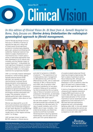
Issue No 21 April 2013
In this edition of Clinical Vision Dr. Di Stasi from A. Gemelli Hospital in
Rome, Italy focuses on: Uterine Artery Embolization the radiological-
gynecological approach to fibroid management.
Part of the team at A.Gemelli Hospital
2. Patient background
This was a 30-year-old woman,
gravida 1 para 0, with a large
subserosal-intramural-submucosal
fibroid who complained of
menorrhagia, pelvic pain, bulk-
related symptoms and infertility.
Procedure
Pre-procedure sagittal (Fig. 1A and 1B), axial (Fig.
2) and coronal (Fig. 3) T2-weighted RM images
show the uterus markedly enlarged and the
uterine cavity distorted by a 92 x 64 mm mass of
low heterogeneous T2 - signal intensity.
Enhanced MR shows the heterogeneous
vascularization of the fibroid compared with the
normal myometrium on axial T1-weighted fat-
saturated images (Fig. 4)
Digital subtraction angiogram with selective
injection via the left internal iliac artery (Fig. 5)
shows a hypertrophic uterine artery. Selective
injection via the left uterine artery before (Fig.
6) and after (Fig. 7) embolization with 250-355
μm Contour™ Embolization particles (Boston
Scientific).
Digital subtraction angiogram with selective
injection via the right internal iliac artery (Fig. 8).
Selective injection via the right uterine artery
before (Fig. 9) and after (Fig. 10) embolization with
250-355 μm Contour™ Embolization particles
(Boston Scientific).
Outcome
Post-embolization (6 months) sagittal (Fig. 11A
and 11B), axial (Fig. 12) and coronal (Fig. 13)
T2-weighted RM images show the fibroids to
be decreased in volume (69 x 50 mm) and with
low-signal intensity. Axial (Fig. 14) and sagittal
(Fig. 15) T1-weighted fat-saturated enhanced MR
images show fibroid infarction with complete
devascularization.
Clinical Vision Issue No 21
Embolization of a large subserosal-intramural-submucosal fibroid
OUTCOME IMAGESPROCEDURAL IMAGES
Fig. 1a Fig. 1b
Fig. 6Fig. 5
Fig. 11a Fig. 11b
Fig. 14
Fig. 15
Fig. 7
Fig. 9
Fig. 8
Fig. 10
Fig. 2 Fig. 3
Fig. 4
2 3
Fig. 13Fig. 12
3. Patient background
A 40-year-old woman, gravida 0,
with a history of bicornuate bicollis
uterus associated with multiple
congenital anomalies presented
with multiple symptomatic fibroids
involving both uterine horns and
secondary hydronephrosis.
Procedure
Pre-embolization coronal (Fig. 1) and axial (Fig. 2
and Fig 3) T2-weighted MR images show four
intramural/subserosal fibroids, of which two
originated from the right (Fig. 2) and two from the
left horn of the uterus (Fig. 3), and dilatation of
the pelvicaliceal system of the right kidney, which
was presumably caused by ureteric obstruction
secondary to pressure from the right-horn fibroids
at the pelvic brim.
On axial (Fig. 4 and Fig. 5) T1-weighted fat-
saturated enhanced MR images all fibroids
demonstrate homogeneous vascularization
compared with the normal myometrium .
Digital subtraction angiogram with selective
injection via the left internal iliac artery (Fig. 6)
shows a thin uterine artery.
Selective injection via the left uterine artery before
(Fig. 7) and after (Fig. 8) embolization with 250-
355 μm Contour™ Embolization particles (Boston
Scientific). Digital subtraction angiogram with
selective injection via the right internal iliac artery
(Fig. 9).
Selective injection via the right uterine artery
before (Fig. 10) and after (Fig. 11) embolization
with 250-355 μm Contour™ Embolization particles
(Boston Scientific).
Outcome
Post-procedure (6 months) coronal (Fig. 12)
and axial (Fig. 13 and Fig. 14) T2-weighted MR
images show the fibroids to be decreased in size
and the hydronephrosis improved. T1-weighted
fat-saturated enhanced MR images (Fig. 15 and
Fig. 16) show fibroid infarction with complete
devascularization.
Clinical Vision Issue No 21
Embolization of intramural/subserosal fibroids
Fig. 1 Fig. 6 Fig. 12
Fig. 13
Fig. 14
Fig. 15
Fig. 16
Fig. 8
Fig. 10
Fig. 7
Fig. 9
Fig. 11
Fig. 2
Fig. 3
Fig. 4
Fig. 5
4 5
OUTCOME IMAGESPROCEDURAL IMAGES
4. Patient background
A 32-year-old woman, gravida 2,
para 2, presented with a 6-month
history of pelvic pain and pressure
and US diagnosis of a single anterior
fibroid.
Procedure
Pre-procedure trans-vaginal color-Doppler US
scans show an intramural/subserosal hypoechoic
fibroid (Fig. 1), with peripheral arterial flow
(perifibroid plexus) (Fig. 2).
Digital subtraction angiogram with selective
injection via the left internal iliac artery (Fig. 3).
Selective injection via the left uterine artery before
(Fig. 4) and after (Fig. 5) embolization with 250-
355 μm Contour™ Embolization particles (Boston
Scientific).
Digital subtraction angiogram with selective
injection via the right internal iliac artery (Fig. 6).
Selective injection via the right uterine artery
before (Fig. 7) and after (Fig. 8) embolization with
250-355 μm Contour™ Embolization particles
(Boston Scientific).
Outcome
Six months post-procedure trans-vaginal color-
doppler US images (Fig. 9 and Fig.10 ) show
a volume reduction of the fibroid (maximum
diameter less than 1.5 cm) and the lack of
vascularization.
Clinical Vision Issue No 21
Fig. 1 Fig. 5 Fig. 9
Fig. 10
Fig. 6
Fig. 8
Fig. 7
Fig. 2
Fig. 3
Fig. 4
6 7
Embolization of a single anterior fibroid
OUTCOME IMAGESPROCEDURAL IMAGES