1. Sarah Fortney1
, Aaron Ermel1
, James Williams1
, and Kenneth Fife1, 2, 3
Departments of Medicine1
, Microbiology and Immunology2
, and Pathology3
, Indiana University School of Medicine
Validation of a Real Time PCR Assay for the Detection of Ureaplasma urealyticum
ABSTRACT
BACKGROUND
METHODS
Background: Ureaplasma urealyticum (UU) is a bacterium that
occurs naturally in the genital flora of sexually active men and
women. In high concentrations this bacterium can become
pathogenic causing urethritis. Detection of UU by nucleic acid
amplification is not widely practiced in the US, and culture
isolation requires a specialized lab. The aim is to validate an
assay utilizing a Real Time PCR (RT-PCR) for the detection of
UU in our laboratory, and to determine the prevalence of UU in
men and women attending a local STI clinic.
Methods: A previously published RT-PCR assay was utilized to
amplify a 152 base pair fragment of a highly conserved region of
UU. The 152 base pair fragment of UU was amplified from an
isolate and cloned into a TOPO-PCRII plasmid. The plasmid was
used to develop quantitative standards of known concentrations;
then to optimize and confirm the assay parameters, and
determine the limit of detection for the assay. Assay performance
in clinical matrix by spiked clinical specimens will be used to
confirm the limit of detection when using residual samples.
Results: The UU fragment was successfully cloned and purified
into a plasmid, which was verified by restriction enzyme digestion
and PCR followed by gel electrophoresis. The plasmid containing
the UU fragment was quantified using spectrophotometry and
contained 7.27x10¹⁰
copies/µL. This plasmid was utilized in
optimizing the RT-PCR assay, including primer concentrations
and annealing temperature. Standards were developed through a
dilution series of the purified plasmid from 1x10¹⁰
-1x101
copies/µL. An increase in reaction efficiency was noted when the
probe concentration was decreased from 0.2µM to 0.15 µM.
Conclusions: Preliminary results show that the assay can
detect down to ~10 copies/µl using purified plasmid. Thus far the
assay has been optimized for our laboratory, and a set of
standards to determine the limit of detection is being developed.
• The Ureaplasma genus was officially designated in
1974, and the species consist of small prokaryotic cells.
• 2 species in the genus, U. urealyticum (UU) and U.
parvum (UP)
• UU is commonly found in the urethra of men, and the
vaginal canal of women. Past studies have found that
patients who show signs of urethritis and infertility are
frequently infected with UU.
• Currently identified by culture (76% sensitivity), but RT-
PCR (96% sensitivity) is more sensitive
• The aim of this study is to validate the RT-PCR Assay
for the detection of UU in our laboratory and to
determine a limit of detection for clinical samples.
Optimization
•Used previously published University of Alabama at Birmingham multiplex RT-PCR for UU and
UP1
, in a single RT-PCR focusing specifically on the 152 base pair region of UU using the Roche
Light Cycler 2.0. Assay parameters in Table 1.
•Specifically optimized primer and probe concentrations and annealing temperature using gradient
PCR and gel electrophoresis
Cloning
•Cloned 152 base pair fragment of UU into a plasmid
•Transformed into E. coli (TOPO TA Cloning Kit and One Shot Competent E. coli, Life
Technologies, Carlsbad, California)
•Plasmid was confirmed through PCR amplification and EcoR1 digest, which includes 100 base
pairs more than the UU fragment.
•Purified plasmid concentration was determined through NanoDrop Spectrophotometry, and the
plasmid was stored at -70° C.
Standardization
•Purified plasmid was diluted in TE buffer from 1010
-101
copies/µL
•1µL aliquots of each dilution from 101
-106
were run in triplicate on RT-PCR
Specificity
•UU serovar (4, 5, 10, 13) and UP serovar (3, 6, 14) isolates were run by the RT-PCR to confirm
specificity.
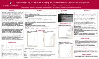
CLONING RESULTS
REFERENCE
CONCLUSION
OPTIMIZATION
Figure 1: Primer Concentration
Confirmation
Figure 2: Probe Concentration
Confirmation
Figure 3: Plasmid Insert Confirmation
STANDARDIZATION
SPECIFICITY
Figure 5: UU and UP Specificity
UU Serovars 4, 5, 10, 13 and UP Serovars
3, 6, 14
Figure 4: Preliminary Standard Curve
Optimization
•Thermocycling conditions were confirmed by gradient PCR.
•Tested concentrations of forward and reverse primers to be
symmetric, compared to 0.2µM(forward) and 0.5µM(reverse).
Increased efficiency was shown in symmetric compared to
asymmetric (Figure 1).
•Tested concentrations of probes at 0.2µM, 0.15µM, and
0.1µM. Increased efficiency of 0.15µM probe (Figure 2).
Cloning
•UU7 was amplified and cloned into E. coli, and purified
•10 colonies were picked and purified using LB Media
Plasmid confirmation through PCR and EcoR1 digest (Figure 3,
red circles indicate plasmid band)
•Determined to have a concentration of 7.27x10¹⁰
copies/µL.
Standardization
•Preliminary amplification standard curve found with 106
copies/reaction crossing at 24 cycles and 101
copies/reaction
crossing at 41 cycles.
•Final standard curve is yet to be determined through RT-PCR
and known concentrations
Specificity
•The RT-PCR correctly identified all UU serovars (4, 5, 10, 13)
while all UP serovars (3. 6. 14) were negative. (Figure 5)
• RT-PCR Assay was successfully optimized for our lab.
• 152 base pair fragment was successfully cloned and purified.
• Assay specificity was determined to detect UU and not UP.
• Future Directions
• Determine Limit of Detection in clinical samples
• Determine prevalence in local STI clinic population
1
Xiao L, Glass JI, Paralanov V, et al. Detection and Characterization of human
Ureaplasma Species and Serovars by Real-Time PCR. J Clin. Microbiol. 2010;48:
2715-23.
Primers UU 127#1F: 5’-GGATTTGTTAGATATCGTCAAGG-3’
UU127#1R: 5’-TCATCTTTTAAAGCTCCACATTATTAGT-3’
Probes UU127 1: 5’-AAACACGAGTATGGATGAATACAAAATCATCAAA/36-FAM/-3’
UU127 2: 5’/5Cy55/AATAACGGTGGTTCAGCTATTTGAGTATGAGC/3Phos/-3’
Master Mix 10X Multiplex DNA Master HybProbe Buffer (Roche Diagnostics, Indianapolis, Indiana)
Reaction
Mixture
0.2 µM UU127#1F, 0.2 µM UU127#1R, 0.2 µM each UU probe, 0.5 U of uracil-DNA glycosylase
(UNG), 3 mM of MgCl2, and 2 µL of 10x Multiplex DNA Master HybProbe buffer
Amplification
Parameters
40°C, then 95°C for 10 min; 45 cycles at 95°C for 15s, 55° for 10s, and 72°C for 9s; melting curve
95°C for 0s, 65°C for 30s, and 95°C; 40°C for 30s.
ACKNWLDGEMENTS
• UU and UP isolates provided by Pat Totten, University of Washington
• Brahim Qadadri for technical assistance
Table 1: Assay Parameters
Ladder PCREcoR1