Understanding Chest X Rays - Dr Khan Health Blogs
•
39 recomendaciones•2,392 vistas

http://www.drkhanblogs.com/2015/06/understanding-chest-x-rays-chest-x-rays.html Dr Khan Health Blogs.

Recomendados
Más contenido relacionado
La actualidad más candente
La actualidad más candente (20)
Similar a Understanding Chest X Rays - Dr Khan Health Blogs
Similar a Understanding Chest X Rays - Dr Khan Health Blogs (20)
Último
Último (20)
Understanding Chest X Rays - Dr Khan Health Blogs
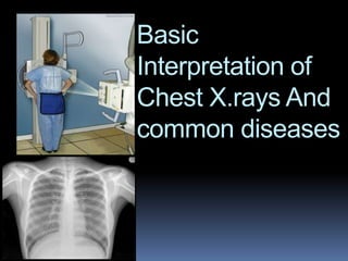
- 1. Basic Interpretation of Chest X.rays And common diseases
- 3. Five Radiographic Opacities Air Fat Soft tissue Bone Metal least opaque to most opaque most lucent to least lucent Black to White
- 4. ANATOMY
- 5. Lobes • Right upper lobe:
- 6. • Right middle lobe:
- 7. • Right lower lobe:
- 8. • Left upper lobe with Lingula:
- 9. • Left lower lobe:
- 20. Portable (AP or Antero- posterior) FILM
- 24. PA vs AP views PA view Scapula is seen in periphery of thorax Clavicles project over lung fields Posterior ribs are distinct AP view Scapulae are over lung fields Clavicles are above the apex of lung fields Anterior ribs are distinct
- 29. Rotation
- 31. 9
- 135. • Lobar consolidation: – Alveolar space filled with inflammatory exudate – Interstitium and architecture remain intact – The airway is patent – Radiologically: • A density corresponding to a segment or lobe • Air bronchogram, and • No significant loss of lung volume Consolidation
- 136. Consolidation
- 168. COMMON CHEST X.RAYS
- 169. Congestive Heart Failure Increased heart size: cardiothoracic ratio >0.5
- 170. CongestiveHeartFailure Alveolar edema (Bat’s wings) Kerley B lines (Interstitial edema) Cardiomegaly Dilated prominent upper lobe vessels Pleural effusion
- 172. ARDS Congestion Interstitial and alveolar edema Collapsed or distended alveoli Bilateral
- 173. Pneumothorax
- 175. Pleuraleffusion
- 177. RLL Pneumonia
- 179. A single, 3cm relatively thin-walled cavity is noted in the left midlung.This finding is most typical of squamous cell carcinoma (SCC). One-third of SCC masses show cavitation
- 180. ???????????????
- 181. Right Middle and Left Upper Lobe Pneumonia
- 182. ????????????
- 183. Cavitation : cystic changes in the area of consolidation due to the bacterial destruction of lung tissue. Notice air fluid level.
- 184. Cavitation
- 185. ????????????
- 186. Tuberculosis
- 187. ??????????
- 188. COPD: increase in heart diameter, flattening of the diaphragm, and increase in the size of the retrosternal air space. In addition the upper lobes will become hyperlucent due to destruction of the lung tissue.
- 189. ????????
- 190. CHF:a great deal of accentuated interstitial markings, Curly lines, and an enlarged heart. Normally indistinct upper lobe vessels are prominent but are also masked by interstitial edema.
- 191. 24 hours after diuretic therapy
- 193. Chest wall lesion: arising off the chest wall and not the lung
- 194. Pleural effusion: Note loss of left hemidiaphragm. Fluid drained via thoracentesis
- 196. Lung Mass
- 198. Small Pneumothorax : LUL
- 200. Right Middle Lobe Pneumothorax: complete lobar collapse
- 201. Post chest tube insertion and re-expansion
- 205. Tuberculosis
- 206. Pleural Effusion
- 207. Pulmonary Fibrosis
- 208. Cavitating lesion
- 209. Miliary shadowing
- 210. 5. 65 yo male admitted for sepsis. CHF or ARDS?