1. Axilla & Axillary Vessels
Dr. Diwakar Kumar Shah
Assistant Professor
Department of Anatomy
Nobel Medical College, Biratnagar
2.
3. Introduction
• The axilla or armpit is a fat-filled pyramid-shaped space, between the
upper part of the arm and the side of the chest wall.
• It contains the brachial plexus, axillary vessels, and lymph nodes.
• It also acts as a funnel shaped tunnel for neurovascular structures to pass
from the root of the neck to the upper limb and vice versa.
• Groups of lymph nodes within it drain the upper limb and the breast.
• Axillary lymph nodes are often enlarged and hence routinely palpated
during physical examination of the patient.
• Abscess in this region is also common.
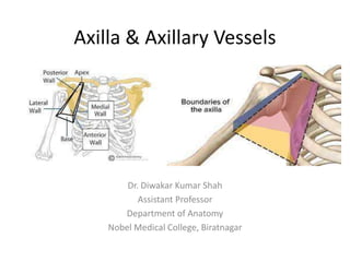
4. BOUNDARIES
• The axilla resembles a truncated four-sided pyramid and presents an apex,
a base and four walls (anterior, posterior, medial, and lateral).
Apex/cervico-axillary canal:
• It is a passageway between the neck and axilla.
• It is directed upwards and medially into the root of the neck and
corresponds to the triangular space bounded in front by the clavicle,
behind by the upper border of the scapula and medially by the outer
border of the first rib.
• The axillary artery and brachial plexus enter the axilla from neck through
this gap, hence it is also termed cervico-axillary canal.
• The axillary vein enters the neck from axilla into the neck through this
canal.
5.
6.
7.
8. Base/floor
• It is at the lower end of the axilla and directed
downwards.
• It is formed by the axillary fascia.
• The base corresponds to the hollow bounded
– In front by the anterior axillary fold, formed by the lower
border of the pectoralis major muscle
– Behind by the posterior axillary fold formed by the tendon
of latissimus dorsi and teres major muscles
– Medially by the lateral aspect of the chest wall
9. • Anterior wall:
– It is formed by the pectoralis major, subclavius, and pectoralis minor
muscle.
• Posterior wall:
– It is formed by the subscapular muscle above and latissimus dorsi and
teres major muscle below.
• Medial wall:
– It is formed by the upper four or five ribs, and corresponding
intercostal spaces covered by the serratus anterior muscle
• Lateral wall:
– It is formed by tendon of biceps brachii in the bicipital groove of
humerus, coracobrachialis and short head of biceps brachii.
– The lateral wall is extremely narrow because anterior and posterior
walls of the axilla converge at this site.
10. Contents of Axilla
• Axillary artery and its branches
• Axillary vein and its tributaries
• Cords of brachial plexus
• Axillary lymph nodes
• Fibrofatty tissue
• Axillary tail of breast
• Long thoracic and intercostobrachial nerves
11. Axillary artery
• It is the main artery of the upper limb.
• It begins at the outer border of the first rib as the
continuation of subclavian artery and ends by becoming
brachial artery at the lower border of teres major.
• In axilla, it runs from its apex to the base along the lateral wall
nearer to the anterior wall than the posterior wall.
• During its course through axilla, it is crossed on its superficial
aspect by the pectoralis minor muscle, which divides it into
three parts.
• Axillary vein is medial to the artery and the cords of brachial
plexus are arranged around the second part of the artery (i.e.,
part deep to the pectoralis minor); the lateral cord being
lateral, the medial cord medial, and posterior cord behind.
12. Parts
• First part, superior (or proximal) to the muscle
• Second part, posterior (or deep) to the muscle
• Third part, inferior (or distal) to the muscle
13. Relations
• The axillary vein lies medial to the axillary artery
throughout its course, but the relationship of cords
of brachial plexus and their branches are different for
each of the three parts of the artery.
14.
15. Branches of Axillary Artery
• From First Part
– Superior thoracic artery, a very small branch, arises near the
subclavius, passes between the pectoralis major and minor muscle,
and supplies these muscles and medial wall of the axilla
16. From Second part
• Thoraco-acromial artery (acromiothoracic artery), emerges at the upper
border of pectoralis minor, pierces clavipectoral fascia and soon breaks up
into four branches:
• Pectoral branch
• Deltoid branch
• Acromial branch
• Clavicular branch.
– These branches radiate at right angle to each other.
– The pectoral branch supplies pectoral muscles, deltoid branch, ends by
joining anastomosis over the acromion, clavicular branch supplied
sternoclavicular joint.
• Lateral thoracic artery, emerges at and runs along the inferior border of
pectoralis minor, supplying the branches to pectoralis major and minor
and serratus anterior muscles.
– In the females, the lateral thoracic artery is large and provides
important supply to the breast through its lateral mammary branches.
17.
18. From Third part
• Subscapular artery, the largest branch of axillary
artery, runs along the lower border of the
subscapularis and ends near the inferior angle of the
scapula.
– It gives a large branch, the circumflex scapular artery,
which passes through upper triangular intermuscular
space, winds round the lateral border of scapula to enter
infraspinous fossa.
– In addition, it gives numerous small branches.
19. • Anterior circumflex humeral artery, a small branch, passes in
front of surgical neck of humerus and anastomoses with the
posterior circumflex humeral artery to form an arterial circle
around the surgical neck of humerus.
– It gives an ascending branch, which runs upwards into the
intertubercular sulcus of humerus to supply the head of humerus and
shoulder joint.
• Posterior circumflex humeral artery, larger than the anterior
circumflex humeral artery, passes backwards, along with
axillary nerve through the quadrangular intermuscular space,
crosses the posterior aspect of surgical neck of humerus to
anastomose with the anterior circumflex humeral artery.
– It supplies the deltoid muscle and shoulder joint.
20. Arterial Anastomosis Around Scapula
• The arterial anastomosis around scapula is principally formed between the
branches of the first part of the subclavian and the third part of the
axillary arteries.
• The scapular anastomosis takes place at two sites: around the body of
scapula and over the acromion process of the scapula.
• Around the body of scapula: it occurs between the
– Suprascapular artery, a branch of the thyrocervical trunk from the first part of
the subclavian artery
– Circumflex scapular artery, a branch of the subscapular artery from the third
part of the axillary artery
– Deep branch of the transverse cervical artery, a branch of the thyrocervical
trunk
• Over the acromion process: it occurs between the
– Acromial branch of the thoraco-acromial artery
– Acromial branch of the suprascapular artery
– Acromial branch of the posterior circumflex humeral artery
21. Clinical Correlation
• Collateral circulation through scapular anastomosis:
– If the subclavian and axillary arteries are blocked anywhere between
1st part of subclavian artery and 3rd part of axillary artery, the
scapular anastomosis serves as a potential pathway (collateral
circulation) between the first part of the subclavian artery and the
third part of the axillary artery, to ensure the adequate circulation to
the upper limb.
22.
23. Axillary Vein
• It is formed at the lower border of teres major muscle by the
union of basilic vein and venae comitantes of the brachial
artery.
• It runs upwards along the medial side of the axillary artery
and ends at the outer border of the first rib
• Tributaries:
– Veins which correspond to the branches of axillary artery, namely,
lateral thoracic vein and subscapular vein.
– Cephalic vein, which joins it after piercing the clavipectoral fascia
24. Clinical Correlation
• Spontaneous thrombosis of the axillary vein:
– Occasionally, a muscular band- the axillary arch, overlies
the vein.
– It may compress the vein, following excessive and
unaccustomed movements of the arm at the shoulder
joint and cause spontaneous thrombosis of the axillary
vein