
Recomendados
Más contenido relacionado
La actualidad más candente
La actualidad más candente (20)
Similar a MASTITIS & GYNECOMASTIA_074508.pptx
Similar a MASTITIS & GYNECOMASTIA_074508.pptx (20)
Más de ShubhrimaKhan
Más de ShubhrimaKhan (20)
Último
Último (20)
MASTITIS & GYNECOMASTIA_074508.pptx
- 1. MASTITIS
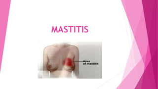
- 2. DEFINITION An acute inflammation of the interlobular connective tissue within the mammary gland that results in breast pain, swelling, warmth and redness of the breast.
- 3. TYPES PUERPERAL MASTITIS – It is the inflammation of the breast in connection with pregnancy and lactation. It is caused by blocked milk ducts or excess milk. NONPUERPERAL MASTITIS – The term describes inflammatory lesions of the breast occurring unrelated to pregnancy and breastfeeding.
- 4. ETIOLOGY INFECTION MILK STATIS BREAST ABSCESS PREDISPOSING FACTORS
- 5. INFECTION Most common pathogen is staphylococcus aureus causes deeper walled off infection or abscess. Sometimes streptococcal is also detected leading to a superficial small area of inflammation. Cracks or break in the skin of the nipple. Impaired immune system.
- 6. MILK STATIS Milk is being made faster than it is removed. Complication of delivery. Inability to breastfeed soon after birth. Feeding to a strict routine. Clogged ducts due to hormonal changes. Stagnant milk increases pressure in breast leakage in surrounding breast tissue and causes an inflammatory response.
- 7. BREAST ABSCESS Collection of pus that develops into the breast. Most common within six weeks after delivery. 5 – 11 % of mastitis causes.
- 8. PREDISPOSING FACTORS Maternal stress or fatigue. Wearing a tight fitting bra. Women with diabetes, chronic illness, AIDS. Trauma to the breast. Previous history of mastitis. Genetic
- 9. CLINICAL MANIFESTATIONS INFECTION Pain and swelling of the breast. Tenderness and warm to touch. Redness often in a wedge shaped pattern. Fever of 101⁰F or greater. Burning sensation continuously or while breast feeding. General malaise & fatigue.
- 10. CONT.. ABSCESS Tender lump in the breast. Mass may be moveable or compressible. Discharge from nipple. Persistent fever. No improvement of symptoms within 48 – 72 hours of treatment.
- 11. DIAGNOSTIC STUDIES Physical examination Breast ultrasound Mammography Culture Breast biopsies
- 12. MANAGEMENT MEDICAL Analgesic: acetaminophen or ibuprofen safe while breastfeeding. Antibiotic: Cephalexin or dicloxacillin are recommended. Minimum treatment 10 – 14 days. Anti inflammatory agents sometimes recommended.
- 13. CONT.. SURGERY Percutaneous aspiration Surgical incision and drainage.
- 14. CONT.. NURSING Encourage frequent breast feeding. Frequent emptying of the breast by using breast pump or expressing milk between feedings. A warm bath or warm compression before and after feeding may provide some relief. If heat is ineffective, ice packs applied after feeding. Encourage the patient to drink plenty of water.
- 15. CONT.. A well balanced diet (500 extra calories) should be taken. If discharge present instruct patient to wash the nipple gently. Wear a supportive bra that does not compress the breast. Use of cabbage leaves also effective.
- 16. GYNECOMASTIA
- 17. DEFINITION GYNECOMASTIA – It is a benign enlargement of the male breast caused by proliferation of glandular breast tissue. In response to too much estrogen or too little testosterone the glandular tissue of the breast swells and forms a breast bud. PSEUDOGYNECOMASTIA – Enlargement of the male breast as a result of increased fat deposition is called pseudogynecomastia.
- 18. PREVALENCE Gynecomastia has three peaks. Neonates: 60 – 90 % transient gynecomastia due to high maternal estrogen. Normally regresses over 2 – 3 week period. Puberty: transient gynecomastia may occur in up to 60% boys. It may first appear as early as 10 years of age, with a peak onset between 13 – 14 years followed by an involution that is generally complete by 16 – 17 years. Old age: 24 – 65% with highest prevalence in the 50 – 80 years of age.
- 19. ETIOLOGY Idiopathic – 25% Certain drugs – steroids, chemotherapy drugs, medicine used to treat epilepsy, ulcer. Cirrhosis and malnutrition Primary or secondary hypogonadism Hyperthyroidism Testicular tumors Chronic renal insufficiency Lung and liver cancer
- 20. GRADES OF GYNECOMASTIA GRADE 1 A localized button of tissue that is concentrated around the areola. Severity: very mild
- 21. CONT.. GRADE 2 Moderate breast enlargement exceeding areola boundaries with edges that are indistinct from the chest. Severity: mild to high
- 22. CONT.. GRADE 3 Moderate breast enlargement exceeding areola boundaries with edges that are distinct from the chest with skin redundancy present. Severity: high and identifiable.
- 23. CONT.. GRADE 4 Marked breast enlargement with skin redundancy and feminization of the breast. severity: severe and visibly feminine.
- 25. DIAGNOSTIC STUDIES Medical history and physical examination. Blood test Mammogram CT scan MRI Testicular ultrasound Biopsy
- 26. MANAGEMENT MEDICATIONS: tamoxifen and raloxifene to treat breast cancer or other condition. SURGERY: Liposuction Mastectomy