SlideShare una empresa de Scribd logo
1 de 103
INTRODUCTION
• Skin is the largest organ of your body. It is a soft outer covering of an animal, in particular
a vertebrate.
• The skin functions in thermoregulation, protection, metabolic functions and sensation.
• Skin is composed of three primary layers:
1. The epidermis, which serves as a barrier to infection
2. The dermis, which serves as a location for the appendages of skin
3. The hypodermis (subcutaneous adipose layer)
• Its function is to protect from infection. Sometimes the skin itself becomes infected.
• Skin infections are caused by a wide variety of germs, and symptoms can vary from mild
to serious. Mild infections may be treatable with over-the-counter medications and home
remedies, whereas other infections may require medical attention
TYPES OF SKIN INFECTIONS
• Bacterial skin infections
• Viral skin infections
• Fungal skin infections
• Parasitic skin infections
BACTERIAL SKIN INFECTIONS
• Gram positive staphylococcus aureus and beta hemolytic streptococci cause
bacterial infections.
• Primary infection occur if break present in skin present.
• Secondary infection occurs if it followed after already damaged skin or as sign of
systematic infection.
• Predisposing Factors: Moisture, Obesity, Skin disease, Systematic corticosteroids,
Antibiotics, Chronic diseases like diabetes mellitus increase the incidence of
infection.
IMPETIGO
• Impetigo is superficial skin infection caused by staphylococci, streptococci or
multiple bacteria,
• It involves exposed areas of body, face, hands, neck and extremities. It is often
called school sores because it most often affects children. It is quite contagious.
• Primary impetigo occurs due to poor hygiene and malnutrition. Secondary
impetigo occurs due to pediculosis capitis (Head lice scabies, herpes simplex,
insect bite or eczema).
CLINICAL MANIFESTATIONS
• Impetigo presents with pustules and round, oozing patches which grow larger day
by day
• There may be clear blisters or golden yellow crusts.
CONT..
TREATMENT
• Soak moist or crusted areas: Soak a clean cloth in a mixture of half a cup of white
vinegar in a liter of tepid water. Apply the compress to moist areas for about ten
minutes several times a day. Gently wipe off the crusts.
• Antiseptic or antibiotic ointment: apply it two or three times a day to the affected
areas and surrounding skin.
• Oral antibiotics: Oral antibiotics are recommended if infection is extensive,
proving slow to respond to topical antibiotics, or if impetigo is recurrent. The
preferred antibiotic is penicillin antibiotic. The complete course should be taken,
usually at least 7 days.
FOLLICULITIS
• Folliculitis means an inflammation or infection of the hair follicles of the skin. It is a
problem that is not usually serious.
• Tiny pus-filled spots (pustules) develop at the hair where friction, moisture, rubbing
or oil is more.
• Common sites for folliculitis are the beard, arms, legs, armpits.
CONT..
CLINICAL MANIFESTATIONS
Superficial folliculitis, which affects the upper part of the hair follicle, may cause:
• Clusters of small red or pus-filled bumps that develop around hair follicles
• Red and inflamed skin
• Itchiness or tenderness
Deep folliculitis starts deeper in the skin surrounding the hair follicle and affects the
entire hair follicle. Signs and symptoms include:
• A large swollen bump or mass
• Pus-filled blisters that break open and crust over
• Pain
CONT..
TREATMENT
• Mild folliculitis usually heals on its own in about 2 weeks.
• Warm compresses made with white vinegar solution ease itching & helps healing.
• Medicated shampoo can be used to treat folliculitis on the scalp or beard.
• Antibiotics applied to the skin (mupirocin) or taken by mouth (dicloxacillin).
• Gently wash the infected skin twice a day with antibacterial soap.
• Avoid shaving irritated skin. If must shave, use an electric razor rather than a blade
and apply a soothing aftershave lotion when finished. Also, shave in the direction of
hair growth rather than against it.
• Don't share towels or washcloths
FURUNCLE
• Furuncle is another word for "boil." A boil is
a bacterial infection of a hair follicle.
• The infected hair follicle can be on any part
of body. Furuncles mostly occur on body
areas prone to excessive perspiration,
friction and irritation such as back of neck,
thigh, axillae, and perineum or on buttocks.
CONT..
CLINICAL MANIFESTATIONS
• Deep, firm, red, raised bump on skin which is 1 – 5 cm in diameter,
• Pain which gets worse as it fills with pus and dead tissue.
• White or yellow centers (pustules)
• Spreading to other skin areas or joining with other boils
• Weeping, oozing, or crusting
• Fatigue
• Fever and general ill-feeling
• Itching before the boil develops
• Skin redness around the boil
CONT..
TREATMENT
• Put warm, moist compresses on the boil several times a day to speed draining and
healing.
• Cleanse surrounding skin with antibacterial soap, followed by application of
antibacterial ointment.
• Surgical incision and drainage may be performed.
• Systemic antibiotic therapy is instituted for carbuncles or spreading furuncles.
Analgesia and antipyretics are ordered as necessary.
• Cover mattress and pillows with plastic and wipe daily with a disinfectant.
• Wash all linens, towels, and clothing after each use.
• Strict hand washing is maintained to prevent cross-contamination.
CARBUNCLES
• A carbuncle is a red, swollen, and painful cluster of boils that are connected to
each other under the skin. A carbuncle occurs when a group of hair follicles next
to each other become infected. It is like a multiple boil. Furuncles may progress to
carbuncles.
• Carbuncles found on back of neck, upper arm, buttocks and lateral thighs.
CONT..
CLINICAL MANIFESTATIONS
• The boils that collect to form carbuncles usually start as red, painful bumps.
• The carbuncle fills with pus and develops white or yellow tips that weep, ooze, or
crust.
• Untreated carbuncles rupture, discharging a creamy white or pink fluid.
• Superficial carbuncles which have multiple openings on the skin's surface are less
likely to leave a deep scar.
• Deep carbuncles are more likely to cause significant scarring.
• Other carbuncle symptoms include chills, fever, fatigue, and a feeling of general
sickness.
• Swelling may occur in nearby tissue and lymph nodes, especially lymph nodes in the
neck, armpit, or groin.
CELLULITIS
• Cellulitis is an inflammation of the skin and subcutaneous tissue resulting from a
generalized bacterial infection.
• Cellulitis appears as a swollen, red area of skin that feels hot and tender.
CAUSES AND RISK FACTORS
• Staphylococcus and streptococcus bacteria
• Cracks or peeling skin between the toes
• Weakened immune system
• Injury or trauma with a break in the skin (skin wounds)
• Insect bites, animal bites.
• Ulcers from certain diseases, including diabetes and vascular disease.
• Use of corticosteroid medications or medications that suppress the immune system
• Intravenous drug use
• Obesity
• Wound from a recent surgery
CONT..
CLINICAL MANIFESTATIONS
• Pain or tenderness in the affected area
• Skin redness or inflammation (Swelling)
• Warm skin in the area of redness
• Fever with chills
• Malaise
• Headache
• Swollen lymph nodes
• Tight, glossy, stretched appearance of the skin
• Hair loss at the site of infection
• Joint stiffness caused by swelling of the tissue over the joint
CONT..
TREATMENT
• Topical/systemic antibiotics are prescribed according to culture and sensitivity test
results.
• Debridement of nonviable tissue is necessary if there is an open wound.
• Systemic antibiotics are indicated if fever and lymphadenopathy are present.
• Warm, moist compresses may be ordered.
• Immobilization and raise the infected area higher than heart to reduce swelling
• Wash wound daily with soap and water
• Moisturizing and lubricating skin regularly helps prevent cracking and peeling.
• Wear appropriate footwear and gloves.
• Regularly check feet for signs of injury.
• In severe cases: Hospitalization required
DIAGNOSIS OFALL BACTERIAL INFECTIONS
Lesion drainage and blood culture tested to identify causative organism. Drug
sensitivity also checked.
In case of repeated bacterial infections. Culture is taken from external nares to
determine carriers of bacteria (MRSA).
VIRAL SKIN INFECTIONS
• Viral infections are common and can occur anywhere in body.
• They range from the common to the rare, from the mild to the severe and
from localized skin infection to systemic disease.
• The principle site of attack in the skin is the epidermis.
ETIOLOGY
• Birth control pills and corticosteroids have immune-suppressive properties.
• Antibiotics also kill normal skin flora of bacteria that serve as resistance
against virus.
WARTS
• Warts are small, usually painless growths on the skin. Most of the time
warts are harmless.
• They are caused by a virus called human papillomavirus (HPV).
• Warts found on mucous membrane and genital skin of humans.
• All warts can spread from one part of your body to another. Warts may be
spread from person to person but this is uncommon.
CONT..
TYPES
1. Common wart (Verruca vulgaris): most commonly appears on fingers. They
grow above the skin surface and they are usually dome shaped.
2. Planter warts (Verruca plantaris): occur on bottom surface of foot. They grow
inwards as pressure of shoes or walking prevents its outward growth. These
warts extend deeper beneath the skin
3. Flat wart (verruca plana): They are flat lesion present on forehead or dorsum
of the hand
4. Condylomata acuminata or venereal warts: occur in moist areas - Glans of
penis, anal region, on vulva & cervix. They are pink or purple in color and have
cauliflower appearance.
CONT..
CLINICAL MANIFESTATIONS
• Most warts are raised with a rough surface. They may be round or oval.
• The spot where wart grows may be lighter or darker than the other skin. Rarely,
warts are black.
• Some warts have smooth or flat surfaces (very rare).
• Some warts may cause pain.
CONT..
TREATMENT
1. Chemicals:
• Typical chemicals like salicylic acid, trichloroacetic acid, 5-
• fluorouracil, cantharidin, tretinoin etc. are used.
• Flat warts are often treated with peeling agents such as tretinoin or salicylic
acid. Imiquimod can be used for the treatment of genital warts.
• Chemicals usually require multiple applications over several weeks to months.
• Applied every 12-24 hours and warts disappear within 2-3 days.
CONT..
TREATMENT
2. Freezing (Cryotherapy): Freezing is safe. Warts may be frozen with various
commercial freezing probes or with liquid nitrogen sprayed on or applied with a
cotton swab. Cryotherapy is often used for plantar warts, and warts under the
fingernails.
3. Burning and Cutting: These methods are effective but are more painful and
usually leave a scar. A laser or electrical current is used to burn warts off.
CONT..
PREVENTION
Avoiding cross-contamination can reduce the risk of spreading warts.
• Don't bite fingernails: Warts occur more often in skin that has been broken.
Nibbling the skin around fingernails opens the door for the virus.
• Groom with care: In order to avoid spreading the virus, don't brush, clip, comb or
shave areas that have warts.
• Don't pick at warts: Picking may spread the virus. Consider covering warts with an
adhesive bandage to discourage picking.
• Keep hands dry: Warts are more difficult to control in a moist environment.
HERPES SIMPLEX
Herpes simplex virus infection is a common viral infection Of the skin that tends to
recur repeatedly. It is also called fever blister or cold sore.
There are two types of herpes simplex:
1. Type I virus (HSV-I), which occurs above the waist on lips, face and mouth and
causes a fever blister or cold sore.
2. Type Il virus (HSVII), which occurs below the waist and causes genital herpes.
MODES OF TRANSMISSION
• It is transmitted by direct contact, respiratory droplet, or contact with virus containing
fluid-saliva or cervical secretions in the form of kissing and sexual activity.
• Trauma, sunlight, menses, stress and systematic infection often cause reoccurrence of
infection.
CONT..
CLINICAL MANIFESTATIONS
• Burning and tingling sensation at the site for a few hours before eruption.
• Area becomes erythematous and swollen
• Vesicles and pustules erupt in 1 to 2 days
• Lesions can burn, itch, and be painful
• Vesicles become pustules, ulcers and crusting until healing occurs in 10-14 days
• Fever, sore throat are systematic manifestations of initial infection
• The patient is contagious until scabs are formed
CONT..
DIAGNOSTIC EVALUATIONS
• Assess the appearance of lesions.
• Tzanck smear identifies herpes virus.
• Vesicle fluid culture differentiate herpes virus from bacterial infections.
CONT..
TREATMENT
• There is no complete cure for herpes simplex.
• Topical acyclovir (Zovirax) ointment is the drug of choice for primary lesions, to
suppress the multiplication of vesicles.
• Oral acyclovir may be recommended for severe attacks or immunosuppressed
patients. Lotions, creams, and ointments may be prescribed to accelerate drying and
healing of lesions (e.g., camphor, phenol, alcohol).
• Antibiotics may be indicated for secondary infections.
HERPES ZOSTER (SHINGLES)
• Herpes zoster, or shingles, is an acute inflammatory viral infection that produces a
painful vesicular eruption along the distribution of nerves from one or more posterior
ganglia. This eruption follows the course of the cutaneous sensory nerve and is almost
always unilateral.
• It's caused by the varicella-zoster virus, which is the same virus that causes
chickenpox. The rash most often appears as a band of rashes or blisters in one area of
the body.
Where does shingles come from?
When you have chickenpox as a child, your body fights off the varicella-zoster virus
and the physical signs of chickenpox fade away, but the virus always remains in your
body. In adulthood, sometimes the virus becomes active again. This time, the varicella-
zoster virus makes its second appearance in the form of shingles.
CAUSES AND RISK FACTORS
• Previously suffered from chicken pox
• Most common in the elderly people above 60 years.
• With a weakened immune system (such as people with cancer, HIV, organ transplant
recipients or those receiving chemotherapy).
• Patient injury to the spine or a cranial nerve
• Who are under stress.
PATHOPHYSIOLOGY
• Herpes virus reactivate Varicella virus (latent infection present in spinal nerve
root called the dorsal root ganglion)
• When latent infection reactivated — Virus travel by way of peripheral nerves
to skin
• Virus multiples and create blisters
STAGES
• First, a few days before the rash appears, there may pain in an area
on the skin. The pain is described as itching, burning, stabbing or
shooting.
• Next, the raised rash appears as a band or a patch, usually on one
side of body. The rash usually appears around waistline or on one
side of face, neck, or on the trunk (chest/abdomen/back), but not
always. It can occur in other areas including arms and legs.
• Within three to four days, the rash develops into red, fluid-filled,
painful, open blisters.
• Usually, these blisters begin to dry out and crust over within about
10 days.
• The scabs clear up about two to three weeks later.
• It can take three to five weeks from the time begin to feel
symptoms until the rash totally disappears.
CLINICAL MANIFESTATIONS
Early symptoms of shingles may include:
• Fever.
• Chills.
• Headache.
• Feeling tired.
• Sensitivity to light.
• Stomach upset.
Other signs and symptoms that appear a few days after the early symptoms include:
• An itching, tingling or burning feeling in an area of skin.
• Redness on skin in the affected area.
• Raised rash in a small area of skin.
• Fluid-filled blisters that break open then scab over.
• Mild to severe pain in the area of skin affected.
DIAGNOSIS AND TESTS
Distribution or appearance of the rash: The blisters of a shingles rash usually
appear in a band on one side of your body.
Shingles also may be diagnosed in a laboratory using scrapings or a swab of the
fluid from the blisters.
TREATMENT
There is no cure for shingles but there are treatments for managing the symptoms.
Treatment is aimed at controlling the outbreak, reducing pain and discomfort, and
preventing complications.
• Antiviral drugs: such as acyclovir (Zovirax), famciclovir (Famvir), and valacyclovir
(Valtrex), interfere with viral replication.
• Pain management: aspirin, acetaminophen, NSAIDs, opioids are useful during the
acute stage.
• Topical steroids should not be applied if a secondary infection is present because they
suppress the immune system.
• Anti-inflammatory drugs like prednisone may be prescribed if shingles affects the
eyes or other parts of the face.
• Antihistamines are administered to control itching.
• Antibiotics are prescribed for secondary bacterial infections.
NURSING MANAGEMENT
Nursing Diagnosis:
1. Acute Pain may be related to presence of localized inflammation and open
lesions, possibly evidenced by verbal reports, distraction behaviors, and
restlessness.
2. Risk for [secondary] Infection: risk factors may include broken/traumatized
tissue, altered immune response, and untreated infection/ treatment failure.
3. Risk for ineffective sexuality pattern: risk factors may include lack of
knowledge, and/or fear of transmitting the disease.
CONT..
Nursing interventions
• Monitor location, duration and intensity of pain. Monitor for adverse effects of
pain medications.
• Assess patient's level of discomfort and medicate as prescribed.
• Encourage distraction techniques such as music therapy.
• Teach relaxation techniques, such as deep breathing, progressive muscle
relaxation, and imagery, to help control pain.
• Administer antiviral medication in dosage prescribed (usually high dose).
CONT..
• Apply antibacterial ointments (after acute stage) as prescribed, to soften and
separate adherent crusts and prevent secondary infection.
• Teach patient to use proper hand-washing technique, to avoid secondary infection.
• Advice the patient to observe signs of systematic infection i.e. fever, malaise,
headache, increased redness, formation & drainage of pustules.
• Maintain strict isolation.
• Wear gown & gloves if contact has to be established with patient.
• Keep patient’s room cool and avoid heavy clothes and bedding.
COMPLICATIONS
• Postherpetic neuralgia: After the shingles rash has disappeared, there might
continue to have nerve pain in that same area, last for months or years and become
quite severe. More than 10% of people who get shingles develop postherpetic
neuralgia. It may be that nerves become more sensitive or that the virus may be
invading and damaging the central nervous system.
• Other complications include:
• Other types of nerve issues like numbness or itching.
• A bacterial infection of the shingles rash.
• Eye and ear inflammation if the rash is near these organs.
FUNGAL SKIN INFECTIONS
• Fungi are free living plant like organism that live in the soil, on animals and
plants.
• Fungus disorder also called mycoses.
• Fungi that cause superficial skin infections are called dermatophytes.
• Tinea is the term used to describe fungal skin infections which is also called
ringworm because of its characteristic appearance of ring.
• Fungal infections are common in skin folds, between the toes and in mouth.
CONT..
MODE OF TRANSMISSION
• Direct skin to skin contact with infected persons or animals
• Contact with inanimate object such as combs, pillow cover, towel and hats.
PREDISPOSING FACTORS
• Moisture is an important factor in the development of fungal infection.
• Prolonged use of antibiotics
• Diabetes mellitus
• Immunodeficiency
• Nutritional deficiencies
• Increasing age
• Iron deficiency diseases
TINEA PEDIS
• Tinea pedis is a foot infection due to a dermatophyte fungus.
• It is fungal infection of the soles of the feet, space between toes or toe nails.
• It is also called Ringworm of feet or Athlete's foot. Athlete's foot is contagious.
• Athlete’s foot is caused by a microscopic fungus called trichophyton rubrum.
SIGNS AND SYMPTOMS
• Redness, blisters, swelling, itching or burning
• Scaly or peeling skin
• Bad smelling feet
• Thick, dark skin on the bottoms or sides of feet
• Thick, abnormal toenails
TINEA CORPORIS (RING WORM OF BODY)
• Tinea corporis (ringworm) is the infection of the trunk, legs or arms with a
dermatophyte fungus. It's characterized by a red circular rash with clearer skin in
the middle.
• Tinea corporis affects face, neck, trunk, extremities and caused by several
different fungi.
• Most commonly found in a child younger than 15 years old.
• Heat and humidity aggravate the infection.
SIGNS AND SYMPTOMS
• Red ring-shaped patches with a raised scaly border ranging from I to 10 cm.
• The central area may be clear of any redness.
• The border of the affected skin may contain blisters, bumps, or scabs.
• Itching or burning sensation
TINEA CAPITIS
• Tinea capitis is a scalp infection caused by a fungus. It is also called ringworm of
the scalp or head.
• It is a highly contagious infection, is most common in toddlers and school-age
children.
• The most common species of fungi affecting humans are Microsporum canis and
Tricophyton verrucosum.
SIGNS AND SYMPTOMS
• Hair loss, fragile hair
• Raised scaly skin,
• Itchy scalp
• Tender or painful areas on the scalp
TINEA CORPORIS TINEA CAPITIS
TINEA VERSICOLOR
• Tinea versicolor is a long-term (chronic) fungal infection of the skin, also called
pityriasis versicolor
• It is caused by a type of yeast that naturally lives on skin, called Pityrosporum
ovale. When the yeast grows out of control, the skin disease results, which appears
as a rash.
• A number of factors may trigger this growth, including:
• Hot, humid weather
• Excessive sweating
• Oily skin
• Hormonal changes
• Weakened immune system
• Certain medicines, such as steroids or birth control pills.
• It is not spread from one person to another.
CONT..
CLINICAL MANIFESTATIONS
• Patches of discolored skin: white, pink or dark brown, located on the back, chest,
neck and upper arms
• Slow-growing, scaly and mildly itchy
• More noticeable after sun exposure
• Increased sweating and Itching
TINEA CRURIS
• Jock itch (tinea cruris) is a fungal infection that affects the skin
of genitals, inner thighs and buttocks. Jock itch causes an itchy,
red, often ring-shaped rash in these warm, moist areas of body.
• Jock itch gets its name because it is common in people who
sweat a lot, as do athletes.
• Men who play sports and use a male athletic guard are
particularly prone to develop this tinea infection.
TINEA UNGUIUM
• Fungal nail infections is a condition in which a fungal or yeast infection occurs in the
nail plate that causes the nails to be thick and brittle. It is caused by Trichophyton
species or candida albicans.
RISK FACTORS
• Diminished blood circulation
• Prolonged exposure to fungi
• Perspiring heavily
• Working in a humid or moist environment
• AIDS and diabetes
SIGNS & SYMPTOMS
• Occasionally, it can cause pain or discomfort
• The nail usually is brittle, thickened, dull, and easily breakable
• Debris accumulates under nail plate and nail plate separates
• Sometimes, surrounding skin can become infected and cause cellulitis
(inflammation)
CANDIDIASIS
• Candidiasis is an infection caused by a yeast-like fungus called Candida albicans.
• Candidiasis normally found on mucous membranes, on skin, in vagina and in
gastrointestinal tract.
• Oral infections, called oral thrush, are most common in infants, elderly people, and
those with a weakened immune system.
PREDISPOSING FACTORS
• Broad-spectrum antibiotics, Corticosteroid treatment
• Central venous catheters or parenteral nutrition
• Pregnancy, Use of birth control pills
• Poor nutrition
• Diabetes mellitus
• Malignancies of the blood
• GI tract surgery
• Renal replacement surgery
CONT..
CLINICAL MANIFESTATION
Oral Candidiasis (Thrush):
• Creamy white (curd - like) patches in the mouth or on the throat
• Redness and difficulty swallowing
• Painful cracks at the corners of the mouth
Candidiasis of the Genitals:
• Extreme itching in the vaginal area, redness
• White, clumpy vaginal discharge
• Painful intercourse
• Red rash on penis
• Itching or burning on the tip of the penis
Skin Infection: Skin rashes, patches, and blisters found most commonly in the groin,
between fingers and toes, and under the breasts.
NURSING MANAGEMENT FOR FUNGAL INFECTION
1. Wash hands and teach other caregivers to wash hands before contact with patient,
and between procedures with patient.
2. Encourage intake of protein- and calorie-rich foods to maintain optimal nutritional
status.
3. Encourage fluid intake of 2000 ml to 3000 ml of water per day to promote diluted
urine and frequent emptying of bladder
4. Advice the person not to sharing the linen and personal items like towel, comb etc.
Use a clean towel each day.
5. Carefully dry all skin folds including under breast, under arms & between toes. Wear
clean under clothes and it should not be very tight fitted.
6. Do not wear same shoes everyday. Do not wear rubber or plastic soled shoes.
7. Use talcum powder or antifungal agents (clotrimazole) twice a day.
8. For Candidiasis, advice the patient that sexual partners should also be treated with
antifungal medicines. Use condom or abstinence.
SKIN CANCER
Introduction
• Skin cancer is the most commonly diagnosed cancer.
• Most skin cancers start in the top layer of skin, called the epidermis. There are 3 main
types of cells in this layer
1. Squamous cells: These are flat cells in the upper (outer) part of the epidermis, which
are constantly shed as new ones form. When these cells grow out of control, they can
develop into squamous cell skin cancer (also called squamous cell carcinoma).
2. Basal cells: These cells are in the lower part of the epidermis. They constantly divide
to form new cells to replace the squamous cells. As these cells move up in the
epidermis, they get flatter, eventually becoming squamous cells. Skin cancers that
start in the basal cell layer are called basal cell skin cancers or basal cell carcinomas.
Cont…
3. Melanocytes: These cells make the brown pigment called melanin, which gives the
skin its tan or brown color. Melanin acts as the body’s natural sunscreen, protecting
the deeper layers of the skin from some of the harmful effects of the sun. Melanoma
skin cancer starts in these cells.
The epidermis is separated from the deeper layers of skin by the basement
membrane. When a skin cancer becomes more advanced, it generally grows through
this barrier and into the deeper layers.
Types of skin cancers
The most common types of skin cancer are:
1. Basal Cell Carcinoma (BCC)
2. Squamous cell (epidermoid) carcinoma (SCC).
3. Malignant melanoma
Risk Factors for Skin Cancer
• Damage in the ozone layer
• Fair-skinned and fair-haired people due to insufficient skin pigmentation
• Prolonged exposure to sun light.
• Exposure to chemical pollutants(arsenic, nitrates, coal, pitch, oils and paraffins)
• History of x-ray therapy for acne or benign lesions
• Scars from severe burns
• Chronic skin irritations
• Immunosuppression
• Genetic factors
Basal cell carcinoma
• Basal cell carcinoma is most common type of skin cancer. About 8 out of 10 skin
cancers are basal cell carcinomas.
• These cancers usually develop on sun-exposed areas, especially the face, head, and
neck.
• BCC is a locally invasive malignancy and tend to grow slowly. But if it's left
untreated, basal cell cancer can grow into nearby areas and invade the bone or other
tissues beneath the skin.
• It rarely metastasizes, but recurrence is common.
• BCC usually begins as a small, waxy nodule. As it grows, it undergoes central
ulceration and sometimes crusting.
Squamous cell carcinoma
• About 2 out of 10 skin cancers are squamous cell carcinomas. SCC is less common
than BCC.
• It frequently occurs on sun-exposed skin or at the base of skin lesion. They can also
develop in scars or chronic skin sores elsewhere.
• Squamous cell cancers can usually be removed completely, although they are more
likely than basal cell cancers to grow into deeper layers of skin and spread to other
parts of the body.
• SCC appears as a rough, thickened, scaly tumor. The border of an SCC lesion may be
wider, more infiltrated, and more inflammatory than that of a BCC lesion.
Kaposi’s sarcoma
• Kaposi’s sarcoma (KS) has received renewed attention since its association with HIV
infection and AIDS.
• Most patients have nodules or plaques on the lower extremities that rarely metastasize
beyond the lower extremities.
• This KS is chronic, relatively benign, and rarely fatal.
Malignant Melanoma
• Melanoma is a very serious form of skin cancer. It is cancer of the melanocytes.
• When skin is exposed to sunlight, melanocytes produce more pigment, causing the
skin to tan.
• Sometimes, clusters of melanocytes form noncancerous (benign) growths called moles
(nevus). Moles can be either flat or raised, round or oval, generally harmless, but can
become cancerous
• A malignant melanoma is a cancerous neoplasm in which abnormal melanocytes are
present in the epidermis and the dermis (and sometimes the subcutaneous cells).
• Most lethal of all the skin cancers
Causes of Melanoma
• 90% of all melanomas are linked to UV radiation. (Sun exposure)
• 8% are due to chromosomal abnormalities
• About 2% are unknown
Risk Factors
• Family history of melanoma
• Dysplastic nevi (noncancerous, but unusual- looking moles)
• Previous melanoma
• Many nevi (ordinary moles): more than 50
• Severe, blistering sunburns
• Fair skin
Histomorphology types of Melanoma
1. Superficial Spreading Melanoma
2. Nodular Melanoma
3. Lentigo Maligna Melanoma
4. Acral Lentiginous Melanoma
Cont..
1. Superficial Spreading Melanoma
• Most commonly present (about 70% of all cases).
• Common between 25-50 years age group.
• Common sites: Male — back; Female — legs.
• Diameter 2 cm.
• Appear as a flat, pigmented lesion growing in the
radial pattern.
Cont..
2. Nodular Melanoma
• Second most common type (15% – 20%)
• Raised and dome shaped lesion
• Lesion's diameter is about 1 to 2 cm.
• Vertical growth pattern
• Worst prognosis based on a higher
average tumor thickness.
Cont..
3. Lentigo Maligna Melanoma
• Lesions are flat, large and darkly
pigmented
• History of slow development
• Commonly occurs in face
Cont..
4. Acral Lentiginous Melanoma
• Commonly present in individual with dark
coloured skin
• Lesions are present in soles, palm and nail
beds
• Diameter of lesion is about 3 mm.
Signs and symptoms of melanoma
• Melanoma can appear suddenly as a new mole, or it can develop slowly in or near
an existing mole.
• In men, melanomas are often found between the shoulders and hips, or the head and
neck area.
• In women, melanoma often develops on the lower legs as well as between the
shoulders and hips.
• It may also appear under the fingernails or toenails or on the palms or soles
Indicators of melanoma
A is for Asymmetry:
– One half of a mole or birthmark does not match the other.
B is for Border:
– The edges are irregular, ragged, notched, or blurred.
C is for Color:
– The color is not the same all over and may include shades of brown or black, or
sometimes with patches of pink, red, white or blue.
D is for Diameter:
– The spot is larger than 6 millimeters across (about ¼ inch – the size of a pencil
eraser)
E is for Evolving:
– The mole is changing in size, shape, or color.
Stage (Clark’s level or Breslow Depth)
Clark Classification (Level of Invasion)
• Level I: Lesions involving only the epidermis (in situ melanoma); not an
invasive lesion.
• Level II: Invasion of the papillary dermis but does not reach the papillary-
reticular dermal interface.
• Level III: Invasion fills and expands the papillary dermis but does not
penetrate the reticular dermis.
• Level IV: Invasion into the reticular dermis but not into the subcutaneous
tissue.
• Level V: Invasion through the reticular dermis into the subcutaneous tissue.
Breslow level of invasion
• Current stage system is based on depth of invasion
• Measured using ocular micrometer
• Currently Breslow's level of < 1mm, 1 to 4mm and > 4 mm is used for TNM
staging
TNM staging
The American Joint Committee on Cancer (AJCC) has designated staging by
TNM.
• T = tumor size/ thickness
• N = lymph node involvement /regional metastasis
• M = distal metastasis
CONT..
Primary Tumor (T)
T0: No evidence of primary tumor
Tis: Melanoma in situ
T1: Melanomas 1.0 mm or less in thickness
T2: Melanomas 1.01 – 2.0 mm
T3: Melanomas 2.01 – 4.0 mm
T4: Melanomas more that 4.0 mm
CONT..
Regional Lymph Nodes (N)
N0: No regional metastases detected
N1: 1 node
N2: 2-3 nodes
N3: 4 or more metastatic nodes, or matted nodes.
CONT..
Distant Metastasis (M)
M0: No detectable evidence of distant metastases
M1a: Metastases to skin, subcutaneous, or distant lymph nodes
M1b: Metastases to lung
M1c: Metastases to all other visceral sites or distant metastases to any site
combined with an elevated serum LDH
CONT..
Stage Primary tumor (T) Nodal involvement (N) Metastasis (M)
0 Tis N0 M0
I A T1 N0 M0
I B T (1 – 2) N0 M0
II A T (2 – 3) N1 M0
II B T (3 – 4) N2 M0
II C T4 N3 M0
III A T (1 – 4) N1 M0
III B T (1 – 4) N2 M0
III C Any T N (2 – 3) M0
Diagnostic study
• History taking including risk factors.
• Physical examination.
• Imaging techniques
• Chest x ray
• CT Chest and abdomen
• PET scan
• MRI brain
• Biopsy:
• Incisional biopsy
• Excisional biopsy
• Ultrasound core biopsy
Management of skin cancer
The management includes
• Surgical excision
• Mohs’ micrographic surgery
• Electrosurgery
• Cryosurgery
• Radiation therapy
• Chemotherapy
Cont..
Surgical excision
• The primary goal is to remove the tumor entirely.
• When the tumor is large, reconstructive surgery with use of a skin flap
or skin grafting may be required.
• A pressure dressing applied over the wound provides support.
Cont..
Mohs’ Micrographic Surgery:
• The procedure removes the tumor layer by layer.
• The first layer excised includes all evident tumor and a small margin of
normal-appearing tissue.
• The specimen is frozen and analyzed by section to determine if all the
tumor has been removed.
• If not, additional layers of tissue are shaved and examined until all tissue
margins are tumor free.
• In this manner, only the tumor and a safe, normal-tissue margin are
removed
Cont..
Electro surgery
• Electro surgery is the destruction or removal of tissue by electrical
energy.
• The current is converted to heat, which then passes to the tissue from an
electrode.
Cont..
Cryosurgery
• Cryosurgery destroys the tumor by deep freezing the tissue.
• A thermocouple needle apparatus is inserted into the skin, and liquid
nitrogen is directed to the center of the tumor until the tumor base is
−40°C to −60°C.
Cont..
Radiation Therapy
• Expose the tumor surface to ionizing and non-ionizing radiation to kill
the cancerous cells
Chemotherapy
• Chemotherapy may be used for metastatic melanoma.
Lymphadenectomy
• Complete dissection of the lymph nodes to rule out metastasis.
Nursing diagnoses
• Acute pain related to surgical excision and grafting
• Anxiety and depression related to possible life-threatening
consequences of melanoma
• Deficient knowledge related to early signs of melanoma
• Risk for infection related to break in the skin barrier
Everything You Need to Know About Bacterial and Viral Skin Infections

Más contenido relacionado

Similar a Everything You Need to Know About Bacterial and Viral Skin Infections

Common Dermatological Problems
Common Dermatological ProblemsCommon Dermatological Problems
Common Dermatological ProblemsMustafa AlWard
 
4_6010284531448287624.pptx
4_6010284531448287624.pptx4_6010284531448287624.pptx
4_6010284531448287624.pptxGetanehLiknaw
 
Part 2 skin disorders lecture-pictures(kirin)
Part 2 skin disorders lecture-pictures(kirin)Part 2 skin disorders lecture-pictures(kirin)
Part 2 skin disorders lecture-pictures(kirin)aiannanggansalim
 
Soft tissue infections in children
Soft tissue infections in childrenSoft tissue infections in children
Soft tissue infections in childrenkannan sekar
 
Fungal infection of the skin by avina ravaliya.pptx
Fungal infection of the skin by avina ravaliya.pptxFungal infection of the skin by avina ravaliya.pptx
Fungal infection of the skin by avina ravaliya.pptxNiharParmar6
 
Ben ten Baby lotion for Diaper Rash
Ben ten Baby lotion for Diaper RashBen ten Baby lotion for Diaper Rash
Ben ten Baby lotion for Diaper RashAhmed AliKasem
 
skin diseases, disorders, and prevention.pptx
skin diseases, disorders, and prevention.pptxskin diseases, disorders, and prevention.pptx
skin diseases, disorders, and prevention.pptxMARYQUISHASINORO
 
Integumentary disorders 1
Integumentary disorders 1Integumentary disorders 1
Integumentary disorders 1Richie Chacko
 
Cellulitis
CellulitisCellulitis
CellulitisPriya
 
Week 6_Dermatology nursing science notes
Week 6_Dermatology nursing science notesWeek 6_Dermatology nursing science notes
Week 6_Dermatology nursing science notesPalesaLebenya
 
COMMON SKIN INFECTIONS IN CHILDREN.pptx
COMMON SKIN INFECTIONS IN CHILDREN.pptxCOMMON SKIN INFECTIONS IN CHILDREN.pptx
COMMON SKIN INFECTIONS IN CHILDREN.pptxLordInnoz
 
Psoriasis & Acne vulgaris.pptx (Integumentary System)
Psoriasis & Acne vulgaris.pptx (Integumentary System)Psoriasis & Acne vulgaris.pptx (Integumentary System)
Psoriasis & Acne vulgaris.pptx (Integumentary System)PatelVedanti
 
Bacterial skin infections
Bacterial skin infectionsBacterial skin infections
Bacterial skin infectionsMEEQAT HOSPITAL
 

Similar a Everything You Need to Know About Bacterial and Viral Skin Infections (20)

Common Dermatological Problems
Common Dermatological ProblemsCommon Dermatological Problems
Common Dermatological Problems
 
GROUP NO 2 PPT.pptx
GROUP NO 2 PPT.pptxGROUP NO 2 PPT.pptx
GROUP NO 2 PPT.pptx
 
4_6010284531448287624.pptx
4_6010284531448287624.pptx4_6010284531448287624.pptx
4_6010284531448287624.pptx
 
Part 2 skin disorders lecture-pictures(kirin)
Part 2 skin disorders lecture-pictures(kirin)Part 2 skin disorders lecture-pictures(kirin)
Part 2 skin disorders lecture-pictures(kirin)
 
integumentary system.pdf
integumentary system.pdfintegumentary system.pdf
integumentary system.pdf
 
Soft tissue infections in children
Soft tissue infections in childrenSoft tissue infections in children
Soft tissue infections in children
 
Impetigo
Impetigo Impetigo
Impetigo
 
Fungal infection of the skin by avina ravaliya.pptx
Fungal infection of the skin by avina ravaliya.pptxFungal infection of the skin by avina ravaliya.pptx
Fungal infection of the skin by avina ravaliya.pptx
 
Fungal skin infection
Fungal skin infectionFungal skin infection
Fungal skin infection
 
Ben ten Baby lotion for Diaper Rash
Ben ten Baby lotion for Diaper RashBen ten Baby lotion for Diaper Rash
Ben ten Baby lotion for Diaper Rash
 
skin diseases, disorders, and prevention.pptx
skin diseases, disorders, and prevention.pptxskin diseases, disorders, and prevention.pptx
skin diseases, disorders, and prevention.pptx
 
Integumentary disorders 1
Integumentary disorders 1Integumentary disorders 1
Integumentary disorders 1
 
Cellulitis
CellulitisCellulitis
Cellulitis
 
Week 6_Dermatology nursing science notes
Week 6_Dermatology nursing science notesWeek 6_Dermatology nursing science notes
Week 6_Dermatology nursing science notes
 
Eczema and scabies
Eczema and scabiesEczema and scabies
Eczema and scabies
 
COMMON SKIN INFECTIONS IN CHILDREN.pptx
COMMON SKIN INFECTIONS IN CHILDREN.pptxCOMMON SKIN INFECTIONS IN CHILDREN.pptx
COMMON SKIN INFECTIONS IN CHILDREN.pptx
 
Psoriasis & Acne vulgaris.pptx (Integumentary System)
Psoriasis & Acne vulgaris.pptx (Integumentary System)Psoriasis & Acne vulgaris.pptx (Integumentary System)
Psoriasis & Acne vulgaris.pptx (Integumentary System)
 
Pustular lesions
Pustular lesionsPustular lesions
Pustular lesions
 
Impetigo.pdf
Impetigo.pdfImpetigo.pdf
Impetigo.pdf
 
Bacterial skin infections
Bacterial skin infectionsBacterial skin infections
Bacterial skin infections
 

Más de ShubhrimaKhan

HIV AIDS_124217.pptx
HIV AIDS_124217.pptxHIV AIDS_124217.pptx
HIV AIDS_124217.pptxShubhrimaKhan
 
Plural effusion_092054.pptx
Plural effusion_092054.pptxPlural effusion_092054.pptx
Plural effusion_092054.pptxShubhrimaKhan
 
PNEUMOTHORAX_075811.pptx
PNEUMOTHORAX_075811.pptxPNEUMOTHORAX_075811.pptx
PNEUMOTHORAX_075811.pptxShubhrimaKhan
 
ARDS , RESPIRATORY FAILURE_085830.pptx
ARDS , RESPIRATORY FAILURE_085830.pptxARDS , RESPIRATORY FAILURE_085830.pptx
ARDS , RESPIRATORY FAILURE_085830.pptxShubhrimaKhan
 
Lung abscess_102210.pptx
Lung abscess_102210.pptxLung abscess_102210.pptx
Lung abscess_102210.pptxShubhrimaKhan
 
Spinal cord injury_040036.pptx
Spinal cord injury_040036.pptxSpinal cord injury_040036.pptx
Spinal cord injury_040036.pptxShubhrimaKhan
 
GB syndrome_015823.pptx
GB syndrome_015823.pptxGB syndrome_015823.pptx
GB syndrome_015823.pptxShubhrimaKhan
 
Plural effusion, PE & lung abscess, pneumothorax_014402.pptx
Plural effusion, PE & lung abscess, pneumothorax_014402.pptxPlural effusion, PE & lung abscess, pneumothorax_014402.pptx
Plural effusion, PE & lung abscess, pneumothorax_014402.pptxShubhrimaKhan
 
multiple sclerosis_063233.pptx
multiple sclerosis_063233.pptxmultiple sclerosis_063233.pptx
multiple sclerosis_063233.pptxShubhrimaKhan
 
Gatekeeper training programme on mental health royal college of nursing.pptx
Gatekeeper training programme on mental health royal college of nursing.pptxGatekeeper training programme on mental health royal college of nursing.pptx
Gatekeeper training programme on mental health royal college of nursing.pptxShubhrimaKhan
 
UPPER RESPIRATORY TRACT INFECTIONS_015624.pptx
UPPER RESPIRATORY TRACT INFECTIONS_015624.pptxUPPER RESPIRATORY TRACT INFECTIONS_015624.pptx
UPPER RESPIRATORY TRACT INFECTIONS_015624.pptxShubhrimaKhan
 
EXPERIMENTAL DESIGN.pptx
EXPERIMENTAL DESIGN.pptxEXPERIMENTAL DESIGN.pptx
EXPERIMENTAL DESIGN.pptxShubhrimaKhan
 
Respiratory_Tract_Infection
Respiratory_Tract_Infection Respiratory_Tract_Infection
Respiratory_Tract_Infection ShubhrimaKhan
 

Más de ShubhrimaKhan (20)

HIV AIDS_124217.pptx
HIV AIDS_124217.pptxHIV AIDS_124217.pptx
HIV AIDS_124217.pptx
 
Plural effusion_092054.pptx
Plural effusion_092054.pptxPlural effusion_092054.pptx
Plural effusion_092054.pptx
 
PNEUMOTHORAX_075811.pptx
PNEUMOTHORAX_075811.pptxPNEUMOTHORAX_075811.pptx
PNEUMOTHORAX_075811.pptx
 
ASTHMA_012154.pptx
ASTHMA_012154.pptxASTHMA_012154.pptx
ASTHMA_012154.pptx
 
ARDS , RESPIRATORY FAILURE_085830.pptx
ARDS , RESPIRATORY FAILURE_085830.pptxARDS , RESPIRATORY FAILURE_085830.pptx
ARDS , RESPIRATORY FAILURE_085830.pptx
 
ASTHMA_012154.pptx
ASTHMA_012154.pptxASTHMA_012154.pptx
ASTHMA_012154.pptx
 
Lung abscess_102210.pptx
Lung abscess_102210.pptxLung abscess_102210.pptx
Lung abscess_102210.pptx
 
Spinal cord injury_040036.pptx
Spinal cord injury_040036.pptxSpinal cord injury_040036.pptx
Spinal cord injury_040036.pptx
 
COPD
COPDCOPD
COPD
 
GB syndrome_015823.pptx
GB syndrome_015823.pptxGB syndrome_015823.pptx
GB syndrome_015823.pptx
 
Plural effusion, PE & lung abscess, pneumothorax_014402.pptx
Plural effusion, PE & lung abscess, pneumothorax_014402.pptxPlural effusion, PE & lung abscess, pneumothorax_014402.pptx
Plural effusion, PE & lung abscess, pneumothorax_014402.pptx
 
multiple sclerosis_063233.pptx
multiple sclerosis_063233.pptxmultiple sclerosis_063233.pptx
multiple sclerosis_063233.pptx
 
LRTIs_025720.pptx
LRTIs_025720.pptxLRTIs_025720.pptx
LRTIs_025720.pptx
 
Gatekeeper training programme on mental health royal college of nursing.pptx
Gatekeeper training programme on mental health royal college of nursing.pptxGatekeeper training programme on mental health royal college of nursing.pptx
Gatekeeper training programme on mental health royal college of nursing.pptx
 
UPPER RESPIRATORY TRACT INFECTIONS_015624.pptx
UPPER RESPIRATORY TRACT INFECTIONS_015624.pptxUPPER RESPIRATORY TRACT INFECTIONS_015624.pptx
UPPER RESPIRATORY TRACT INFECTIONS_015624.pptx
 
EXPERIMENTAL DESIGN.pptx
EXPERIMENTAL DESIGN.pptxEXPERIMENTAL DESIGN.pptx
EXPERIMENTAL DESIGN.pptx
 
Webiner.pptx
Webiner.pptxWebiner.pptx
Webiner.pptx
 
Dissertation
DissertationDissertation
Dissertation
 
Respiratory_Tract_Infection
Respiratory_Tract_Infection Respiratory_Tract_Infection
Respiratory_Tract_Infection
 
pain_103744.pptx
pain_103744.pptxpain_103744.pptx
pain_103744.pptx
 

Último

Call Girls in Dwarka Mor Delhi Contact Us 9654467111
Call Girls in Dwarka Mor Delhi Contact Us 9654467111Call Girls in Dwarka Mor Delhi Contact Us 9654467111
Call Girls in Dwarka Mor Delhi Contact Us 9654467111Sapana Sha
 
The byproduct of sericulture in different industries.pptx
The byproduct of sericulture in different industries.pptxThe byproduct of sericulture in different industries.pptx
The byproduct of sericulture in different industries.pptxShobhayan Kirtania
 
Accessible design: Minimum effort, maximum impact
Accessible design: Minimum effort, maximum impactAccessible design: Minimum effort, maximum impact
Accessible design: Minimum effort, maximum impactdawncurless
 
Sanyam Choudhary Chemistry practical.pdf
Sanyam Choudhary Chemistry practical.pdfSanyam Choudhary Chemistry practical.pdf
Sanyam Choudhary Chemistry practical.pdfsanyamsingh5019
 
Web & Social Media Analytics Previous Year Question Paper.pdf
Web & Social Media Analytics Previous Year Question Paper.pdfWeb & Social Media Analytics Previous Year Question Paper.pdf
Web & Social Media Analytics Previous Year Question Paper.pdfJayanti Pande
 
CARE OF CHILD IN INCUBATOR..........pptx
CARE OF CHILD IN INCUBATOR..........pptxCARE OF CHILD IN INCUBATOR..........pptx
CARE OF CHILD IN INCUBATOR..........pptxGaneshChakor2
 
Arihant handbook biology for class 11 .pdf
Arihant handbook biology for class 11 .pdfArihant handbook biology for class 11 .pdf
Arihant handbook biology for class 11 .pdfchloefrazer622
 
1029-Danh muc Sach Giao Khoa khoi 6.pdf
1029-Danh muc Sach Giao Khoa khoi  6.pdf1029-Danh muc Sach Giao Khoa khoi  6.pdf
1029-Danh muc Sach Giao Khoa khoi 6.pdfQucHHunhnh
 
SOCIAL AND HISTORICAL CONTEXT - LFTVD.pptx
SOCIAL AND HISTORICAL CONTEXT - LFTVD.pptxSOCIAL AND HISTORICAL CONTEXT - LFTVD.pptx
SOCIAL AND HISTORICAL CONTEXT - LFTVD.pptxiammrhaywood
 
1029 - Danh muc Sach Giao Khoa 10 . pdf
1029 -  Danh muc Sach Giao Khoa 10 . pdf1029 -  Danh muc Sach Giao Khoa 10 . pdf
1029 - Danh muc Sach Giao Khoa 10 . pdfQucHHunhnh
 
APM Welcome, APM North West Network Conference, Synergies Across Sectors
APM Welcome, APM North West Network Conference, Synergies Across SectorsAPM Welcome, APM North West Network Conference, Synergies Across Sectors
APM Welcome, APM North West Network Conference, Synergies Across SectorsAssociation for Project Management
 
Nutritional Needs Presentation - HLTH 104
Nutritional Needs Presentation - HLTH 104Nutritional Needs Presentation - HLTH 104
Nutritional Needs Presentation - HLTH 104misteraugie
 
social pharmacy d-pharm 1st year by Pragati K. Mahajan
social pharmacy d-pharm 1st year by Pragati K. Mahajansocial pharmacy d-pharm 1st year by Pragati K. Mahajan
social pharmacy d-pharm 1st year by Pragati K. Mahajanpragatimahajan3
 
Ecosystem Interactions Class Discussion Presentation in Blue Green Lined Styl...
Ecosystem Interactions Class Discussion Presentation in Blue Green Lined Styl...Ecosystem Interactions Class Discussion Presentation in Blue Green Lined Styl...
Ecosystem Interactions Class Discussion Presentation in Blue Green Lined Styl...fonyou31
 
JAPAN: ORGANISATION OF PMDA, PHARMACEUTICAL LAWS & REGULATIONS, TYPES OF REGI...
JAPAN: ORGANISATION OF PMDA, PHARMACEUTICAL LAWS & REGULATIONS, TYPES OF REGI...JAPAN: ORGANISATION OF PMDA, PHARMACEUTICAL LAWS & REGULATIONS, TYPES OF REGI...
JAPAN: ORGANISATION OF PMDA, PHARMACEUTICAL LAWS & REGULATIONS, TYPES OF REGI...anjaliyadav012327
 
A Critique of the Proposed National Education Policy Reform
A Critique of the Proposed National Education Policy ReformA Critique of the Proposed National Education Policy Reform
A Critique of the Proposed National Education Policy ReformChameera Dedduwage
 
Paris 2024 Olympic Geographies - an activity
Paris 2024 Olympic Geographies - an activityParis 2024 Olympic Geographies - an activity
Paris 2024 Olympic Geographies - an activityGeoBlogs
 
Z Score,T Score, Percential Rank and Box Plot Graph
Z Score,T Score, Percential Rank and Box Plot GraphZ Score,T Score, Percential Rank and Box Plot Graph
Z Score,T Score, Percential Rank and Box Plot GraphThiyagu K
 

Último (20)

Call Girls in Dwarka Mor Delhi Contact Us 9654467111
Call Girls in Dwarka Mor Delhi Contact Us 9654467111Call Girls in Dwarka Mor Delhi Contact Us 9654467111
Call Girls in Dwarka Mor Delhi Contact Us 9654467111
 
The byproduct of sericulture in different industries.pptx
The byproduct of sericulture in different industries.pptxThe byproduct of sericulture in different industries.pptx
The byproduct of sericulture in different industries.pptx
 
Accessible design: Minimum effort, maximum impact
Accessible design: Minimum effort, maximum impactAccessible design: Minimum effort, maximum impact
Accessible design: Minimum effort, maximum impact
 
Sanyam Choudhary Chemistry practical.pdf
Sanyam Choudhary Chemistry practical.pdfSanyam Choudhary Chemistry practical.pdf
Sanyam Choudhary Chemistry practical.pdf
 
Web & Social Media Analytics Previous Year Question Paper.pdf
Web & Social Media Analytics Previous Year Question Paper.pdfWeb & Social Media Analytics Previous Year Question Paper.pdf
Web & Social Media Analytics Previous Year Question Paper.pdf
 
CARE OF CHILD IN INCUBATOR..........pptx
CARE OF CHILD IN INCUBATOR..........pptxCARE OF CHILD IN INCUBATOR..........pptx
CARE OF CHILD IN INCUBATOR..........pptx
 
Arihant handbook biology for class 11 .pdf
Arihant handbook biology for class 11 .pdfArihant handbook biology for class 11 .pdf
Arihant handbook biology for class 11 .pdf
 
1029-Danh muc Sach Giao Khoa khoi 6.pdf
1029-Danh muc Sach Giao Khoa khoi  6.pdf1029-Danh muc Sach Giao Khoa khoi  6.pdf
1029-Danh muc Sach Giao Khoa khoi 6.pdf
 
SOCIAL AND HISTORICAL CONTEXT - LFTVD.pptx
SOCIAL AND HISTORICAL CONTEXT - LFTVD.pptxSOCIAL AND HISTORICAL CONTEXT - LFTVD.pptx
SOCIAL AND HISTORICAL CONTEXT - LFTVD.pptx
 
1029 - Danh muc Sach Giao Khoa 10 . pdf
1029 -  Danh muc Sach Giao Khoa 10 . pdf1029 -  Danh muc Sach Giao Khoa 10 . pdf
1029 - Danh muc Sach Giao Khoa 10 . pdf
 
APM Welcome, APM North West Network Conference, Synergies Across Sectors
APM Welcome, APM North West Network Conference, Synergies Across SectorsAPM Welcome, APM North West Network Conference, Synergies Across Sectors
APM Welcome, APM North West Network Conference, Synergies Across Sectors
 
Nutritional Needs Presentation - HLTH 104
Nutritional Needs Presentation - HLTH 104Nutritional Needs Presentation - HLTH 104
Nutritional Needs Presentation - HLTH 104
 
social pharmacy d-pharm 1st year by Pragati K. Mahajan
social pharmacy d-pharm 1st year by Pragati K. Mahajansocial pharmacy d-pharm 1st year by Pragati K. Mahajan
social pharmacy d-pharm 1st year by Pragati K. Mahajan
 
Advance Mobile Application Development class 07
Advance Mobile Application Development class 07Advance Mobile Application Development class 07
Advance Mobile Application Development class 07
 
Ecosystem Interactions Class Discussion Presentation in Blue Green Lined Styl...
Ecosystem Interactions Class Discussion Presentation in Blue Green Lined Styl...Ecosystem Interactions Class Discussion Presentation in Blue Green Lined Styl...
Ecosystem Interactions Class Discussion Presentation in Blue Green Lined Styl...
 
JAPAN: ORGANISATION OF PMDA, PHARMACEUTICAL LAWS & REGULATIONS, TYPES OF REGI...
JAPAN: ORGANISATION OF PMDA, PHARMACEUTICAL LAWS & REGULATIONS, TYPES OF REGI...JAPAN: ORGANISATION OF PMDA, PHARMACEUTICAL LAWS & REGULATIONS, TYPES OF REGI...
JAPAN: ORGANISATION OF PMDA, PHARMACEUTICAL LAWS & REGULATIONS, TYPES OF REGI...
 
A Critique of the Proposed National Education Policy Reform
A Critique of the Proposed National Education Policy ReformA Critique of the Proposed National Education Policy Reform
A Critique of the Proposed National Education Policy Reform
 
Código Creativo y Arte de Software | Unidad 1
Código Creativo y Arte de Software | Unidad 1Código Creativo y Arte de Software | Unidad 1
Código Creativo y Arte de Software | Unidad 1
 
Paris 2024 Olympic Geographies - an activity
Paris 2024 Olympic Geographies - an activityParis 2024 Olympic Geographies - an activity
Paris 2024 Olympic Geographies - an activity
 
Z Score,T Score, Percential Rank and Box Plot Graph
Z Score,T Score, Percential Rank and Box Plot GraphZ Score,T Score, Percential Rank and Box Plot Graph
Z Score,T Score, Percential Rank and Box Plot Graph
 

Everything You Need to Know About Bacterial and Viral Skin Infections

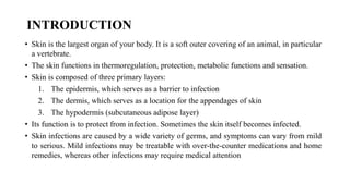
  • 1. INTRODUCTION • Skin is the largest organ of your body. It is a soft outer covering of an animal, in particular a vertebrate. • The skin functions in thermoregulation, protection, metabolic functions and sensation. • Skin is composed of three primary layers: 1. The epidermis, which serves as a barrier to infection 2. The dermis, which serves as a location for the appendages of skin 3. The hypodermis (subcutaneous adipose layer) • Its function is to protect from infection. Sometimes the skin itself becomes infected. • Skin infections are caused by a wide variety of germs, and symptoms can vary from mild to serious. Mild infections may be treatable with over-the-counter medications and home remedies, whereas other infections may require medical attention
  • 2.
  • 3. TYPES OF SKIN INFECTIONS • Bacterial skin infections • Viral skin infections • Fungal skin infections • Parasitic skin infections
  • 4. BACTERIAL SKIN INFECTIONS • Gram positive staphylococcus aureus and beta hemolytic streptococci cause bacterial infections. • Primary infection occur if break present in skin present. • Secondary infection occurs if it followed after already damaged skin or as sign of systematic infection. • Predisposing Factors: Moisture, Obesity, Skin disease, Systematic corticosteroids, Antibiotics, Chronic diseases like diabetes mellitus increase the incidence of infection.
  • 5. IMPETIGO • Impetigo is superficial skin infection caused by staphylococci, streptococci or multiple bacteria, • It involves exposed areas of body, face, hands, neck and extremities. It is often called school sores because it most often affects children. It is quite contagious. • Primary impetigo occurs due to poor hygiene and malnutrition. Secondary impetigo occurs due to pediculosis capitis (Head lice scabies, herpes simplex, insect bite or eczema). CLINICAL MANIFESTATIONS • Impetigo presents with pustules and round, oozing patches which grow larger day by day • There may be clear blisters or golden yellow crusts.
  • 6.
  • 7. CONT.. TREATMENT • Soak moist or crusted areas: Soak a clean cloth in a mixture of half a cup of white vinegar in a liter of tepid water. Apply the compress to moist areas for about ten minutes several times a day. Gently wipe off the crusts. • Antiseptic or antibiotic ointment: apply it two or three times a day to the affected areas and surrounding skin. • Oral antibiotics: Oral antibiotics are recommended if infection is extensive, proving slow to respond to topical antibiotics, or if impetigo is recurrent. The preferred antibiotic is penicillin antibiotic. The complete course should be taken, usually at least 7 days.
  • 8. FOLLICULITIS • Folliculitis means an inflammation or infection of the hair follicles of the skin. It is a problem that is not usually serious. • Tiny pus-filled spots (pustules) develop at the hair where friction, moisture, rubbing or oil is more. • Common sites for folliculitis are the beard, arms, legs, armpits.
  • 9. CONT.. CLINICAL MANIFESTATIONS Superficial folliculitis, which affects the upper part of the hair follicle, may cause: • Clusters of small red or pus-filled bumps that develop around hair follicles • Red and inflamed skin • Itchiness or tenderness Deep folliculitis starts deeper in the skin surrounding the hair follicle and affects the entire hair follicle. Signs and symptoms include: • A large swollen bump or mass • Pus-filled blisters that break open and crust over • Pain
  • 10. CONT.. TREATMENT • Mild folliculitis usually heals on its own in about 2 weeks. • Warm compresses made with white vinegar solution ease itching & helps healing. • Medicated shampoo can be used to treat folliculitis on the scalp or beard. • Antibiotics applied to the skin (mupirocin) or taken by mouth (dicloxacillin). • Gently wash the infected skin twice a day with antibacterial soap. • Avoid shaving irritated skin. If must shave, use an electric razor rather than a blade and apply a soothing aftershave lotion when finished. Also, shave in the direction of hair growth rather than against it. • Don't share towels or washcloths
  • 11. FURUNCLE • Furuncle is another word for "boil." A boil is a bacterial infection of a hair follicle. • The infected hair follicle can be on any part of body. Furuncles mostly occur on body areas prone to excessive perspiration, friction and irritation such as back of neck, thigh, axillae, and perineum or on buttocks.
  • 12. CONT.. CLINICAL MANIFESTATIONS • Deep, firm, red, raised bump on skin which is 1 – 5 cm in diameter, • Pain which gets worse as it fills with pus and dead tissue. • White or yellow centers (pustules) • Spreading to other skin areas or joining with other boils • Weeping, oozing, or crusting • Fatigue • Fever and general ill-feeling • Itching before the boil develops • Skin redness around the boil
  • 13. CONT.. TREATMENT • Put warm, moist compresses on the boil several times a day to speed draining and healing. • Cleanse surrounding skin with antibacterial soap, followed by application of antibacterial ointment. • Surgical incision and drainage may be performed. • Systemic antibiotic therapy is instituted for carbuncles or spreading furuncles. Analgesia and antipyretics are ordered as necessary. • Cover mattress and pillows with plastic and wipe daily with a disinfectant. • Wash all linens, towels, and clothing after each use. • Strict hand washing is maintained to prevent cross-contamination.
  • 14. CARBUNCLES • A carbuncle is a red, swollen, and painful cluster of boils that are connected to each other under the skin. A carbuncle occurs when a group of hair follicles next to each other become infected. It is like a multiple boil. Furuncles may progress to carbuncles. • Carbuncles found on back of neck, upper arm, buttocks and lateral thighs.
  • 15. CONT.. CLINICAL MANIFESTATIONS • The boils that collect to form carbuncles usually start as red, painful bumps. • The carbuncle fills with pus and develops white or yellow tips that weep, ooze, or crust. • Untreated carbuncles rupture, discharging a creamy white or pink fluid. • Superficial carbuncles which have multiple openings on the skin's surface are less likely to leave a deep scar. • Deep carbuncles are more likely to cause significant scarring. • Other carbuncle symptoms include chills, fever, fatigue, and a feeling of general sickness. • Swelling may occur in nearby tissue and lymph nodes, especially lymph nodes in the neck, armpit, or groin.
  • 16.
  • 17. CELLULITIS • Cellulitis is an inflammation of the skin and subcutaneous tissue resulting from a generalized bacterial infection. • Cellulitis appears as a swollen, red area of skin that feels hot and tender. CAUSES AND RISK FACTORS • Staphylococcus and streptococcus bacteria • Cracks or peeling skin between the toes • Weakened immune system • Injury or trauma with a break in the skin (skin wounds) • Insect bites, animal bites. • Ulcers from certain diseases, including diabetes and vascular disease. • Use of corticosteroid medications or medications that suppress the immune system • Intravenous drug use • Obesity • Wound from a recent surgery
  • 18. CONT.. CLINICAL MANIFESTATIONS • Pain or tenderness in the affected area • Skin redness or inflammation (Swelling) • Warm skin in the area of redness • Fever with chills • Malaise • Headache • Swollen lymph nodes • Tight, glossy, stretched appearance of the skin • Hair loss at the site of infection • Joint stiffness caused by swelling of the tissue over the joint
  • 19. CONT.. TREATMENT • Topical/systemic antibiotics are prescribed according to culture and sensitivity test results. • Debridement of nonviable tissue is necessary if there is an open wound. • Systemic antibiotics are indicated if fever and lymphadenopathy are present. • Warm, moist compresses may be ordered. • Immobilization and raise the infected area higher than heart to reduce swelling • Wash wound daily with soap and water • Moisturizing and lubricating skin regularly helps prevent cracking and peeling. • Wear appropriate footwear and gloves. • Regularly check feet for signs of injury. • In severe cases: Hospitalization required
  • 20. DIAGNOSIS OFALL BACTERIAL INFECTIONS Lesion drainage and blood culture tested to identify causative organism. Drug sensitivity also checked. In case of repeated bacterial infections. Culture is taken from external nares to determine carriers of bacteria (MRSA).
  • 21. VIRAL SKIN INFECTIONS • Viral infections are common and can occur anywhere in body. • They range from the common to the rare, from the mild to the severe and from localized skin infection to systemic disease. • The principle site of attack in the skin is the epidermis.
  • 22. ETIOLOGY • Birth control pills and corticosteroids have immune-suppressive properties. • Antibiotics also kill normal skin flora of bacteria that serve as resistance against virus.
  • 23. WARTS • Warts are small, usually painless growths on the skin. Most of the time warts are harmless. • They are caused by a virus called human papillomavirus (HPV). • Warts found on mucous membrane and genital skin of humans. • All warts can spread from one part of your body to another. Warts may be spread from person to person but this is uncommon.
  • 24.
  • 25. CONT.. TYPES 1. Common wart (Verruca vulgaris): most commonly appears on fingers. They grow above the skin surface and they are usually dome shaped. 2. Planter warts (Verruca plantaris): occur on bottom surface of foot. They grow inwards as pressure of shoes or walking prevents its outward growth. These warts extend deeper beneath the skin 3. Flat wart (verruca plana): They are flat lesion present on forehead or dorsum of the hand 4. Condylomata acuminata or venereal warts: occur in moist areas - Glans of penis, anal region, on vulva & cervix. They are pink or purple in color and have cauliflower appearance.
  • 26.
  • 27.
  • 28. CONT.. CLINICAL MANIFESTATIONS • Most warts are raised with a rough surface. They may be round or oval. • The spot where wart grows may be lighter or darker than the other skin. Rarely, warts are black. • Some warts have smooth or flat surfaces (very rare). • Some warts may cause pain.
  • 29. CONT.. TREATMENT 1. Chemicals: • Typical chemicals like salicylic acid, trichloroacetic acid, 5- • fluorouracil, cantharidin, tretinoin etc. are used. • Flat warts are often treated with peeling agents such as tretinoin or salicylic acid. Imiquimod can be used for the treatment of genital warts. • Chemicals usually require multiple applications over several weeks to months. • Applied every 12-24 hours and warts disappear within 2-3 days.
  • 30. CONT.. TREATMENT 2. Freezing (Cryotherapy): Freezing is safe. Warts may be frozen with various commercial freezing probes or with liquid nitrogen sprayed on or applied with a cotton swab. Cryotherapy is often used for plantar warts, and warts under the fingernails. 3. Burning and Cutting: These methods are effective but are more painful and usually leave a scar. A laser or electrical current is used to burn warts off.
  • 31.
  • 32. CONT.. PREVENTION Avoiding cross-contamination can reduce the risk of spreading warts. • Don't bite fingernails: Warts occur more often in skin that has been broken. Nibbling the skin around fingernails opens the door for the virus. • Groom with care: In order to avoid spreading the virus, don't brush, clip, comb or shave areas that have warts. • Don't pick at warts: Picking may spread the virus. Consider covering warts with an adhesive bandage to discourage picking. • Keep hands dry: Warts are more difficult to control in a moist environment.
  • 33. HERPES SIMPLEX Herpes simplex virus infection is a common viral infection Of the skin that tends to recur repeatedly. It is also called fever blister or cold sore. There are two types of herpes simplex: 1. Type I virus (HSV-I), which occurs above the waist on lips, face and mouth and causes a fever blister or cold sore. 2. Type Il virus (HSVII), which occurs below the waist and causes genital herpes. MODES OF TRANSMISSION • It is transmitted by direct contact, respiratory droplet, or contact with virus containing fluid-saliva or cervical secretions in the form of kissing and sexual activity. • Trauma, sunlight, menses, stress and systematic infection often cause reoccurrence of infection.
  • 34. CONT.. CLINICAL MANIFESTATIONS • Burning and tingling sensation at the site for a few hours before eruption. • Area becomes erythematous and swollen • Vesicles and pustules erupt in 1 to 2 days • Lesions can burn, itch, and be painful • Vesicles become pustules, ulcers and crusting until healing occurs in 10-14 days • Fever, sore throat are systematic manifestations of initial infection • The patient is contagious until scabs are formed
  • 35. CONT.. DIAGNOSTIC EVALUATIONS • Assess the appearance of lesions. • Tzanck smear identifies herpes virus. • Vesicle fluid culture differentiate herpes virus from bacterial infections.
  • 36. CONT.. TREATMENT • There is no complete cure for herpes simplex. • Topical acyclovir (Zovirax) ointment is the drug of choice for primary lesions, to suppress the multiplication of vesicles. • Oral acyclovir may be recommended for severe attacks or immunosuppressed patients. Lotions, creams, and ointments may be prescribed to accelerate drying and healing of lesions (e.g., camphor, phenol, alcohol). • Antibiotics may be indicated for secondary infections.
  • 37. HERPES ZOSTER (SHINGLES) • Herpes zoster, or shingles, is an acute inflammatory viral infection that produces a painful vesicular eruption along the distribution of nerves from one or more posterior ganglia. This eruption follows the course of the cutaneous sensory nerve and is almost always unilateral. • It's caused by the varicella-zoster virus, which is the same virus that causes chickenpox. The rash most often appears as a band of rashes or blisters in one area of the body.
  • 38.
  • 39.
  • 40. Where does shingles come from? When you have chickenpox as a child, your body fights off the varicella-zoster virus and the physical signs of chickenpox fade away, but the virus always remains in your body. In adulthood, sometimes the virus becomes active again. This time, the varicella- zoster virus makes its second appearance in the form of shingles.
  • 41. CAUSES AND RISK FACTORS • Previously suffered from chicken pox • Most common in the elderly people above 60 years. • With a weakened immune system (such as people with cancer, HIV, organ transplant recipients or those receiving chemotherapy). • Patient injury to the spine or a cranial nerve • Who are under stress.
  • 42. PATHOPHYSIOLOGY • Herpes virus reactivate Varicella virus (latent infection present in spinal nerve root called the dorsal root ganglion) • When latent infection reactivated — Virus travel by way of peripheral nerves to skin • Virus multiples and create blisters
  • 43.
  • 44. STAGES • First, a few days before the rash appears, there may pain in an area on the skin. The pain is described as itching, burning, stabbing or shooting. • Next, the raised rash appears as a band or a patch, usually on one side of body. The rash usually appears around waistline or on one side of face, neck, or on the trunk (chest/abdomen/back), but not always. It can occur in other areas including arms and legs. • Within three to four days, the rash develops into red, fluid-filled, painful, open blisters. • Usually, these blisters begin to dry out and crust over within about 10 days. • The scabs clear up about two to three weeks later. • It can take three to five weeks from the time begin to feel symptoms until the rash totally disappears.
  • 45. CLINICAL MANIFESTATIONS Early symptoms of shingles may include: • Fever. • Chills. • Headache. • Feeling tired. • Sensitivity to light. • Stomach upset. Other signs and symptoms that appear a few days after the early symptoms include: • An itching, tingling or burning feeling in an area of skin. • Redness on skin in the affected area. • Raised rash in a small area of skin. • Fluid-filled blisters that break open then scab over. • Mild to severe pain in the area of skin affected.
  • 46. DIAGNOSIS AND TESTS Distribution or appearance of the rash: The blisters of a shingles rash usually appear in a band on one side of your body. Shingles also may be diagnosed in a laboratory using scrapings or a swab of the fluid from the blisters.
  • 47. TREATMENT There is no cure for shingles but there are treatments for managing the symptoms. Treatment is aimed at controlling the outbreak, reducing pain and discomfort, and preventing complications. • Antiviral drugs: such as acyclovir (Zovirax), famciclovir (Famvir), and valacyclovir (Valtrex), interfere with viral replication. • Pain management: aspirin, acetaminophen, NSAIDs, opioids are useful during the acute stage. • Topical steroids should not be applied if a secondary infection is present because they suppress the immune system. • Anti-inflammatory drugs like prednisone may be prescribed if shingles affects the eyes or other parts of the face. • Antihistamines are administered to control itching. • Antibiotics are prescribed for secondary bacterial infections.
  • 48. NURSING MANAGEMENT Nursing Diagnosis: 1. Acute Pain may be related to presence of localized inflammation and open lesions, possibly evidenced by verbal reports, distraction behaviors, and restlessness. 2. Risk for [secondary] Infection: risk factors may include broken/traumatized tissue, altered immune response, and untreated infection/ treatment failure. 3. Risk for ineffective sexuality pattern: risk factors may include lack of knowledge, and/or fear of transmitting the disease.
  • 49. CONT.. Nursing interventions • Monitor location, duration and intensity of pain. Monitor for adverse effects of pain medications. • Assess patient's level of discomfort and medicate as prescribed. • Encourage distraction techniques such as music therapy. • Teach relaxation techniques, such as deep breathing, progressive muscle relaxation, and imagery, to help control pain. • Administer antiviral medication in dosage prescribed (usually high dose).
  • 50. CONT.. • Apply antibacterial ointments (after acute stage) as prescribed, to soften and separate adherent crusts and prevent secondary infection. • Teach patient to use proper hand-washing technique, to avoid secondary infection. • Advice the patient to observe signs of systematic infection i.e. fever, malaise, headache, increased redness, formation & drainage of pustules. • Maintain strict isolation. • Wear gown & gloves if contact has to be established with patient. • Keep patient’s room cool and avoid heavy clothes and bedding.
  • 51. COMPLICATIONS • Postherpetic neuralgia: After the shingles rash has disappeared, there might continue to have nerve pain in that same area, last for months or years and become quite severe. More than 10% of people who get shingles develop postherpetic neuralgia. It may be that nerves become more sensitive or that the virus may be invading and damaging the central nervous system. • Other complications include: • Other types of nerve issues like numbness or itching. • A bacterial infection of the shingles rash. • Eye and ear inflammation if the rash is near these organs.
  • 52. FUNGAL SKIN INFECTIONS • Fungi are free living plant like organism that live in the soil, on animals and plants. • Fungus disorder also called mycoses. • Fungi that cause superficial skin infections are called dermatophytes. • Tinea is the term used to describe fungal skin infections which is also called ringworm because of its characteristic appearance of ring. • Fungal infections are common in skin folds, between the toes and in mouth.
  • 53. CONT.. MODE OF TRANSMISSION • Direct skin to skin contact with infected persons or animals • Contact with inanimate object such as combs, pillow cover, towel and hats. PREDISPOSING FACTORS • Moisture is an important factor in the development of fungal infection. • Prolonged use of antibiotics • Diabetes mellitus • Immunodeficiency • Nutritional deficiencies • Increasing age • Iron deficiency diseases
  • 54. TINEA PEDIS • Tinea pedis is a foot infection due to a dermatophyte fungus. • It is fungal infection of the soles of the feet, space between toes or toe nails. • It is also called Ringworm of feet or Athlete's foot. Athlete's foot is contagious. • Athlete’s foot is caused by a microscopic fungus called trichophyton rubrum. SIGNS AND SYMPTOMS • Redness, blisters, swelling, itching or burning • Scaly or peeling skin • Bad smelling feet • Thick, dark skin on the bottoms or sides of feet • Thick, abnormal toenails
  • 55. TINEA CORPORIS (RING WORM OF BODY) • Tinea corporis (ringworm) is the infection of the trunk, legs or arms with a dermatophyte fungus. It's characterized by a red circular rash with clearer skin in the middle. • Tinea corporis affects face, neck, trunk, extremities and caused by several different fungi. • Most commonly found in a child younger than 15 years old. • Heat and humidity aggravate the infection. SIGNS AND SYMPTOMS • Red ring-shaped patches with a raised scaly border ranging from I to 10 cm. • The central area may be clear of any redness. • The border of the affected skin may contain blisters, bumps, or scabs. • Itching or burning sensation
  • 56. TINEA CAPITIS • Tinea capitis is a scalp infection caused by a fungus. It is also called ringworm of the scalp or head. • It is a highly contagious infection, is most common in toddlers and school-age children. • The most common species of fungi affecting humans are Microsporum canis and Tricophyton verrucosum. SIGNS AND SYMPTOMS • Hair loss, fragile hair • Raised scaly skin, • Itchy scalp • Tender or painful areas on the scalp
  • 58. TINEA VERSICOLOR • Tinea versicolor is a long-term (chronic) fungal infection of the skin, also called pityriasis versicolor • It is caused by a type of yeast that naturally lives on skin, called Pityrosporum ovale. When the yeast grows out of control, the skin disease results, which appears as a rash. • A number of factors may trigger this growth, including: • Hot, humid weather • Excessive sweating • Oily skin • Hormonal changes • Weakened immune system • Certain medicines, such as steroids or birth control pills. • It is not spread from one person to another.
  • 59. CONT.. CLINICAL MANIFESTATIONS • Patches of discolored skin: white, pink or dark brown, located on the back, chest, neck and upper arms • Slow-growing, scaly and mildly itchy • More noticeable after sun exposure • Increased sweating and Itching
  • 60. TINEA CRURIS • Jock itch (tinea cruris) is a fungal infection that affects the skin of genitals, inner thighs and buttocks. Jock itch causes an itchy, red, often ring-shaped rash in these warm, moist areas of body. • Jock itch gets its name because it is common in people who sweat a lot, as do athletes. • Men who play sports and use a male athletic guard are particularly prone to develop this tinea infection.
  • 61. TINEA UNGUIUM • Fungal nail infections is a condition in which a fungal or yeast infection occurs in the nail plate that causes the nails to be thick and brittle. It is caused by Trichophyton species or candida albicans. RISK FACTORS • Diminished blood circulation • Prolonged exposure to fungi • Perspiring heavily • Working in a humid or moist environment • AIDS and diabetes SIGNS & SYMPTOMS • Occasionally, it can cause pain or discomfort • The nail usually is brittle, thickened, dull, and easily breakable • Debris accumulates under nail plate and nail plate separates • Sometimes, surrounding skin can become infected and cause cellulitis (inflammation)
  • 62. CANDIDIASIS • Candidiasis is an infection caused by a yeast-like fungus called Candida albicans. • Candidiasis normally found on mucous membranes, on skin, in vagina and in gastrointestinal tract. • Oral infections, called oral thrush, are most common in infants, elderly people, and those with a weakened immune system. PREDISPOSING FACTORS • Broad-spectrum antibiotics, Corticosteroid treatment • Central venous catheters or parenteral nutrition • Pregnancy, Use of birth control pills • Poor nutrition • Diabetes mellitus • Malignancies of the blood • GI tract surgery • Renal replacement surgery
  • 63. CONT.. CLINICAL MANIFESTATION Oral Candidiasis (Thrush): • Creamy white (curd - like) patches in the mouth or on the throat • Redness and difficulty swallowing • Painful cracks at the corners of the mouth Candidiasis of the Genitals: • Extreme itching in the vaginal area, redness • White, clumpy vaginal discharge • Painful intercourse • Red rash on penis • Itching or burning on the tip of the penis Skin Infection: Skin rashes, patches, and blisters found most commonly in the groin, between fingers and toes, and under the breasts.
  • 64.
  • 65. NURSING MANAGEMENT FOR FUNGAL INFECTION 1. Wash hands and teach other caregivers to wash hands before contact with patient, and between procedures with patient. 2. Encourage intake of protein- and calorie-rich foods to maintain optimal nutritional status. 3. Encourage fluid intake of 2000 ml to 3000 ml of water per day to promote diluted urine and frequent emptying of bladder 4. Advice the person not to sharing the linen and personal items like towel, comb etc. Use a clean towel each day. 5. Carefully dry all skin folds including under breast, under arms & between toes. Wear clean under clothes and it should not be very tight fitted. 6. Do not wear same shoes everyday. Do not wear rubber or plastic soled shoes. 7. Use talcum powder or antifungal agents (clotrimazole) twice a day. 8. For Candidiasis, advice the patient that sexual partners should also be treated with antifungal medicines. Use condom or abstinence.
  • 67. Introduction • Skin cancer is the most commonly diagnosed cancer. • Most skin cancers start in the top layer of skin, called the epidermis. There are 3 main types of cells in this layer 1. Squamous cells: These are flat cells in the upper (outer) part of the epidermis, which are constantly shed as new ones form. When these cells grow out of control, they can develop into squamous cell skin cancer (also called squamous cell carcinoma). 2. Basal cells: These cells are in the lower part of the epidermis. They constantly divide to form new cells to replace the squamous cells. As these cells move up in the epidermis, they get flatter, eventually becoming squamous cells. Skin cancers that start in the basal cell layer are called basal cell skin cancers or basal cell carcinomas.
  • 68. Cont… 3. Melanocytes: These cells make the brown pigment called melanin, which gives the skin its tan or brown color. Melanin acts as the body’s natural sunscreen, protecting the deeper layers of the skin from some of the harmful effects of the sun. Melanoma skin cancer starts in these cells. The epidermis is separated from the deeper layers of skin by the basement membrane. When a skin cancer becomes more advanced, it generally grows through this barrier and into the deeper layers.
  • 69. Types of skin cancers The most common types of skin cancer are: 1. Basal Cell Carcinoma (BCC) 2. Squamous cell (epidermoid) carcinoma (SCC). 3. Malignant melanoma
  • 70. Risk Factors for Skin Cancer • Damage in the ozone layer • Fair-skinned and fair-haired people due to insufficient skin pigmentation • Prolonged exposure to sun light. • Exposure to chemical pollutants(arsenic, nitrates, coal, pitch, oils and paraffins) • History of x-ray therapy for acne or benign lesions • Scars from severe burns • Chronic skin irritations • Immunosuppression • Genetic factors
  • 71. Basal cell carcinoma • Basal cell carcinoma is most common type of skin cancer. About 8 out of 10 skin cancers are basal cell carcinomas. • These cancers usually develop on sun-exposed areas, especially the face, head, and neck. • BCC is a locally invasive malignancy and tend to grow slowly. But if it's left untreated, basal cell cancer can grow into nearby areas and invade the bone or other tissues beneath the skin. • It rarely metastasizes, but recurrence is common. • BCC usually begins as a small, waxy nodule. As it grows, it undergoes central ulceration and sometimes crusting.
  • 72.
  • 73. Squamous cell carcinoma • About 2 out of 10 skin cancers are squamous cell carcinomas. SCC is less common than BCC. • It frequently occurs on sun-exposed skin or at the base of skin lesion. They can also develop in scars or chronic skin sores elsewhere. • Squamous cell cancers can usually be removed completely, although they are more likely than basal cell cancers to grow into deeper layers of skin and spread to other parts of the body. • SCC appears as a rough, thickened, scaly tumor. The border of an SCC lesion may be wider, more infiltrated, and more inflammatory than that of a BCC lesion.
  • 74.
  • 75.
  • 76. Kaposi’s sarcoma • Kaposi’s sarcoma (KS) has received renewed attention since its association with HIV infection and AIDS. • Most patients have nodules or plaques on the lower extremities that rarely metastasize beyond the lower extremities. • This KS is chronic, relatively benign, and rarely fatal.
  • 77. Malignant Melanoma • Melanoma is a very serious form of skin cancer. It is cancer of the melanocytes. • When skin is exposed to sunlight, melanocytes produce more pigment, causing the skin to tan. • Sometimes, clusters of melanocytes form noncancerous (benign) growths called moles (nevus). Moles can be either flat or raised, round or oval, generally harmless, but can become cancerous • A malignant melanoma is a cancerous neoplasm in which abnormal melanocytes are present in the epidermis and the dermis (and sometimes the subcutaneous cells). • Most lethal of all the skin cancers
  • 78. Causes of Melanoma • 90% of all melanomas are linked to UV radiation. (Sun exposure) • 8% are due to chromosomal abnormalities • About 2% are unknown Risk Factors • Family history of melanoma • Dysplastic nevi (noncancerous, but unusual- looking moles) • Previous melanoma • Many nevi (ordinary moles): more than 50 • Severe, blistering sunburns • Fair skin
  • 79. Histomorphology types of Melanoma 1. Superficial Spreading Melanoma 2. Nodular Melanoma 3. Lentigo Maligna Melanoma 4. Acral Lentiginous Melanoma
  • 80. Cont.. 1. Superficial Spreading Melanoma • Most commonly present (about 70% of all cases). • Common between 25-50 years age group. • Common sites: Male — back; Female — legs. • Diameter 2 cm. • Appear as a flat, pigmented lesion growing in the radial pattern.
  • 81. Cont.. 2. Nodular Melanoma • Second most common type (15% – 20%) • Raised and dome shaped lesion • Lesion's diameter is about 1 to 2 cm. • Vertical growth pattern • Worst prognosis based on a higher average tumor thickness.
  • 82. Cont.. 3. Lentigo Maligna Melanoma • Lesions are flat, large and darkly pigmented • History of slow development • Commonly occurs in face
  • 83. Cont.. 4. Acral Lentiginous Melanoma • Commonly present in individual with dark coloured skin • Lesions are present in soles, palm and nail beds • Diameter of lesion is about 3 mm.
  • 84. Signs and symptoms of melanoma • Melanoma can appear suddenly as a new mole, or it can develop slowly in or near an existing mole. • In men, melanomas are often found between the shoulders and hips, or the head and neck area. • In women, melanoma often develops on the lower legs as well as between the shoulders and hips. • It may also appear under the fingernails or toenails or on the palms or soles
  • 85. Indicators of melanoma A is for Asymmetry: – One half of a mole or birthmark does not match the other. B is for Border: – The edges are irregular, ragged, notched, or blurred. C is for Color: – The color is not the same all over and may include shades of brown or black, or sometimes with patches of pink, red, white or blue. D is for Diameter: – The spot is larger than 6 millimeters across (about ¼ inch – the size of a pencil eraser) E is for Evolving: – The mole is changing in size, shape, or color.
  • 86. Stage (Clark’s level or Breslow Depth)
  • 87.
  • 88. Clark Classification (Level of Invasion) • Level I: Lesions involving only the epidermis (in situ melanoma); not an invasive lesion. • Level II: Invasion of the papillary dermis but does not reach the papillary- reticular dermal interface. • Level III: Invasion fills and expands the papillary dermis but does not penetrate the reticular dermis. • Level IV: Invasion into the reticular dermis but not into the subcutaneous tissue. • Level V: Invasion through the reticular dermis into the subcutaneous tissue.
  • 89. Breslow level of invasion • Current stage system is based on depth of invasion • Measured using ocular micrometer • Currently Breslow's level of < 1mm, 1 to 4mm and > 4 mm is used for TNM staging
  • 90. TNM staging The American Joint Committee on Cancer (AJCC) has designated staging by TNM. • T = tumor size/ thickness • N = lymph node involvement /regional metastasis • M = distal metastasis
  • 91. CONT.. Primary Tumor (T) T0: No evidence of primary tumor Tis: Melanoma in situ T1: Melanomas 1.0 mm or less in thickness T2: Melanomas 1.01 – 2.0 mm T3: Melanomas 2.01 – 4.0 mm T4: Melanomas more that 4.0 mm
  • 92. CONT.. Regional Lymph Nodes (N) N0: No regional metastases detected N1: 1 node N2: 2-3 nodes N3: 4 or more metastatic nodes, or matted nodes.
  • 93. CONT.. Distant Metastasis (M) M0: No detectable evidence of distant metastases M1a: Metastases to skin, subcutaneous, or distant lymph nodes M1b: Metastases to lung M1c: Metastases to all other visceral sites or distant metastases to any site combined with an elevated serum LDH
  • 94. CONT.. Stage Primary tumor (T) Nodal involvement (N) Metastasis (M) 0 Tis N0 M0 I A T1 N0 M0 I B T (1 – 2) N0 M0 II A T (2 – 3) N1 M0 II B T (3 – 4) N2 M0 II C T4 N3 M0 III A T (1 – 4) N1 M0 III B T (1 – 4) N2 M0 III C Any T N (2 – 3) M0
  • 95. Diagnostic study • History taking including risk factors. • Physical examination. • Imaging techniques • Chest x ray • CT Chest and abdomen • PET scan • MRI brain • Biopsy: • Incisional biopsy • Excisional biopsy • Ultrasound core biopsy
  • 96. Management of skin cancer The management includes • Surgical excision • Mohs’ micrographic surgery • Electrosurgery • Cryosurgery • Radiation therapy • Chemotherapy
  • 97. Cont.. Surgical excision • The primary goal is to remove the tumor entirely. • When the tumor is large, reconstructive surgery with use of a skin flap or skin grafting may be required. • A pressure dressing applied over the wound provides support.
  • 98. Cont.. Mohs’ Micrographic Surgery: • The procedure removes the tumor layer by layer. • The first layer excised includes all evident tumor and a small margin of normal-appearing tissue. • The specimen is frozen and analyzed by section to determine if all the tumor has been removed. • If not, additional layers of tissue are shaved and examined until all tissue margins are tumor free. • In this manner, only the tumor and a safe, normal-tissue margin are removed
  • 99. Cont.. Electro surgery • Electro surgery is the destruction or removal of tissue by electrical energy. • The current is converted to heat, which then passes to the tissue from an electrode.
  • 100. Cont.. Cryosurgery • Cryosurgery destroys the tumor by deep freezing the tissue. • A thermocouple needle apparatus is inserted into the skin, and liquid nitrogen is directed to the center of the tumor until the tumor base is −40°C to −60°C.
  • 101. Cont.. Radiation Therapy • Expose the tumor surface to ionizing and non-ionizing radiation to kill the cancerous cells Chemotherapy • Chemotherapy may be used for metastatic melanoma. Lymphadenectomy • Complete dissection of the lymph nodes to rule out metastasis.
  • 102. Nursing diagnoses • Acute pain related to surgical excision and grafting • Anxiety and depression related to possible life-threatening consequences of melanoma • Deficient knowledge related to early signs of melanoma • Risk for infection related to break in the skin barrier