SlideShare una empresa de Scribd logo
1 de 24
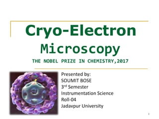
Cryo-Electron
Microscopy
THE NOBEL PRIZE IN CHEMISTRY,2017
1
Presented by:
SOUMIT BOSE
3rd Semester
Instrumentation Science
Roll-04
Jadavpur University
2
What is Microscopy?
Microscopy is the technical field of using microscopes to view object that cannot
be seen with the naked eye. There are three well known branches of microscopy:
1. Optical Microscopy
2. Electron Microscopy
3. Scanning probe Microscopy
Optical and electron microscopy involves the diffraction , reflection or refraction
of electromagnetic radiations or electron beam interacting with the specimen
Scanning Probe Microscopy involves the interaction of a scanning probe with the
surface of the object of interest.
Light Microscopy Electron Microscopy
Can study live
Color imaging
Relatively fast
cells Can study ultra-structure
Need to kill and ‘fix’ cells
Difficult and time consuming
Expensive
HIGH Resolution (upto 2 nm)
HIGH Magnification upto
100000x(SEM) and
250000x(TEM)
and easy
Relatively cheap
LOW Resolution (Limit
200 nm)
LOW Magnification upto
1000x
3
How Do We Study Cells?
Electron Microscopy
Two types of the Electron Microscope:
Transmission Electron Microscope (TEM): A beam of
electrons interacts with the specimen to form an image

Scanning Electron Microscope (SEM): A beam of electrons
scans the sample surface to create image

4
Cryo-Electron Microscopy
A Form of TEM; sample is studied at Cryogenic
Temperatures

Native state of specimen is maintained ;
Specimens are observed in vitreous ice
Cryo-fixation; Rapid freezing of sample
Specimens are not stained or fixed

5
Automated 3D image to get high resolution images
Origin and Development
6
In 1975, Joachim Frank began work on the algorithms that would analyze
fuzzy 2D images and reconstruct them into sharp 3D structures.
In the early 1980s, Jacques Dubochet succeeded in vitrifying water, which
allowed the biomolecules to retain their shape even in high vacuum.
In 1990, Richard Henderson was the first to use an electron microscope to
generate a 3D image of a protein at atomic resolution.
Why Use It?
7
Native state of sample is maintained – Cryo electron microscopy was meant to fight
radiation damage for biological specimens. The amount of radiation required to collect an
image of a specimen in the electron Microscope (EM) is high enough to damage delicate
structures of Specimen .In addition, the High vacuum required on the column of an
electron microscope makes the environment for the sample quite harsh.
Use of Vitreous ice to preserve biological sample – The biological sample is preserved by
Use of vitreous/amorphous ice. Common ice is a crystalline material where the molecules
are regularly arranged in a hexagonal lattice whereas amorphous ice is distinguished by a
lack of long-range order in its molecular arrangement. Amorphous ice is produced
by rapid cooling of liquid water (so the molecules do not have enough time to form a
crystal lattice) at low temperatures.
High resolution and Magnification Power- Resolution (~2nm) can be achieved with magnification
upto atomic resolution level .
3D Reconstruction can be achieved- Cryo electron tomography process( CET) is an imaging
technique used to produce high-resolution (~2 nm) three-dimensional views of samples, typically
biological Macromolecules and cells.
Schematic Diagram of Cryo-EM 8
9
Procedures
Involved in Cryo-EM
Specimen Preparation
Two methods of specimen preparation are:
Thin Film: Specimen is placed on EM grid and is
rapidly frozen without crystallizing it

Vitreous Sections: Larger samples are vitrified
by high pressure freezing, cut thinly and placed
on the EM grid

10
Vitrification
Rapid Cooling is required to avoid the
formation of ice;

Rapid cooling traps the water in a vitrified
state in which it does not crystallize
Vitrified state is maintained by keeping it at
liquid nitrogen temperature(b.p -195.79 c)
Vitrified state can be maintained for long
periods
Sample is placed on carbon grid and dipped
into a bath of ethane held in a container of
liquid nitrogen




11
Cryo-Sectioning
Applicable for whole cells and tissues are too
thick to be spread layer

First vitrify sample and then cut into thin sections using diamond
knives

Sectioning is a difficult task, distortions are made in sample
These distortions cause a loss in order of the structure and
makes it difficult for images to form.

12
Cryo-EM Grids
The grid on which the sample is placed is made from
carbon

High quality carbon grid is used to get better results
Grids are:
A Carbon GridContinuous Films: Enable the sample to cover
the surface as a regular, thin layer.

13
Cryogens
Cryogens are used for Chilling and freezing purpose
Type of cryogen used affects the rate of freezing
Common cryogens are Liquid Nitrogen, Ethane or Propane
Nitrogen is not directly used; It can make crystals due to slow
cooling

14
Observation of the Specimen
The contrast of the specimen depends on:
Specimen itself
Defocus value of the objective lens
Thickness of the ice
There are three methods of observing and recording images:
Fluorescent Screen
Photographic Film
CCD Cameras
15
3D Reconstruction
16
3D images of biological sample is obtained by a process called
Cryo-Electron Tomography(CET),where a 3D reconstruction of a
sample is created from tilted 2D images.
Raw Images from different projections are recorded and at last
they are stitched together to form the required 3D structures
using computer software.
Images of sample
17
Structure of Hepatitis B Virus Solved by
Cryo-EM
18
Example
X RAY CRYSTALLOGRAPHY VS NUCLEUR MAGNETIC
RESONANCE (NMR)VS CRYO ELECTRON MICROSCOPE
19
Pros and Cons
20
ADVANTAGE : Structure remain in native state and no dehydration is
required. Thus the almost exact nature of water based molecules are
obtained.
Atomic resolution level (~2nm) is achieved.
Can be used for examining delicate sample structures .
One of the biggest advantages of cryo-electron microscopy is that it is
applicable for both larger and smaller specimens. Compared to other
microscopy techniques, cry-electron microscopy still produces good
images (as long as the sample is in good condition).
No requirement of staining or fixation of sample.
LIMITATION: It takes time to generate the sample.
Fully hydrated specimen have been shown to be electron-beam sensitive.
It is expensive .
Applications
Nanoparticle Research
Pharmaceutical Drug Research
3D Structure Visualization of:
Single Particles such as Ribosome, tRNA


Viruses
Proteins
Macromolecules; Lipid Vesicles
21
Conclusions
Cryo-EM is a form of Transmission Electron Microscopy (TEM)
where the sample is studied in its native state at cryogenic
temperatures

Used for 3D visualization of biological molecules
Resolution of Cryo-EM is not high enough but it is improving using
different computer techniques

With the advancement of technology, this technique will certainly
Improve.

22
23
ACKNOWLEDGEMENT
I am very grateful for the strong support and guidance
provided to me by Prof. Radhaballabh Bhar Sir who helped
me a lot in preparing this seminar
I am very thankful to him for same !
24

Más contenido relacionado

La actualidad más candente

Fplc(fast protein liquid chromatography )
Fplc(fast protein liquid chromatography )Fplc(fast protein liquid chromatography )
Fplc(fast protein liquid chromatography )Safina Kouser
 
transmission Electron Microscopy (Tem)
transmission Electron Microscopy (Tem)transmission Electron Microscopy (Tem)
transmission Electron Microscopy (Tem)Sivasangari Shanmugam
 
Transmission electron microscope
Transmission electron microscopeTransmission electron microscope
Transmission electron microscopeSenthil Arasan
 
Gel electrophoresis native, denaturing&reducing
Gel electrophoresis native, denaturing&reducingGel electrophoresis native, denaturing&reducing
Gel electrophoresis native, denaturing&reducingLovnish Thakur
 
Quantum Dots and its applications
Quantum Dots and its applicationsQuantum Dots and its applications
Quantum Dots and its applicationsArun Kumar
 
X ray crystallography
X ray crystallographyX ray crystallography
X ray crystallographyBhaskar Das
 
Pulse Field Gel Electrophoresis
Pulse Field Gel ElectrophoresisPulse Field Gel Electrophoresis
Pulse Field Gel ElectrophoresisPankaj Gaonkar
 
Characterization of nanopartical
Characterization of nanoparticalCharacterization of nanopartical
Characterization of nanoparticalAmany EL-Hallaq
 
Mass Spectrometry: Protein Identification Strategies
Mass Spectrometry: Protein Identification StrategiesMass Spectrometry: Protein Identification Strategies
Mass Spectrometry: Protein Identification StrategiesMichel Dumontier
 
Scanning electron microscopy
Scanning electron microscopyScanning electron microscopy
Scanning electron microscopyJessa Ariño
 

La actualidad más candente (20)

Fplc(fast protein liquid chromatography )
Fplc(fast protein liquid chromatography )Fplc(fast protein liquid chromatography )
Fplc(fast protein liquid chromatography )
 
transmission Electron Microscopy (Tem)
transmission Electron Microscopy (Tem)transmission Electron Microscopy (Tem)
transmission Electron Microscopy (Tem)
 
Dna microarray mehran
Dna microarray  mehranDna microarray  mehran
Dna microarray mehran
 
microscopy
 microscopy microscopy
microscopy
 
ICAT Technique in Proteomics
ICAT Technique in ProteomicsICAT Technique in Proteomics
ICAT Technique in Proteomics
 
Quantum Dots PPT
Quantum Dots PPTQuantum Dots PPT
Quantum Dots PPT
 
Electron microscope
Electron microscopeElectron microscope
Electron microscope
 
Proteomics ppt
Proteomics pptProteomics ppt
Proteomics ppt
 
Transmission electron microscope
Transmission electron microscopeTransmission electron microscope
Transmission electron microscope
 
Scanning electron microscopy (sem)
Scanning electron microscopy (sem)Scanning electron microscopy (sem)
Scanning electron microscopy (sem)
 
CYTOPHOTOMETRY
CYTOPHOTOMETRYCYTOPHOTOMETRY
CYTOPHOTOMETRY
 
Gel electrophoresis native, denaturing&reducing
Gel electrophoresis native, denaturing&reducingGel electrophoresis native, denaturing&reducing
Gel electrophoresis native, denaturing&reducing
 
Quantum Dots and its applications
Quantum Dots and its applicationsQuantum Dots and its applications
Quantum Dots and its applications
 
Protein protein interactions
Protein protein interactionsProtein protein interactions
Protein protein interactions
 
X ray crystallography
X ray crystallographyX ray crystallography
X ray crystallography
 
Pulse Field Gel Electrophoresis
Pulse Field Gel ElectrophoresisPulse Field Gel Electrophoresis
Pulse Field Gel Electrophoresis
 
Characterization of nanopartical
Characterization of nanoparticalCharacterization of nanopartical
Characterization of nanopartical
 
Mass Spectrometry: Protein Identification Strategies
Mass Spectrometry: Protein Identification StrategiesMass Spectrometry: Protein Identification Strategies
Mass Spectrometry: Protein Identification Strategies
 
Scanning electron microscopy
Scanning electron microscopyScanning electron microscopy
Scanning electron microscopy
 
Maldi tof
Maldi tofMaldi tof
Maldi tof
 

Similar a Cryo electron microscopy

CRYO ELECTRON MICROSCOPY.pptx
CRYO ELECTRON MICROSCOPY.pptxCRYO ELECTRON MICROSCOPY.pptx
CRYO ELECTRON MICROSCOPY.pptxMuskanWahab
 
20231122(Cryo-EM).pdf
20231122(Cryo-EM).pdf20231122(Cryo-EM).pdf
20231122(Cryo-EM).pdfelsarazavi1
 
Microscopy
MicroscopyMicroscopy
Microscopytrixie19
 
Recent advancement in microscopic techniques cryoem and super
Recent advancement in microscopic techniques  cryoem and superRecent advancement in microscopic techniques  cryoem and super
Recent advancement in microscopic techniques cryoem and superDanishNigar
 
Electron Microscopy Principle and Application
Electron Microscopy Principle and ApplicationElectron Microscopy Principle and Application
Electron Microscopy Principle and ApplicationKARTHIK REDDY C A
 
Electron microscope - SEM and TEM
Electron microscope - SEM and TEMElectron microscope - SEM and TEM
Electron microscope - SEM and TEMgaurav raja
 
5. Microsocope ELECTRON MICROSCOPE (TEM & SEM ) - Basics
5. Microsocope   ELECTRON MICROSCOPE (TEM & SEM ) - Basics5. Microsocope   ELECTRON MICROSCOPE (TEM & SEM ) - Basics
5. Microsocope ELECTRON MICROSCOPE (TEM & SEM ) - BasicsNethravathi Siri
 
scanning electron microscope
scanning electron microscopescanning electron microscope
scanning electron microscopeDrAkhilaCNV
 
Transmission Electron Microscopy
Transmission Electron MicroscopyTransmission Electron Microscopy
Transmission Electron MicroscopyBijit Nath
 
Cryo_app._note
Cryo_app._noteCryo_app._note
Cryo_app._noteLloyd Peto
 
Electron microscopy ameena
Electron microscopy ameenaElectron microscopy ameena
Electron microscopy ameenaAnnakurian9
 
Lec 3; Microscopy ALI.pptx
Lec 3; Microscopy ALI.pptxLec 3; Microscopy ALI.pptx
Lec 3; Microscopy ALI.pptxalihaider64675
 
Principle of transmission electron microscope.
Principle of transmission electron microscope.Principle of transmission electron microscope.
Principle of transmission electron microscope.naren
 
Principle of transmission electron microscope.
Principle of transmission electron microscope.Principle of transmission electron microscope.
Principle of transmission electron microscope.naren
 
electron microscopy tem
electron microscopy   temelectron microscopy   tem
electron microscopy temkarthi keyan
 
Scanning electron microscope ppt.pptx
Scanning electron microscope ppt.pptxScanning electron microscope ppt.pptx
Scanning electron microscope ppt.pptxkavinkumars23mech
 
Transmission Electron Microscope
Transmission Electron MicroscopeTransmission Electron Microscope
Transmission Electron Microscope20was
 
Scanning Electron Microscope (SEM)
Scanning Electron Microscope (SEM)Scanning Electron Microscope (SEM)
Scanning Electron Microscope (SEM)Amna Jalil
 

Similar a Cryo electron microscopy (20)

Poster
PosterPoster
Poster
 
CRYO ELECTRON MICROSCOPY.pptx
CRYO ELECTRON MICROSCOPY.pptxCRYO ELECTRON MICROSCOPY.pptx
CRYO ELECTRON MICROSCOPY.pptx
 
20231122(Cryo-EM).pdf
20231122(Cryo-EM).pdf20231122(Cryo-EM).pdf
20231122(Cryo-EM).pdf
 
Microscopy
MicroscopyMicroscopy
Microscopy
 
Recent advancement in microscopic techniques cryoem and super
Recent advancement in microscopic techniques  cryoem and superRecent advancement in microscopic techniques  cryoem and super
Recent advancement in microscopic techniques cryoem and super
 
Electron Microscopy Principle and Application
Electron Microscopy Principle and ApplicationElectron Microscopy Principle and Application
Electron Microscopy Principle and Application
 
Electron microscope - SEM and TEM
Electron microscope - SEM and TEMElectron microscope - SEM and TEM
Electron microscope - SEM and TEM
 
5. Microsocope ELECTRON MICROSCOPE (TEM & SEM ) - Basics
5. Microsocope   ELECTRON MICROSCOPE (TEM & SEM ) - Basics5. Microsocope   ELECTRON MICROSCOPE (TEM & SEM ) - Basics
5. Microsocope ELECTRON MICROSCOPE (TEM & SEM ) - Basics
 
scanning electron microscope
scanning electron microscopescanning electron microscope
scanning electron microscope
 
Transmission Electron Microscopy
Transmission Electron MicroscopyTransmission Electron Microscopy
Transmission Electron Microscopy
 
Cryo_app._note
Cryo_app._noteCryo_app._note
Cryo_app._note
 
Electron microscopy ameena
Electron microscopy ameenaElectron microscopy ameena
Electron microscopy ameena
 
Lec 3; Microscopy ALI.pptx
Lec 3; Microscopy ALI.pptxLec 3; Microscopy ALI.pptx
Lec 3; Microscopy ALI.pptx
 
Principle of transmission electron microscope.
Principle of transmission electron microscope.Principle of transmission electron microscope.
Principle of transmission electron microscope.
 
Principle of transmission electron microscope.
Principle of transmission electron microscope.Principle of transmission electron microscope.
Principle of transmission electron microscope.
 
electron microscopy tem
electron microscopy   temelectron microscopy   tem
electron microscopy tem
 
Electron microscope
Electron microscopeElectron microscope
Electron microscope
 
Scanning electron microscope ppt.pptx
Scanning electron microscope ppt.pptxScanning electron microscope ppt.pptx
Scanning electron microscope ppt.pptx
 
Transmission Electron Microscope
Transmission Electron MicroscopeTransmission Electron Microscope
Transmission Electron Microscope
 
Scanning Electron Microscope (SEM)
Scanning Electron Microscope (SEM)Scanning Electron Microscope (SEM)
Scanning Electron Microscope (SEM)
 

Último

ICT role in 21st century education and its challenges
ICT role in 21st century education and its challengesICT role in 21st century education and its challenges
ICT role in 21st century education and its challengesrafiqahmad00786416
 
Biography Of Angeliki Cooney | Senior Vice President Life Sciences | Albany, ...
Biography Of Angeliki Cooney | Senior Vice President Life Sciences | Albany, ...Biography Of Angeliki Cooney | Senior Vice President Life Sciences | Albany, ...
Biography Of Angeliki Cooney | Senior Vice President Life Sciences | Albany, ...Angeliki Cooney
 
Polkadot JAM Slides - Token2049 - By Dr. Gavin Wood
Polkadot JAM Slides - Token2049 - By Dr. Gavin WoodPolkadot JAM Slides - Token2049 - By Dr. Gavin Wood
Polkadot JAM Slides - Token2049 - By Dr. Gavin WoodJuan lago vázquez
 
Architecting Cloud Native Applications
Architecting Cloud Native ApplicationsArchitecting Cloud Native Applications
Architecting Cloud Native ApplicationsWSO2
 
Repurposing LNG terminals for Hydrogen Ammonia: Feasibility and Cost Saving
Repurposing LNG terminals for Hydrogen Ammonia: Feasibility and Cost SavingRepurposing LNG terminals for Hydrogen Ammonia: Feasibility and Cost Saving
Repurposing LNG terminals for Hydrogen Ammonia: Feasibility and Cost SavingEdi Saputra
 
Cloud Frontiers: A Deep Dive into Serverless Spatial Data and FME
Cloud Frontiers:  A Deep Dive into Serverless Spatial Data and FMECloud Frontiers:  A Deep Dive into Serverless Spatial Data and FME
Cloud Frontiers: A Deep Dive into Serverless Spatial Data and FMESafe Software
 
TrustArc Webinar - Unlock the Power of AI-Driven Data Discovery
TrustArc Webinar - Unlock the Power of AI-Driven Data DiscoveryTrustArc Webinar - Unlock the Power of AI-Driven Data Discovery
TrustArc Webinar - Unlock the Power of AI-Driven Data DiscoveryTrustArc
 
Exploring Multimodal Embeddings with Milvus
Exploring Multimodal Embeddings with MilvusExploring Multimodal Embeddings with Milvus
Exploring Multimodal Embeddings with MilvusZilliz
 
AXA XL - Insurer Innovation Award Americas 2024
AXA XL - Insurer Innovation Award Americas 2024AXA XL - Insurer Innovation Award Americas 2024
AXA XL - Insurer Innovation Award Americas 2024The Digital Insurer
 
Exploring the Future Potential of AI-Enabled Smartphone Processors
Exploring the Future Potential of AI-Enabled Smartphone ProcessorsExploring the Future Potential of AI-Enabled Smartphone Processors
Exploring the Future Potential of AI-Enabled Smartphone Processorsdebabhi2
 
MS Copilot expands with MS Graph connectors
MS Copilot expands with MS Graph connectorsMS Copilot expands with MS Graph connectors
MS Copilot expands with MS Graph connectorsNanddeep Nachan
 
DEV meet-up UiPath Document Understanding May 7 2024 Amsterdam
DEV meet-up UiPath Document Understanding May 7 2024 AmsterdamDEV meet-up UiPath Document Understanding May 7 2024 Amsterdam
DEV meet-up UiPath Document Understanding May 7 2024 AmsterdamUiPathCommunity
 
Web Form Automation for Bonterra Impact Management (fka Social Solutions Apri...
Web Form Automation for Bonterra Impact Management (fka Social Solutions Apri...Web Form Automation for Bonterra Impact Management (fka Social Solutions Apri...
Web Form Automation for Bonterra Impact Management (fka Social Solutions Apri...Jeffrey Haguewood
 
Corporate and higher education May webinar.pptx
Corporate and higher education May webinar.pptxCorporate and higher education May webinar.pptx
Corporate and higher education May webinar.pptxRustici Software
 
Manulife - Insurer Transformation Award 2024
Manulife - Insurer Transformation Award 2024Manulife - Insurer Transformation Award 2024
Manulife - Insurer Transformation Award 2024The Digital Insurer
 
Emergent Methods: Multi-lingual narrative tracking in the news - real-time ex...
Emergent Methods: Multi-lingual narrative tracking in the news - real-time ex...Emergent Methods: Multi-lingual narrative tracking in the news - real-time ex...
Emergent Methods: Multi-lingual narrative tracking in the news - real-time ex...Zilliz
 
Navigating the Deluge_ Dubai Floods and the Resilience of Dubai International...
Navigating the Deluge_ Dubai Floods and the Resilience of Dubai International...Navigating the Deluge_ Dubai Floods and the Resilience of Dubai International...
Navigating the Deluge_ Dubai Floods and the Resilience of Dubai International...Orbitshub
 
Spring Boot vs Quarkus the ultimate battle - DevoxxUK
Spring Boot vs Quarkus the ultimate battle - DevoxxUKSpring Boot vs Quarkus the ultimate battle - DevoxxUK
Spring Boot vs Quarkus the ultimate battle - DevoxxUKJago de Vreede
 
"I see eyes in my soup": How Delivery Hero implemented the safety system for ...
"I see eyes in my soup": How Delivery Hero implemented the safety system for ..."I see eyes in my soup": How Delivery Hero implemented the safety system for ...
"I see eyes in my soup": How Delivery Hero implemented the safety system for ...Zilliz
 
Apidays New York 2024 - Passkeys: Developing APIs to enable passwordless auth...
Apidays New York 2024 - Passkeys: Developing APIs to enable passwordless auth...Apidays New York 2024 - Passkeys: Developing APIs to enable passwordless auth...
Apidays New York 2024 - Passkeys: Developing APIs to enable passwordless auth...apidays
 

Último (20)

ICT role in 21st century education and its challenges
ICT role in 21st century education and its challengesICT role in 21st century education and its challenges
ICT role in 21st century education and its challenges
 
Biography Of Angeliki Cooney | Senior Vice President Life Sciences | Albany, ...
Biography Of Angeliki Cooney | Senior Vice President Life Sciences | Albany, ...Biography Of Angeliki Cooney | Senior Vice President Life Sciences | Albany, ...
Biography Of Angeliki Cooney | Senior Vice President Life Sciences | Albany, ...
 
Polkadot JAM Slides - Token2049 - By Dr. Gavin Wood
Polkadot JAM Slides - Token2049 - By Dr. Gavin WoodPolkadot JAM Slides - Token2049 - By Dr. Gavin Wood
Polkadot JAM Slides - Token2049 - By Dr. Gavin Wood
 
Architecting Cloud Native Applications
Architecting Cloud Native ApplicationsArchitecting Cloud Native Applications
Architecting Cloud Native Applications
 
Repurposing LNG terminals for Hydrogen Ammonia: Feasibility and Cost Saving
Repurposing LNG terminals for Hydrogen Ammonia: Feasibility and Cost SavingRepurposing LNG terminals for Hydrogen Ammonia: Feasibility and Cost Saving
Repurposing LNG terminals for Hydrogen Ammonia: Feasibility and Cost Saving
 
Cloud Frontiers: A Deep Dive into Serverless Spatial Data and FME
Cloud Frontiers:  A Deep Dive into Serverless Spatial Data and FMECloud Frontiers:  A Deep Dive into Serverless Spatial Data and FME
Cloud Frontiers: A Deep Dive into Serverless Spatial Data and FME
 
TrustArc Webinar - Unlock the Power of AI-Driven Data Discovery
TrustArc Webinar - Unlock the Power of AI-Driven Data DiscoveryTrustArc Webinar - Unlock the Power of AI-Driven Data Discovery
TrustArc Webinar - Unlock the Power of AI-Driven Data Discovery
 
Exploring Multimodal Embeddings with Milvus
Exploring Multimodal Embeddings with MilvusExploring Multimodal Embeddings with Milvus
Exploring Multimodal Embeddings with Milvus
 
AXA XL - Insurer Innovation Award Americas 2024
AXA XL - Insurer Innovation Award Americas 2024AXA XL - Insurer Innovation Award Americas 2024
AXA XL - Insurer Innovation Award Americas 2024
 
Exploring the Future Potential of AI-Enabled Smartphone Processors
Exploring the Future Potential of AI-Enabled Smartphone ProcessorsExploring the Future Potential of AI-Enabled Smartphone Processors
Exploring the Future Potential of AI-Enabled Smartphone Processors
 
MS Copilot expands with MS Graph connectors
MS Copilot expands with MS Graph connectorsMS Copilot expands with MS Graph connectors
MS Copilot expands with MS Graph connectors
 
DEV meet-up UiPath Document Understanding May 7 2024 Amsterdam
DEV meet-up UiPath Document Understanding May 7 2024 AmsterdamDEV meet-up UiPath Document Understanding May 7 2024 Amsterdam
DEV meet-up UiPath Document Understanding May 7 2024 Amsterdam
 
Web Form Automation for Bonterra Impact Management (fka Social Solutions Apri...
Web Form Automation for Bonterra Impact Management (fka Social Solutions Apri...Web Form Automation for Bonterra Impact Management (fka Social Solutions Apri...
Web Form Automation for Bonterra Impact Management (fka Social Solutions Apri...
 
Corporate and higher education May webinar.pptx
Corporate and higher education May webinar.pptxCorporate and higher education May webinar.pptx
Corporate and higher education May webinar.pptx
 
Manulife - Insurer Transformation Award 2024
Manulife - Insurer Transformation Award 2024Manulife - Insurer Transformation Award 2024
Manulife - Insurer Transformation Award 2024
 
Emergent Methods: Multi-lingual narrative tracking in the news - real-time ex...
Emergent Methods: Multi-lingual narrative tracking in the news - real-time ex...Emergent Methods: Multi-lingual narrative tracking in the news - real-time ex...
Emergent Methods: Multi-lingual narrative tracking in the news - real-time ex...
 
Navigating the Deluge_ Dubai Floods and the Resilience of Dubai International...
Navigating the Deluge_ Dubai Floods and the Resilience of Dubai International...Navigating the Deluge_ Dubai Floods and the Resilience of Dubai International...
Navigating the Deluge_ Dubai Floods and the Resilience of Dubai International...
 
Spring Boot vs Quarkus the ultimate battle - DevoxxUK
Spring Boot vs Quarkus the ultimate battle - DevoxxUKSpring Boot vs Quarkus the ultimate battle - DevoxxUK
Spring Boot vs Quarkus the ultimate battle - DevoxxUK
 
"I see eyes in my soup": How Delivery Hero implemented the safety system for ...
"I see eyes in my soup": How Delivery Hero implemented the safety system for ..."I see eyes in my soup": How Delivery Hero implemented the safety system for ...
"I see eyes in my soup": How Delivery Hero implemented the safety system for ...
 
Apidays New York 2024 - Passkeys: Developing APIs to enable passwordless auth...
Apidays New York 2024 - Passkeys: Developing APIs to enable passwordless auth...Apidays New York 2024 - Passkeys: Developing APIs to enable passwordless auth...
Apidays New York 2024 - Passkeys: Developing APIs to enable passwordless auth...
 

Cryo electron microscopy

  • 1. Cryo-Electron Microscopy THE NOBEL PRIZE IN CHEMISTRY,2017 1 Presented by: SOUMIT BOSE 3rd Semester Instrumentation Science Roll-04 Jadavpur University
  • 2. 2 What is Microscopy? Microscopy is the technical field of using microscopes to view object that cannot be seen with the naked eye. There are three well known branches of microscopy: 1. Optical Microscopy 2. Electron Microscopy 3. Scanning probe Microscopy Optical and electron microscopy involves the diffraction , reflection or refraction of electromagnetic radiations or electron beam interacting with the specimen Scanning Probe Microscopy involves the interaction of a scanning probe with the surface of the object of interest.
  • 3. Light Microscopy Electron Microscopy Can study live Color imaging Relatively fast cells Can study ultra-structure Need to kill and ‘fix’ cells Difficult and time consuming Expensive HIGH Resolution (upto 2 nm) HIGH Magnification upto 100000x(SEM) and 250000x(TEM) and easy Relatively cheap LOW Resolution (Limit 200 nm) LOW Magnification upto 1000x 3 How Do We Study Cells?
  • 4. Electron Microscopy Two types of the Electron Microscope: Transmission Electron Microscope (TEM): A beam of electrons interacts with the specimen to form an image  Scanning Electron Microscope (SEM): A beam of electrons scans the sample surface to create image  4
  • 5. Cryo-Electron Microscopy A Form of TEM; sample is studied at Cryogenic Temperatures  Native state of specimen is maintained ; Specimens are observed in vitreous ice Cryo-fixation; Rapid freezing of sample Specimens are not stained or fixed  5 Automated 3D image to get high resolution images
  • 6. Origin and Development 6 In 1975, Joachim Frank began work on the algorithms that would analyze fuzzy 2D images and reconstruct them into sharp 3D structures. In the early 1980s, Jacques Dubochet succeeded in vitrifying water, which allowed the biomolecules to retain their shape even in high vacuum. In 1990, Richard Henderson was the first to use an electron microscope to generate a 3D image of a protein at atomic resolution.
  • 7. Why Use It? 7 Native state of sample is maintained – Cryo electron microscopy was meant to fight radiation damage for biological specimens. The amount of radiation required to collect an image of a specimen in the electron Microscope (EM) is high enough to damage delicate structures of Specimen .In addition, the High vacuum required on the column of an electron microscope makes the environment for the sample quite harsh. Use of Vitreous ice to preserve biological sample – The biological sample is preserved by Use of vitreous/amorphous ice. Common ice is a crystalline material where the molecules are regularly arranged in a hexagonal lattice whereas amorphous ice is distinguished by a lack of long-range order in its molecular arrangement. Amorphous ice is produced by rapid cooling of liquid water (so the molecules do not have enough time to form a crystal lattice) at low temperatures. High resolution and Magnification Power- Resolution (~2nm) can be achieved with magnification upto atomic resolution level . 3D Reconstruction can be achieved- Cryo electron tomography process( CET) is an imaging technique used to produce high-resolution (~2 nm) three-dimensional views of samples, typically biological Macromolecules and cells.
  • 10. Specimen Preparation Two methods of specimen preparation are: Thin Film: Specimen is placed on EM grid and is rapidly frozen without crystallizing it  Vitreous Sections: Larger samples are vitrified by high pressure freezing, cut thinly and placed on the EM grid  10
  • 11. Vitrification Rapid Cooling is required to avoid the formation of ice;  Rapid cooling traps the water in a vitrified state in which it does not crystallize Vitrified state is maintained by keeping it at liquid nitrogen temperature(b.p -195.79 c) Vitrified state can be maintained for long periods Sample is placed on carbon grid and dipped into a bath of ethane held in a container of liquid nitrogen     11
  • 12. Cryo-Sectioning Applicable for whole cells and tissues are too thick to be spread layer  First vitrify sample and then cut into thin sections using diamond knives  Sectioning is a difficult task, distortions are made in sample These distortions cause a loss in order of the structure and makes it difficult for images to form.  12
  • 13. Cryo-EM Grids The grid on which the sample is placed is made from carbon  High quality carbon grid is used to get better results Grids are: A Carbon GridContinuous Films: Enable the sample to cover the surface as a regular, thin layer.  13
  • 14. Cryogens Cryogens are used for Chilling and freezing purpose Type of cryogen used affects the rate of freezing Common cryogens are Liquid Nitrogen, Ethane or Propane Nitrogen is not directly used; It can make crystals due to slow cooling  14
  • 15. Observation of the Specimen The contrast of the specimen depends on: Specimen itself Defocus value of the objective lens Thickness of the ice There are three methods of observing and recording images: Fluorescent Screen Photographic Film CCD Cameras 15
  • 16. 3D Reconstruction 16 3D images of biological sample is obtained by a process called Cryo-Electron Tomography(CET),where a 3D reconstruction of a sample is created from tilted 2D images. Raw Images from different projections are recorded and at last they are stitched together to form the required 3D structures using computer software.
  • 18. Structure of Hepatitis B Virus Solved by Cryo-EM 18 Example
  • 19. X RAY CRYSTALLOGRAPHY VS NUCLEUR MAGNETIC RESONANCE (NMR)VS CRYO ELECTRON MICROSCOPE 19
  • 20. Pros and Cons 20 ADVANTAGE : Structure remain in native state and no dehydration is required. Thus the almost exact nature of water based molecules are obtained. Atomic resolution level (~2nm) is achieved. Can be used for examining delicate sample structures . One of the biggest advantages of cryo-electron microscopy is that it is applicable for both larger and smaller specimens. Compared to other microscopy techniques, cry-electron microscopy still produces good images (as long as the sample is in good condition). No requirement of staining or fixation of sample. LIMITATION: It takes time to generate the sample. Fully hydrated specimen have been shown to be electron-beam sensitive. It is expensive .
  • 21. Applications Nanoparticle Research Pharmaceutical Drug Research 3D Structure Visualization of: Single Particles such as Ribosome, tRNA   Viruses Proteins Macromolecules; Lipid Vesicles 21
  • 22. Conclusions Cryo-EM is a form of Transmission Electron Microscopy (TEM) where the sample is studied in its native state at cryogenic temperatures  Used for 3D visualization of biological molecules Resolution of Cryo-EM is not high enough but it is improving using different computer techniques  With the advancement of technology, this technique will certainly Improve.  22
  • 23. 23 ACKNOWLEDGEMENT I am very grateful for the strong support and guidance provided to me by Prof. Radhaballabh Bhar Sir who helped me a lot in preparing this seminar I am very thankful to him for same !
  • 24. 24