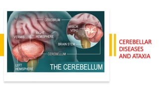
5. CLASSIFICATION
Classification by
phylogenetic & ontogenic
development:
• Archicerebellum
• Paleocerebellum
• Neocerebellum
01
Classification by afferent
connection
• Vestibulo -cerebellum
• Spino-cerebellum
• Ponto-cerebellum
02
Classification by efferent
connection
• Vermis
• Paravermal region
• Cerebellar hemisphere
03
6. NEOCEREBELLUM
• A.k.a lateral zone
• It receives input exclusively from the cerebral cortex (especially
the parietal lobe) via the pontine nuclei (forming the cotico-
ponto-cerebellar pathways)
• It sends output mainly to the ventrolateral thalamus
premotor cortex & primary motor area & to the red nucleus
providing modulation of descending motor systems
coordination of movements.
7. PALEOCEREBELLUM
• It receives proprioception input from the dorsal columns of the spinal
cord(including the spinocerebellar tract)
• It is responsible for maintenance of tone.
8. ARCHICEREBELLUM
• Comprises of the flocculonodular lobe or the vestibulocerebellum
. It has the primary connections with the vestibular nuclei.
• It is responsible for equilibrium.
9. BLOOD SUPPLY
• Superior cerebellar artery (branch of basilar artery)
• Anterior inferior cerebellar artery (branch of basilar artery)
• Posterior inferior cerebellar artery (branch of vertebral
artery)
11. FUNCTIONAL AREAS OF THE CEREBELLUM
• The cortex of the vermis influences the movements of the long axis of the
body, namely, the neck, the shoulders, the thorax, the abdomen, and the hips.
• Immediately lateral to the vermis is a so-called intermediate zone of the
cerebellar hemisphere. This area has been shown to control the muscles of
the distal parts of the limbs, especially the hands and feet.
• The lateral zone of each cerebellar hemisphere appears to be concerned with
the planning of sequential movements of the entire body and is involved with
the conscious assessment of movement errors.
15. HENCE 3 IMPORTANT FUNCTIONS ARE :
1. EQUILIBRIUM
2. MAINTENANCE OF TONE
3. COORDINATION OF MOVEMENT
16. Lesion in one cerebellar hemisphere gives rise to signs and
symptoms that are limited to the same side of the body.
17. GENERAL SIGNS AND SYMPTOMS OF
CEREBELLAR DISORDERS
• Hypotonia
The muscles lose resilience to palpation. There is diminished resistance to passive movements of joints.
Shaking the limb produces excessive movements at the terminal joints. The condition is attributable to
loss of cerebellar influence on the simple stretch reflex.
• Postural Changes and Alteration of Gait
The head is often rotated and flexed, and the shoulder on the side of the lesion is lower than on the
normal side. When the individual walks, he or she lurches and staggers toward the affected side.
• Disturbances of Voluntary Movement (Ataxia)
The muscles contract irregularly and weakly. Tremor occurs when fine movements, such as buttoning
clothes, writing, and shaving, are attempted. Muscle groups fail to work harmoniously . When the
patient is asked to touch the tip of the nose with the index finger, the movements are not properly
coordinated.A similar test can be performed on the lower limbs by asking the patient to place the heel of
one foot on the shin of the opposite leg.
18.
19. • Dysdiadochokinesia
Dysdiadochokinesia is the inability to perform alternating movements regularly
and rapidly. Ask the patient to pronate and supinate the forearms rapidly. On the
side of the cerebellar lesion, the movements are slow, jerky, and incomplete.
• Disturbances of Reflexes
Movement produced by tendon reflexes tends to continue for a longer period of
time than normal. The pendular knee jerk, for example, occurs following tapping
of the patellar tendon. In cerebellar disease, because of loss of influence on the
stretch reflexes, the movement continues as a series of flexion and extension
movements at the knee joint; that is, the leg moves like a pendulum.
20. • Disturbances of Ocular Movement
Nystagmus, which is essentially an ataxia of the ocular muscles, is a rhythmical oscillation of
the eyes. This rhythmic oscillation of the eyes may be of the same rate in both directions
(pendular nystagmus) or quicker in one direction than in the other (jerk nystagmus).
In the latter situation, the movements are referred to as the slow phase away from the visual
object, followed by a quick phase back toward the target.
For example, a patient is said to have a nystagmus to the left if the quick phase is to the left
and the slow phase is to the right.
• Disorders of Speech
Dysarthria occurs in cerebellar disease because of ataxia of the muscles of the larynx.
Articulation is jerky, and the syllables often are separated from one another. Speech tends to
be explosive, and the syllables often are slurred.
21.
22. • Disorders of gait :
• Broad base & reeling gait
• Deviation to the side of lesion
• Truncal ataxia = unsteadiness when seated (SCA)
23. TO SUMMARIZE
Dysmetria Dyssynergia Dysdiadochokinesia Decomposition
Lack of Check Cerebellar Tremor Hypotonia Imbalance
Gait Ataxia Oculomotor Deficits Speech Impairments
Impaired Motor
Learning
24. WITHIN THE LAST 25 YEARS,
RESEARCHERS HAVE EXPOSED A
POSSIBLE ROLE OF THE
CEREBELLUM IN A NUMBER OF
NONMOTOR, COGNITIVE
BEHAVIORS
MUCH OF THE EARLY EVIDENCE FOR CEREBELLAR
INVOLVEMENT IN NONMOTOR TASKS CAME FROM
FUNCTIONAL IMAGING STUDIES SHOWING
INCREASED ACTIVATION WITHIN THE CEREBELLUM
DURING PERFORMANCE OF CERTAIN TASKS WITH A
PREDOMINANT COGNITIVE COMPONENT, SUCH AS
LANGUAGE PROCESSING.
SPECULATION HAS SINCE ARISEN THAT THE
CEREBELLUM MAY BE INVOLVED IN NOT
ONLY LANGUAGE, BUT ALSO WORKING
MEMORY,LEARNING NONMOTOR
ASSOCIATIONS BETWEEN OBJECTS, AND
HIGHER-ORDER EXECUTIVE FUNCTIONS.
25. • Loss of control over emotional behaviors and
certain neurodevelopmental and
neuropsychiatric disorders have also been said to
be linked to cerebellar damage.
• However, interpretation of some investigations
of the relationship between the cerebellum and
cognition is limited in that it is sometimes
difficult to separate the cognitive and motor
components of a task, particularly in imaging
studies in which subjects are instructed to
perform some motor task to indicate a cognitive
• Nevertheless, anatomical studies have shown
clearly that the cerebellum has connections to
brain areas considered relatively purely cognitive
in function, suggesting that a cognitive role for
the cerebellum is likely.
26. CEREBELLAR SYNDROMES
• Vermis Syndrome
The most common cause of vermis syndrome is a medulloblastoma of the vermis in children. Involvement of
the flocculonodular lobe results in signs and symptoms related to the vestibular system. Muscle incoordination
involves the head and trunk and not the limbs.
o tendency to fall forward or backward.
o difficulty in holding the head steady and in an upright position.
o difficulty in holding the trunk erect.
• Cerebellar Hemisphere Syndrome
Tumors of one cerebellar hemisphere may be the cause of cerebellar hemisphere syndrome. The symptoms
and signs are usually unilateral .
o Movements of the limbs, especially the arms, are disturbed.
o Swaying and falling to the side of the lesion.
• Common Diseases Involving the Cerebellum
alcohol poisoning
Tumors
27. CEREBELLAR LOBES LESION MAIN C/F
Flocculonodular lobe
(Archicerebellum =
vestibulocerebellum)
Loss of equilibrium Archicerebellar syndrome:
• Standing : truncal ataxia
(swaying)
• Walking : drunken gait
Anterior
lobe(paleocerebellum =
spinocerebellum)
Impairment of tone Hypotonia & hyporeflexia that
occur in both archicerebellar &
neocerebellar syndromes.
Posterior lobe
(neocerebellum =
cerebrocellum)
Incoordination of
movement
Neocerebellar syndrome
• Nystagmus
• Staccato speech
• Head nodding
• Trunk titubation
• Intention kinetic tremors.
28. POSITIVE TESTS FOR CEREBELLAR FUNCTIONS
FINGER TO NOSE HEEL TO KNEE REBOUND
PHENOMENON
TANDEM GAIT DYSDIADOKOKINESIA
BUTTONING &
UNBUTTONING
29.
30. • Cerebellar ataxia can result from damage to the cerebellum itself or
the pathways to or from it.
• Damage can occur from a number of different causes, such as stroke,
tumor, degenerative disease, trauma, or malformation.
• The cause of cerebellar dysfunction is often an important
consideration when determining a prognosis and developing a
treatment plan.
• Other factors to consider include whether the cerebellar lesion is
static versus progressive, whether it involves only the cerebellum or
multiple neural structures, and whether it was present at birth or
acquired.
• Cerebellar strokes are rarer than cerebral strokes, but not entirely
uncommon.
• They account for less than 5% of all strokes
31. Stroke involving the superior cerebellar artery often leads to
dysmetria of ipsilateral arm movements, unsteadiness in walking,
dysarthric speech, and nystagmus.
Stroke involving the anterior inferior cerebellar artery often causes
both cerebellar and extracerebellar signs (owing to involvement of
the pons) including dysmetria, vestibular signs, and facial sensory
loss.
Finally, stroke involving the posterior inferior cerebellar artery is
usually, in the long run, the most benign, though initially it often
manifests with vertigo, unsteadiness, walking ataxia, and nystagmus.
The best predictor of recovery from cerebellar stroke is whether the
deep cerebellar nuclei are involved: recovery is best when they are
not damaged.
32. Children also typically
recover very well
after cerebellar
damage from tumor
resection and show
little signs of
cerebellar ataxia.
Tumors in adulthood
often are caused by a
more aggressive form
of cancer and
therefore may carry a
poorer prognosis.
33. • Several neurodegenerative diseases can damage the cerebellum .
• One of the more common types of degenerative diseases is a group
of hereditary, autosomal dominant diseases referred to as the
spinocerebellar ataxias (SCAs). Currently there are 30 known distinct
SCAs, which are named by numbers (e.g., SCA1, SCA2).
• Depending on the genetic abnormality, they can cause either purely
cerebellar damage or combined cerebellar and extracerebellar
damage.
• SCAs have onset in midlife and are slowly progressive, which means
that children of an affected parent will likely not know if they are
affected until adulthood.
34. Ataxia is the primary sign of damage to the cerebellum or its input structures.
Ataxia refers generally to uncoordinated or disordered movement, which, though most often associated
with gait (gait ataxia), can also be used to describe uncoordinated arm or leg movements (limb ataxia).
Ataxia is exacerbated by moving multiple joints together and by moving quickly.
In other words incoordination of voluntary motor activity or without disequilibrium in the absence of
motor weakness.
Around the beginning of the twentieth century, Joseph Babinski and Gordon Holmes were two of the
earliest investigators to describe many of these specific features we now discuss here.
35.
36.
37. CLASSIFICATION
• There are over 50 -100 types of ataxia but can be
accurately be classified under 3 broad headings :
• HEREDITARY ATAXIA – inherited genetically
• IDIOPATHIC LATE ONSET CEREBELLAR ATAXIA (ILOA)-
cerebellum is progressively damaged due to an
unexplained cause.
• ACQUIRED ATAXIA – due to stroke or other brain disease.
40. • A related set is the hereditary episodic ataxias, which are rare
autosomal dominant diseases. As the name implies, clients with
episodic ataxia will have periods of ataxia lasting minutes to hours,
brought on by exercise, stress, or excitement. Some of the episodic
ataxias respond well to medications.
• Given the wide range of cerebellar disorders, it is useful for the
clinician to categorize the damage as progressive or nonprogressive.
• Therefore SCAs are classified under progressive types – needind
preodic therapy over life span.
• Non progressive disorders – potential for susbstancial recovery.
47. FRIEDREICH ATAXIA
• This is the rare type of ataxia.
• Symptoms usually begin to appear in
childhood between 8- 15 years.
• BOYS > GIRLS
• USUALLY IN THE FIRST DECADE OF LIFE
• It is transmitted by autosomal recessive
inheritance and is due to an expanded
GAA trinucleotide repeat in a non- coding
region of a gene on chromosome 9.
(MITOCHONDRIAL LOSS &
DEGENERATION)
50. CLINICAL FINDINGS
• Initial symptom is progressive gait ataxia followed by ataxia of all
limbs within 2 years.
• During the same early period , knee & ankle tendon reflexes are lost
along with cerebellar dysarthria.
• Proprioception & vibration are impaired in the legs , typically adding
sensory component to gait ataxia.
• Weakness of legs & less often in arms – is a later development & may
be of UMN & LMN variety or both.
51. • Extensors plantar responses appear during first 5 years.
• Pes cavus is widely recognized.
• Severe progressive kyphoscoliosis contributes to functional
disabilities & may lead to chronic restrictive lung disease.
• Cardiomyopathy is sometimes detectable
• Other abnormalities includes visual impairments including optic
atrophy , nystagmus , paresthesias , peripheral neuropathy (stock &
glove hypothesia) ,tremor , hearing loss , vertigo , spasticity, leg pains
& DM
54. TREATMENT & PROGNOSIS
SUPPORTIVE & REHABILITATION PROGRESSIVE DISEASE , PATIENT
LOOSE AMULANCE AFTER 15
YEARS.
MEAN AGE OF DEATH 40 – 60
YEARS FROM INFECTION OR
CARDIAC DISEASE.
55. ATAXIA -
TELANGIECTASIA
• It is an autosomal recessive
disorder.
• The name is derived from small,
spider like clusters of red blood
vessels in the corner of their eyes
and on their cheeks called
telangiectases.
• This usually co-exists with a weak
immune system making the
children more vulnerable to
infections.
• Rare type of ataxia seen in 1 in
100,000 babies.
56.
57. • Symptoms usually begin in early childhood and progressively
• Pulmonary disease , feeding issues, swallowing & nutrition problems
usually present .
• There may also be endocrine & dermatological manifestations.
• c./f:
• Ataxia
• Abnormal control of eye movement
• Postural instability
58. SENSORY ATAXIA
• Results from disorders that affects the
proprioceptive pathways in peripheral
sensory nerves , sensory roots ,
posterior column of spinal cord or
medial leminiscus.(damage to nerves
in the spinal cord)
• Causes :
• Peripheral neuropathy
• Tabes dorsalis
• Sub acute degeneration of spinal cord
• Brainstem lesions
• Thalamic syndrome
• Parietal lobe lesions
59.
60. CLINICAL
FINDINGS
• Sensory ataxia from polyneuropathy or posterior
column lesions typically affects the gait and legs in
symmetric fashion (arms involved to a lesser extent)
O/E-
• Impaired sensations of joint position and movement
in the affected limbs , and vibratory sense is also
disturbed.
• Vertigo , nystagmus, and dysarthria are
characteristically absent
• KINETIC TREMORS = ON CLOSURE OF EYES
• RHOMBERG’S TEST POSITIVE
• STAMPING GAIT (described as walking on pillows)
• DEEP SENSORY LOSS
• HYPOTONIA
• HYPOREFLEXIA
• TROUBLE WALKING IN DIM LIGHT
61. VESTIBULAR
ATAXIA
• It is ataxia due to lesions of the
vestibular division of the
vestibulocochlear nerve .
Causes:
• Menier’s disease
• Labyrinthitis
• Acoustic neuroma
62. CLINICAL
FINDINGS
• BLURRED VISION & OTHER EYE ISSUES
• NAUSEA + VOMITING
• PROBLEMS ADPATING SITTING OR
STANDING POSTURE
• TROUBLE WALKING IN A SINGLE
STRAIGHT LINE
• VERTIGO OR DIZZINESS
• TINNITUS
• DEAFNESS
• VESTIBULAR NYSTAGMUS
63. ATAXIA DUE TO
VITAMIN – E
DEFICIENCY
• Rare form of ataxia
• Begins in childhood
• Lack of VIT.E = nerve damage
• Symptoms are similar to Friedreich’s
ataxia but symptoms may be
relieved by providing VIT.E as
supplements.
64. CEREBELLAR
ATAXIA
• It is a generalized comprehensive
terminology denoting the cerebellar
dysfunction compromising problems of
posture , movement patterns & gait.
• These are a group of
neurodegenerative disorders
characterized by progressive
degeneration of the cerebellum &
often accompanied by a variety of
neurological & other systemic
symptoms.
• Non – progressive congenital ataxia is a
classical presentation of cerebral
ataxias.
65.
66.
67. INVESTIGATIONS(general)
FIRST LINE :
• CT SCAN & MRI – will usually differentiate between cerebellar tumors , cerebellar
strokes & other forms of cerebellar disease ex. Degeneration & demyelination.
• Presence of antineural antibodies , CSF
• TFT
• VIT.E
• 𝛼-FETOPROTEIN
• LIPID PROFILE
• CXR & ABDOMINAL ULTRASOUND
• VISUAL EVOKED RESPONSE
68. SECOND LINE :
• EMG
• SCA MUTATIONS , MITOCHONDRIAL GENE ANALYSIS
• FUNDAL EXAMINATIONS – angiomas in cerebellar
angioblastoma.
69. MEDICAL MANAGEMENT OF
CEREBELLAR DISORDERS
• The greatest limitation in caring for clients with cerebellar
dysfunction is the lack of any curative treatment for most forms
of cerebellar damage.
• Pharmacological agents have been used, but with limited
success. Often, clients with ataxia and degenerative forms of
cerebellar damage may be prescribed vitamin E, coenzyme Q10,
and/or its synthetic analogs, but the effectiveness of these drugs
in minimizing symptoms or otherwise slowing or halting the
disease is largely unsubstantiated.
• Other drugs that may be used include acetazolamide,
amantadine, 4-aminopyridine, and buspirone chlorhydrate but
these also have questionable effectiveness.
70. • Studies of the effectiveness of medications for cerebellar ataxia have been
significantly limited by inadequate sample sizes, inappropriate outcome
measures, and/or limited follow-up.
• Given the unsatisfactory results from pharmacological interventions thus far,
most clients with cerebellar ataxias must rely primarily or solely on physical
rehabilitation approaches to restore or reduce the symptoms of their disease.
71. REHABILITATION
As is the case with all brain lesions, there
is nearly always some level of natural, or
spontaneous, recovery after damage to
the cerebellum
Several studies have now indicated that
motor recovery from a first-ever ischemic
cerebellar stroke is generally excellent,
with minimal to no residual deficits in up
to 83% of patients. On the other hand,
individuals with degenerative cerebellar
disorders tend to have progressively
worsening clinical signs and symptoms
72. • There are two common standardized rating scales that quantify the
severity of cerebellar ataxia. The more well known is the International
Cooperative Ataxia Rating Scale (ICARS).
• The ICARS measures a client’s ability to perform 19 specific activities
or movements using an ordinal scale. The activities are grouped into
categories based on whether they relate to cerebellar dysfunction
affecting (1) posture and gait, (2) limb movements, (3) speech, or (4)
oculomotor performance.
• A subscore is tallied for each category and a total score is obtained,
ranging from 0 (no ataxia) to 100 (most severe ataxia).
73. REHABILITATION OF PATIENTS HIGHLIGHTS ON
Gait and Balance
Interventions
Aerobic Exercise and
Resistance Training
Compensatory
Strategies
The Controversy of
Weighting
74. Gait and Balance Interventions
• Common interventions include combinations of exercises targeting
gaze, static stance, dynamic stance, gait, and complex gait activities
• Dynamic balance activities performed while sitting, kneeling, and
quadruped have also been advocated. Other interventions specific to
the client’s individual impairments of body structure or function
should be implemented as necessary, for example, stretching of short
or tight ankle plantar flexors and exercises for VOR
• Locomotor training over ground and on treadmills and with and
without body weight support has also been used with some success
in single case examples .
75. • In a recent study in mice, it was suggested that trial-and-error
practice is a requirement for regaining full motor recovery during
cerebellar remyelination.
76. Aerobic Exercise and Resistance Training
• With CEREBELLAR dysfunction, incorporating both aerobic exercise to
improve cardiovascular endurance and submaximal resistive exercise
to improve muscle fatigue resistance appears appropriate.
• Aerobic exercise activities might include walking, dance, recumbent
or stationary cycling, rowing, arm ergometry, swimming and aquatic
exercise, as well as many other possibilities.
77. • Notably, in the literature, gains were reported under conditions of
very frequent (10 hours/week) or very long (6 months) training
schedules. This could be a necessity for clients with health conditions
in which motor learning is impaired
78. COMPENSATORY STRATEGIES
Many clients begin using compensations subconsciously, whereas others need to be taught
these strategies and when to use them.
If the client is not expected to recover premorbid movement patterns, compensation can enable the
individual to regain a certain prior level of activity or societal participation despite an abnormal
movement pattern.
Instruction in compensation can also be valuable in situations when full recovery is expected but the
client would benefit from the use of compensatory strategies for the short term, such as for safety
purposes.
One compensatory strategy that works well for clients with cerebellar dysfunction is the instruction to
simply slow down movements.
80. In some cases, reminding
clients to use visual cues
can also be helpful, for
instance, to use vertical
markings of a doorway to
maintain upright stability,
although for certain types
of cerebellar damage this
is not effective.
For gait, deliberately
widening stance can be
helpful, both for
maintaining balance and
for preventing tripping
over .
81. Gaze and eye movements
• a. VOR: (i) visually fixate on stationary target, slow head movements;
(ii) visually fixate on target moving in opposite direction, slow head
movements; (iii) VOR cancellation, visually fixate on target moving in
same direction, slow head movements. Progression: increase and
vary speed, perform eyes closed, add complexity to background.
• b. Saccades: active eyes alone and combined eye and head
movements between two stationary targets. Progression: increase
and vary speed.
82. Static stance
• a. Feet together, arms across chest, eyes open and closed, with and without slow head
movements. Progression: increase time with eyes closed, increase and vary speed of
head movements.
• b. Stand on foam, feet apart, arms across chest, eyes closed briefly and intermittently.
Progression: narrow base of support, increase time with eyes closed.
• c. Semi-tandem stance, arms across chest, eyes closed briefly and intermittently.
Progression: narrow base to full tandem stance, increase time with eyes closed, perform
semi-tandem stance on foam.
• d. Unilateral stance, arms across chest, eyes open. Progression: perform with
intermittent then longer periods with eyes closed, perform on foam.
83. Dynamic stance
• a. March in place, arms across chest, eyes open and closed. Progression: increase time with eyes
closed, add and incrementally increase pause time in unilateral stance, add head movements.
• b. Standing toe taps, forward, backward, to the side, alternating legs, arms across chest, eyes open
and closed. Progression: increase step distance, increase time with eyes closed, add head movements.
• c. March in place on foam, arms across chest, eyes closed briefly and intermittently. Progression:
increase time with eyes closed, add and incrementally increase pause time in unilateral stance.
• d. Standing 360-degree turn, rightward, leftward, arms across chest, eyes open and closed.
Progression: tighten turns, increase speed.
• e. Standing reaches, feet apart and together, eyes open and closed. Progression: increase time with
eyes closed, increase reach distance, vary directions, narrow base of support to feet together position.
f. Standing bends and squats, feet apart and together, eyes open and closed. Progression: increase time
with eyes closed, narrow base of support to feet together, reach to touch the floor.
• g. Transitions: standing to supine on floor and back up. Progression: transition into and out of all
possible positions, with and without upper-extremity support, eyes open and closed, including kneel,
half-kneel, quadruped, side sit, squat, and so on
84. Gait
1
a. Narrow base of
support, arms at
sides.
Progression:
increase gait
speed
2
b. Normal base of
support, arms at
sides with periodic
head movements.
Progression:
narrow base of
support, increase
and vary speed and
frequency of head
movements..
3
c. Gait with wide
turns, arms at
sides.
Progression:
sharpen angle of
the turn, increase
gait speed, add
head
movements.
4
d.Gait with eyes
closed, arms at
sides.
Progression: add
turns, add head
movements,
increase gait
speed
5
Gait with
perturbations: (i)
self-imposed (e.g.,
large arm
movements); (ii)
external (e.g.,
pushes by
therapist).
Progression:
increase speed and
amplitude of
perturbations,
make external
perturbations
unexpected.
85. Complex gait
a. Sideways and backward gait, eyes open, arms at sides. Progression: narrow base of support, add head movements, perform with
eyes closed, increase gait speed.
b. Incline and decline gait, eyes open, arms at sides. Progression: narrow base of support, add head movements.
c. Gait on foam, padded or other compliant surface, eyes open, arms at sides. Progression: narrow base of support,
add turns, add head movements, perform with eyes closed.
d. Semitandem gait, eyes open, arms at sides. Progression: narrow base to tandem gait, add head movements,
perform with eyes closed.
86. Gait while negotiating obstacles, eyes open, arms at sides, avoid, step onto,
step over. Progression: increase obstacle number and size, vary obstacle
placement. f. Gait while distracted, eyes open, arms at sides. Add cognitive
task as a distracter; start with responding to simple yes-no questions,
progress to difficult tasks (e.g., counting, performing two- or three-digit
addition, subtraction).
87. REFERENCES • UMPHRED’S NEUROLOGICAL
REHABILITATION – DARCY
UMPHRED
• GOLWALA’S TEXTBOOK OF
MEDICINE
• API – TEXTBOOK OF MDICINE