
Recomendados
Más contenido relacionado
Similar a Stroke & TIA Guide
Similar a Stroke & TIA Guide (20)
Más de Sumreen4
Más de Sumreen4 (13)
Último
Último (20)
Stroke & TIA Guide
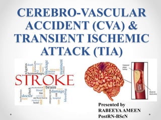
- 1. CEREBRO-VASCULAR ACCIDENT (CVA) & TRANSIENT ISCHEMIC ATTACK (TIA) Presented by RABEEYAAMEEN PostRN-BScN
- 2. OBJECTIVES: By the end of the presentation learners will be able to : • Define Transient Ischemic Attack (TIA) and stroke. • Discuss sign and symptoms of TIA with its management. • Explain the prevalence of stroke. • Examine the types of stroke. o Hemorrhagic stroke o Ischemic stroke • Discuss the sign and symptoms and its causes. • Outline the diagnostic tests. • Discuss the treatment modalities including Pharmacological and Surgical interventions. • Utilize nursing process in caring for the patients with CVA & TIA. • Discuss Rehabilitation and nursing care for stroke patients.
- 3. CEREBROVASCULAR DISORDERS (STROKE AND TIA) Functional inability of the central nervous system that occurs when the normal blood supply to the brain is disrupted.
- 4. TRANSIENT ISCHEMIC ATTACK TIA– is a neurologic deficit lasting less than 24 hours, with most episodes resolving in less than 1 hour. Sudden loss of motor, sensory, or visual function They occur when a blood clot blocks a vessel for a short period of time May serve as a warning sign of stroke.
- 5. SIGN AND SYMPTOMS OF TIA (MINI STROKE) • Weakness, numbness or paralysis in your face, arm or leg, typically on one side of your body. • Slurred speech or difficulty understanding others. • Visual problem or double vision. • Dizziness or loss of balance or coordination. American Stroke Association. (2013)
- 6. MANAGEMENT OF TIA • Antithrombotic Therapy • Daily long-term antiplatelet therapy. • Lower blood pressure to <140/90 mm Hg. • Smoking: Initiate a cessation program. • Lipids: Initiate a goal to control LDL-cholesterol level. Control Diabetes. Physical activity: Recommend ≥10 min of exercise such as walking, bicycling, running, or swimming ≥3 times/week.
- 7. DEFINITION OF STROKE The World Health Organization (WHO) definition of stroke is: “Rapidly developing clinical signs of focal (or global) disturbance of cerebral function, lasting more than 24 hours or leading to death, with no apparent cause other than that of vascular origin” (2013).
- 8. PREVALENCE OF STROKE • In Pakistan, 250 per 100,000 population effect form stroke which means there are 350,000 new stroke patients present every year (2011). • Stroke is the world’s second leading cause of mortality, with a high incidence of severe morbidity (2011). • According to the World Health Organization, 15 million people suffer from stroke worldwide each year. Of these, 5 million die and another 5 million are permanently disabled. • High blood pressure contributes to more than 12.7 million strokes worldwide. • Europe averages approximately 650,000 stroke deaths each year. WHO, (2012)
- 9. TYPES OF STROKE There are two types of stroke: • Ischemic stroke • Hemorrhagic stroke According to American stroke association 87 % of stroke are ischemic and 17% of stroke are hemorrhagic in nature
- 10. ISCHEMIC STROKE According to American stroke association, ischemic stroke is caused by lack of blood supply reaching to the part of the brain.
- 11. TYPES OF ISCHEMIC STROKE There are two types of ischemic strokes. • Thrombotic strokes • Embolic stroke Thrombotic strokes are caused by a blood clot (thrombus) in an artery going to the brain. The clot blocks blood flow to part of the brain Embolic strokes are caused by a wandering clot (embolus) that’s formed elsewhere (usually in the heart or neck arteries). Clots are carried in the bloodstream and block a blood vessel in or leading to the brain.
- 12. HEMORRHAGIC STROKE • According to American stroke association when a blood vessel ruptures in or near the brain, hemorrhagic stroke occurs. • When a hemorrhagic stroke happens, blood collects in the brain tissue. This blood collection is toxic for the brain tissue causing the cells in that area to weaken and die
- 13. TYPES OF HEMORRHAGIC STROKE There are two types of hemorrhagic stroke • Intracerebral hemorrhagic stroke : a blood vessel rupture inside the brain • Subarachnoid hemorrhagic stroke : a blood vessel rupture on the surface of the brain, and the bleeding present at the subarachnoid spaces of the brain
- 14. CAUSES OF STROKE Modifiable risk factor • Obesity • Physical inactivity • Alcohol consumption • Hypertension • Cigarette smoking or exposure to secondhand smoke. • High cholesterol • Diabetes. • Obstructive sleep apnea — a sleep disorder in which the oxygen level intermittently drops during the night. • Cardiovascular disease, including heart failure, heart defects, carotid stenosis, abnormal heart rhythm.
- 15. Cont… Non modifiable risk factor •Personal or family history of stroke, heart attack or transient ischemic attack. •Being age 55 or older. •Race — African-Americans have higher risk of stroke. .
- 16. COMPARISON OF LEFT AND RIGHT HEMISPHERE STROKES LEFT HEMISPHERE • Paralysis or weakness on right side of the body • Right visual field defects • Aphasia • Altered intellectual ability • Slow, cautious behavior RIGHT HEMISPHERE • Paralysis or weakness on left side of the body • Left visual field defects • Perceptual deficits • Increased distractibility • Impulsive behavior and poor judgment • Lack of awareness of deficits.
- 17. SIGNS AND SYMPTOMS OF STROKE • Difficulty walking, dizziness or loss of balance or coordination. • Loss of Sensation and Lack of Reflexes.
- 18. Cont… • Trouble speaking or understanding speech. • Trouble in seeing with one or both eyes.
- 19. Cont… • Sudden numbness or weakness of the face, arm or leg - especially on one side of the body • Sudden severe headache. • Confusion or change in mental status.
- 20. NEUROLOGIC DEFICITS • Visual Field Deficits • Motor Deficits • Sensory Deficits • Verbal Deficits • Cognitive Deficits • Emotional Deficits
- 21. DIAGNOSTIC PROCEDURES • Magnetic resonance imaging (MRI) • Computed tomography (CT Scan)
- 22. MEDICAL MANAGEMENT OF ISCHEMIC STROKE Tissue plasminogen activator The only FDA approved treatment for ischemic strokes is tissue plasminogen activator (tPA, also known as IV rtPA, given through an IV in the arm). tPA works by dissolving the clot and improving blood flow to the part of the brain being deprived of blood flow. It improves the chances of recovering from a stroke and It should be administered within 3 - 4.5 hours (American stroke association, 2015)
- 23. MEDICAL MANAGEMENT OF IS STROKE Pharmacological management : • Antiplatelet agents e.g. Aspirin • Anticoagulants e.g. Warfarin • Antihypertensive e.g. labetelol Other medical interventions includes: • Controlling HTN, • Manage hyperglycemia • Smoking cessation • Manage atrial fibrillation
- 24. CAROTID ENDARTERECTOMY carotid endarterectomy also known as Carotid Artery Surgery, is a procedure in which blood vessel blockage is surgically removed from the carotid artery in the neck to improve blood flow to the brain and reduce the risk of stroke. CAROTID STENTING – less invasive procedure that is used for severe stenosis. SURGICAL MANAGEMENT:
- 25. Cont.. Endovascular Procedures : The endovascular procedure also called mechanical thrombectomy. In which a blood clot which occlude blood flow is removed surgically.
- 26. NURSING INTERVENTION • Place objects within intact field of vision. • Approach the patient from side of intact field of vision • Support patient during the initial ambulation phase. • Provide supportive device for ambulation (walker, cane) • Instruct the patient not to walk without assistance or supportive device. • Test the patient’s pharyngeal reflexes before offering food and fluids. • Assist the patient with meals. • Place food on the unaffected side of the mouth. • Instruct patient that sensation may be altered. • Provide range of motion to affected areas and apply corrective devices as needed • Encourage patient to repeat sounds of the alphabet.
- 27. Cont.. • Explore the patient’s ability to write as an alternative means of communication • Speak slowly and clearly to assist the patient in forming the sounds. • Reorient patient to time, place, and situation, frequently. • Support patient during uncontrollable outburst. • Discuss with the patient and family that the outburst are due to the disease process. • Control stressful situations, if possible. • Provide a safe environment. • Encourage patient to express feelings and frustrations related to disease process.
- 28. Cont.. • Neuro assessment every1 hours. • Keep head straight no neck flexion. • Monitoring and Managing Potential Complications. • Involve family in patient care. • Provide mouth care, back care, NG and Foley's care. • Assess breathing pattern, pulse, BP and temperature. • Observe for airway obstruction. • Monitor glucose level. • Promote early mobilization and physiotherapy. • Other interventions include: o Falls prevention o Skin Care • Promoting Home and Community Based Care.
- 30. REHABILITATION According to WHO “Rehabilitation is a process aimed at enabling them to reach and maintain their optimal physical, sensory, intellectual, psychological and social functional levels. Rehabilitation provides disabled people with the tools they need to attain independence and self-determination.”
- 31. MULTI-DISCIPLINARY TEAM According to National Stroke Association a multidisciplinary team is required for rehabilitation of stroke patient. The team may include some of the following: • Physiatrist. • Neurologist. • Rehabilitation Nurse. • Physical Therapist (PT). • Occupational Therapist (OT). • Speech-Language Pathologists (SLP). • Dietician. • Social Worker. • Case Manager. • Recreation Therapist.
- 34. ASSISTIVE DEVICES TO ENHANCE SELF-CARE OF STROKE PATEINT • Eating devices – nonskid to stabilize plates plate guards to prevent food from being pushed off plate wide-grip utensils to accommodate a weak grasp • Bathing and grooming devices Long-handled bath sponge, grab bars, nonskid mats, hand-held shower heads, electric razors with head at 90 degrees to handle, shower and tub seats, stationary or on wheel.
- 35. Cont... • Toileting aids raised toilet seat grab bars next to toilet • Dressing aids elastic shoelaces loose clothing • Mobility aids canes, walkers, wheelchairs transfer devices such as transfer boards and belts
- 36. SUMMARIZATION
- 37. SMALL QUIZ • What are two main types of stroke • Explain two causes of stroke • List three sign and symptoms of stroke • What is medical Management of ischemic stroke • Explain two Nursing intervention for stroke patient
- 38. ACKNOWLEDGE • Ms. Amina Aijaz • Ms. Zulekha Saleem • Ms.Kiran Shaikh • Ms.Ambreen Allauddin • Ms.Sharifa Lalani • Physiotherapist : Mr.Ali Imran • Occupational therapist: Ms.Fatima
- 39. REFERENCES: Brown, J. A., Lutsep, H. L., Weinand, M., & Cramer, S. C. (2006). Motor cortex stimulation for the enhancement of recovery from stroke: a prospective, multicenter safety study. Neurosurgery, 58(3), 464-473. Cadilhac, D. A., Carter, R., Thrift, A. G., & Dewey, H. M. (2009). Estimating the Long-Term Costs Of Ischemic and Hemorrhagic Stroke for Australia New Evidence Derived From the North East Melbourne Stroke Incidence Study (NEMESIS). Stroke, 40(3), 915- 921. Farooq, M. U., Majid, A., Reeves, M. J., & Birbeck, G. L. (2009). The epidemiology of stroke in Pakistan: past, present, and future. International journal of stroke, 4(5), 381-389. Hacke, W., Kaste, M., Bluhmki, E., Brozman, M., Dávalos, A., Guidetti, D., ... & Toni, D. (2008). Thrombolysis with alteplase 3 to 4.5 hours after acute ischemic stroke. New England Journal of Medicine, 359(13), 1317-1329. Khealani, B. A., Hameed, B., & Mapari, U. U. (2008). Stroke in Pakistan.Journal of the Pakistan Medical Association, 58(7), 400.
- 40. Cont… Party, I. S. W. (2008). National clinical guideline for stroke. Retrieved from http://www.emahsn.ac.uk/emahsn/documents/stroke- idocumentroyalcollegeofphysiciansnationalclinicalguidelineforstroke.pdf Quinn, T. J., Paolucci, S., Sunnerhagen, K. S., Sivenius, J., Walker, M. F., Toni, D., & Lees, K. R. (2009). Evidence-based stroke rehabilitation: an expanded guidance document from the European Stroke Organisation (ESO) guidelines for management of ischaemic stroke and transient ischaemic attack 2008. Journal of Rehabilitation Medicine, 41(2), 99- 111. TIA (Transient ischemic attack). American Stroke Association. http://www.strokeassociation.org/STROKEORG/AboutStroke/TypesofStr oke/TIA/TIA-Transient-Ischemic-Attack_UCM_310942_Article.jsp#. Accessed Dec. 9, 2013. Vanhook, P. (2009). The domains of stroke recovery: a synopsis of the literature. Journal of Neuroscience Nursing, 41(1), 6-17. Kamal, A., Aslam, S., & Khattak, S. (2010). Frequency of Risk Factors in Stroke Patients admitted to DHQ Teaching Hospital, DI Khan. Gomal Journal of Medical Sciences, 8(2).
- 41. Cont… Thomas, T., Stephen, B., & Colin, M. (2000). The global burden of cerebrovascular disease. Global Burden Dis, 21(06), 06. Types of stroke retrieved august, 27 2015 from https://www.nhlbi.nih.gov/health/health-topics/topics/stroke/types Stroke Signs, Symptoms and Prevention retrieved august, 27 2015 from https://www.ohiohealth.com/strokeprevention/ Retrieved august, 27 2015 from http://www.strokecarenow.com/wp-content/uploads/IV-tPA- Screening-Checklist-with-ED-signature.pdf Stroke treatment retrieved august, 27 2015 from http://www.strokeassociation.org/STROKEORG/AboutStro ke/Treatment/Stroke- Treatments_UCM_310892_Article.jsp#i schemic
- 42. Cont… Rehabilitation Therapy after a Stroke retrieved September, 1 2015 from http://www.stroke.org/we-can-help/stroke-survivors/just- experienced-stroke/rehab Rehabilitation retrieved September, 1 2015 from http://www.who.int/topics/rehabilitation/en/ Stroke-rehab.com adaptive equipment.retrieved September, 1 2015 from http://www.stroke-rehab.com/adaptive-equipment.html
Notas del editor
- Nicotine is a highly addictive drug. It causes your blood vessels to narrow, increases your heart rate and raises your blood pressure. Smoking increases the stickiness of special blood cells called platelets. Smoking increases LDL (bad) cholesterol and reduces HDL (good) cholesterol. High levels of LDL cholesterol are a risk for cardiovascular disease and stroke. Diabetes causes atherosclerosis (deposition of fatty material on their inner walls)
- A stroke is caused by the interruption of the blood supply to the brain, usually because a blood vessel bursts or is blocked by a clot. This cuts off the supply of oxygen and nutrients, causing damage to the brain tissue.
- An ischemic stroke occurs if an artery that supplies oxygen-rich blood to the brain becomes blocked. Blood clots often cause the blockages that lead to ischemic strokes.
- The two types of ischemic stroke are thrombotic (throm-BOT-ik) and embolic (em-BOL-ik). In a thrombotic stroke, a blood clot (thrombus) forms in an artery that supplies blood to the brain. In an embolic stroke, a blood clot or other substance (such as plaque, a fatty material) travels through the bloodstream to an artery in the brain. (A blood clot or piece of plaque that travels through the bloodstream is called an embolus.)
- A hemorrhagic stroke occurs if an artery in the brain leaks blood or ruptures (breaks open). The pressure from the leaked blood damages brain cells.
- The two types of hemorrhagic stroke are intracerebral (in-trah-SER-e-bral) and subarachnoid (sub-ah-RAK-noyd). In an intracerebral hemorrhage, a blood vessel inside the brain leaks blood or ruptures. In a subarachnoid hemorrhage, a blood vessel on the surface of the brain leaks blood or ruptures. When this happens, bleeding occurs between the inner and middle layers of the membranes that cover the brain. In both types of hemorrhagic stroke, the leaked blood causes swelling of the brain and increased pressure in the skull. The swelling and pressure damage cells and tissues in the brain.
- The pathophysiology of stroke depends upon the site it affects. The main causes of stroke are stenosis, rupture of blood vessels, occlusion of arteries by atherosclerosis and arteriosclerosis. These causes interrupted blood flow to the brain and results in hypoxia of brain cells, and this ultimately causes neurological deficit. The sign and symptoms of neurological deficits appear according to the brain area affected
- To preserve healthy brain tissue surrounding the blockage. This can be accomplished by removing the blockage and restoring blood flow to the area, or by protecting the surrounding tissue. Inclusion Criteria for IV tPA Age 18 years or older Clinical diagnosis of ischemic stroke with a measurable neurological deficit Time of onset (when patient was last seen as normal) <180 minutes (3 hours) before treatment would begin Exclusion Significant head trauma or prior stroke in previous 3 months Symptoms suggest subarachnoid hemorrhage History of previous intracranial hemorrhage Intracranial neoplasm, arteriovenous malformation, or aneurysm Recent intracranial or intraspinal surgery Arterial puncture at noncompressible site in previous 7 days Elevated blood pressure (systolic >185 mm Hg or diastolic >110 mm Hg) Active internal bleeding Blood glucose concentration <50mg/dl (2.7mmol/L) Acute bleeding diathesisincluding but not limited to Platelet count <100 000/mm³(Inpatients without history of thrombocytopenia, treatment with IV rtPA can be initiated before availability of platelet count but should be discontinued if platelet count is <100 000/mm³.) Heparin received within 48 hours, resulting in abnormally elevated aPTT greater than the upper limit of normal Current use of anticoagulant with INR >1.7 or PT >15 seconds (In patients without recent use of oral anticoagulants or heparin, treatment with IV rtPA can be initiated before availability of coagulation test results but should be discontinued if INR is >1.7 or PT is abnormally elevated by local laboratory standards.) Current use of direct thrombin inhibitors or direct factor Xa inhibitors with elevated sensitive laboratory tests (such as aPTT, INR, platelet count, and ECT; TT; or appropriate factor Xa activity assays) CT demonstrates multilobar infarction (hypodensity >1/3 cerebral hemisphere) CONTRAINDICATIONS: Symptom onset greater than 3 hrs. before admission. Patient with any type of intracranial pathology. Receiving heparin during the past 48 hours with elevated partial thromboplastin time. Platelet count >100,000/mm3 Prior intracranial hemorrhage, neoplasm, arteriovenosus malformation or aneurysm. Major surgical procedures within 14 days. Stroke, serious head injury or intracranial surgery within 3 months. Gastrointestinal or urinary bleeding within 21 days.
- Antiplatelet agents such as aspirin and anticoagulants such as warfarin interfere with the blood’s ability to clot and can play an important role in preventing stroke.
- Another treatment option is an endovascular procedure* called mechanical thrombectomy, strongly recommended, in which trained doctors try removing a large blood clot by sending a wired-caged device called a stent retriever, to the site of the blocked blood vessel in the brain. To remove the brain clot, doctors thread a catheter through an artery in the groin up to the blocked artery in the brain. The stent opens and grabs the clot, allowing doctors to remove the stent with the trapped clot. Special suction tubes may also be used. The procedure should be done within six hours of acute stroke symptoms, and only after a patient receives tPA. Endovascular treatment of stroke is the non-surgical treatment for the sudden loss of brain function due to blood clots. The treatment uses microcatheters (thin tubes visible under X-rays) which are inserted into the blood clot from the groin or the arm. The blood clot is removed from the blood vessel – this procedure is called a thrombectomy. If the blood clot cannot be removed, it is liquefied using drugs delivered through the catheter, in a procedure known as thrombolysis. How does the procedure work? The interventional radiologist will insert a catheter into an artery in your arm or groin and move it towards the blood clot under X-ray guidance. The blood clot is removed in a procedure called a thrombectomy. The blood clot may be removed by trapping it in a stent which is then pulled out with the clot, or the interventional radiologist may suck the clot out through the catheter. If the clot cannot be removed, a medication may be applied through the catheter to liquefy it. If the blood vessel is too narrow, a balloon catheter can be used to restore its original size, in a procedure called an angioplasty. Afterwards, a stent is inserted to hold the blood vessel open.
- Complication of stroke: Aspiration Pneumonia Urinary infection DVT Pulmonary Embolus Shoulder subluxation Depression Malnourishment Pressure sores Falls Seizures
- Physiatrist. Specializes in rehabilitation following injuries, accidents or illness Neurologist. Specializes in the prevention, diagnosis and treatment of stroke and other diseases of the brain and spinal cord Rehabilitation Nurse. Specializes in helping people with disabilities; helps survivors manage health problems that affect stroke (diabetes, high blood pressure) and adjust to life after stroke Physical Therapist (PT). Helps stroke survivors with problems in moving and balance; suggests exercises to strengthen muscles for walking, standing and other activities Occupational Therapist (OT). Helps stroke survivors learn strategies to manage daily activities such as eating, bathing, dressing, writing or cooking Speech-Language Pathologists (SLP). Helps stroke survivors re-learn language skills (talking, reading and writing); shares strategies to help with swallowing problems Dietician. Teaches survivors about healthy eating and special diets (low salt, low fat, low calorie) Social Worker. Helps survivors make decisions about rehab programs, living arrangements, insurance, and support services in the home Neuropsychologist. Diagnoses and treats survivors who may be facing changes in thinking, memory, and behavior after stroke Case Manager. Helps survivors facilitate follow-up to acute care, coordinate care from multiple providers, and link to local services Recreation Therapist. Helps stroke survivors learn strategies to improve the thinking and movement skills needed to join in recreational activities. E.g. dance, cooking, games, cross words and gardening.