Bryophyta.ppt
•Descargar como PPT, PDF•
2 recomendaciones•412 vistas

bryophyta

Recomendados
Más contenido relacionado
La actualidad más candente
La actualidad más candente (20)
Similar a Bryophyta.ppt
Similar a Bryophyta.ppt (20)
Más de Head Department of Botany Govt Degree College Mahabubaba
Más de Head Department of Botany Govt Degree College Mahabubaba (20)
Último
Último (20)
Bryophyta.ppt
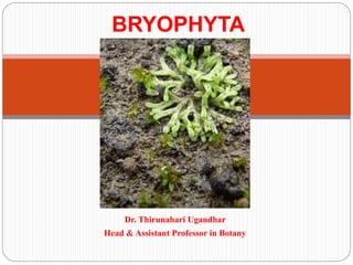
- 1. Dr. Thirunahari Ugandhar Head & Assistant Professor in Botany BRYOPHYTA
- 5. Oldest land plants on earth and have been around for 400 million years or more They do not have true vascular tissue and are therefore non-vascular plants Do not have roots, but have rhizoids, which are relatively simple, sometimes multicellular filaments of thin-walled cells that extend from the photosynthetic tissue into the soil. Composed of haploid cells, containing only one set of chromosomes Have a two-stage life cycle: gametophyte and sporophyte There are about 25,000 species of bryophytes Divided into three: moss, liverworts, and hornworts
- 12. Meaning of Bryophytes: Bryophyta (Gr. Bryon = mass; phyton = plant), a division of kingdom Plantae comprises of mosses, Hornworts and Liverworts. They are groups of green plants which occupy a position between the thallophytes (Algae) and the vascular cryptogams (Pteridophytes). Bryophytes produce embryos but lack seeds and vascular tissues. They are the most simple and primitive group of Embryophyta. They are said to be the first land plants or non- vascular land plants (Atracheata). Presence of swimming antherozoids is an evidence of their aquatic ancestory
- 13. Origin of Bryophytes: Nothing definite is known about the origin of Bryophytes because of the very little fossil record. There are two views regarding the origin of Bryophytes. These are: (i) Algal hypothesis of the origin of Bryophytes. (ii) Pteridophytean hypothesis of the origin of Bryophytes. Algal Hypothesis of Origin: There is no fossil evidence of origin of Bryophytes from algae but Bryophytes resemble with algae in characters like-amphibious nature, presence of flagellated antherozoids and necessity of water for fertilization. This hypothesis was supported by Lignier (1903), Bower (1908), Fritsch (1945) and Smith (1955) etc. According to Fritsch (1945) and Smith (1955) Bryophytes have been originated from the heterotrichous green algae belonging to the order Chaetophorales for e.g.,
- 15. (ii) Pteridophytean Hypothesis of Origin: According to this hypothesis Bryophytes are descendent of Pteridophytes. They are evolved from Pteridophytes by progressive simplification or reduction. This hypothesis is based on certain characters like- presence of type of stomata on the sporogonium of Anthoceros and apophysis of mosses similar to the vascular land plants, similarly in the sporophytes of some Bryophytes (e.g., Anthoceros, Sphagnum, Andreaea) with some members of Psilophytales of Pteridophytes (e.g., Rhynia, Hormophyton etc.) This hypothesis was supported by Scot (1911), Kashyap (1919), Kidston and Lang (1917-21), Haskell (1914) Christensen (1954), Proskaner
- 16. Plant Body of Oedogonium: The thalloid plant body is green, multicellular and filamentous. The filaments are unbranched and cells of each filament are attached end to end and form uniseriate row (Fig. 3.72A). The filament is differentiated into 3 types of cells: 1. Basal cell, 2. Apical and 3. Middle cells. 1. Basal Cell: It is the lowermost cell of the fila- ment. The cell is long, gradually narrowed and towards the basal end it expands to form simple, disc-like, multilobed or finger-shaped structure. The cell is generally colourless, which performs the function of fixation to the substratum and called holdfast.
- 18. Distribution of Bryophytes: Bryophytes are represented by 960 genera and 24,000 species. They are cosmopolitan in distribution and are found growing both in the temperate and tropical regions of the world at an altitude of 4000-8000 feet. In India, Bryophytes are quite abundant in both Nilgiri hills and Himalayas; Kullu, Manali, Shimla, Darjeeling, Dalhousie and Garhwal are some of the hilly regions which also have a luxuriant growth of Bryophytes. Eastern Himalayas have the richest in bryophytic flora. A few species of Riccia, Marchantia and Funaria occur in the plains of U.P., M.P. Rajasthan, Gujarat and South India.
- 19. Habitat of Bryophytes: Bryophytes grow densely in moist and shady places and form thick carpets or mats on damp soils, rocks, bark of trees especially during rainy season. Majority of the species are terrestrial but a few species grow in fresh water (aquatic) e.g., Riccia fluitans, Ricciocarpos natans, Riella etc. Bryophytes are not found in sea but some mosses are found growing in the crevices of rocks and are being regularly bathed by sea water e.g., Grimmia maritima. Some Bryophytes also grow in diverse habitats e.g., Sphagnum-grows in bogs, Dendroceros-epiphytic, Radula protensa. Crossomitrium -epiphyllous, Polytrichum juniperinum-xerophytic, Tortula muralis-on old walls. Tortula desertorum in deserts, Porella platyphylla-on dry rocks, Buxbaumia aphylla (moss), Cryptothallus mirabilis (liverwort) are saprophytic.
- 23. Distribution and Habitat of Marchantia: Marchantia, the most important genus of family Marchantiaceae is represented by about 65 species. The name Marchantia was given in honour of Nicolas Merchant, director of botanical garden of Gaston d’ Orleans in Blois, France. All species are terrestrial and cosmopolitan in distribution. The species prefer to grow in moist and shady places like wet open woodlands, banks of streams, wood rocks or on shaded stub rocks. These grow best after the forest fire in the burnt soil. It is perhaps because of nitrification of soil due to fire (Richard, 1958).
- 24. డిస్ట్రిబ్యూ షన్ అండ్ హ్యూ బిటాట్ ఆఫ్ మచ్చ ంటియా: మార్ింటీయే, కుటంబం యొక్క మార్ింటీయేర యొక్క అతి ముఖ్ూ మైన జాతికి చందినది 65 జాతులు. మార్ ి ంటియా అనే పేరు నికోలస్ మర్చ ంట్ గౌరవారధం ఇవ్వ బడింది, ఫ్రాన్స్ల ోని బ్ ల ో యిస్ల ో ని గాస్ిన్ డి ఓర్లోన్ల బొటానిక్ల్ గార్డెన్ డైర్డక్ ిర్. అనిి జాతులు పంపిణీలో భూమి మర్యు కాస్లో పాలిటన్. ఈ జాతులు తేమతో కూడిన అడవులు, ఫ్రపవాహ్యల ఒడ్డె, క్లప ర్ళ్ళు లేదా షేడ్ె బ్ స్ి్ ర్క్సల వ్ంటి తేమ మర్యు చీక్టి ఫ్రపదేశాలో ో పెరుగుతాయి. దహన మటిిలో అటవీ అగ్ని తర్వ త ఇది బాగా పెరుగుతాయి. బహుశా మంటను నేల నిక్షిపతం చేయడం వ్లో కావ్చ్చచ (ర్చ్ర్ె, 1958).
- 26. In India, Marchantia is represented by about 11 species (Chopra, 1943). Udar (1970) reported only 6 species from different parts of the country. These species are commonly found growing’ in the Himalayan region at an altitude of 4000-8000 feet. Eastern Himalayan region particularly supports the growth of these species. Some species are also found growing in plains of Haryana, Punjab, Uttar Pradesh and hilly regions of South India. Some of the common Indian species are M. palmata, M. polymorpha, M. simlana etc. M. polymorpha is most widely distributed species. M. polymorpha var aquatic grows submerged in swampy meadows. The thalli with gemma cups are found throughout the year whereas the thalli with sex organs occur
- 27. భారతదేశంలో, మార్చ స్ంయా సుమారు 11 జాతుల (చోఫ్రపా, 1943) దావ ర్ సూచంచ్బడ్డతుంది. ఉడార్ (1970) దేశం యొక్క వివిధ ఫ్రపాంతాల నుండి కేవ్లం 6 రకాల మాఫ్రతమే నివేదించంది. ఈ జాతులు 4000-8000 అడ్డగుల ఎతుతలో హిమాలయ ఫ్రపాంతంలో పెరుగుతున్ని యి. తూరుు హిమాలయన్ ఫ్రపాంతం ముఖ్ూ ంగా ఈ జాతుల పెరుగుదలకు మదదతు ఇసుతంది. హర్ూ న్న, పంజా్, ఉతతరఫ్రపదేశ్ మర్యు దక్షిణ భారతదేశంలోని కండఫ్రపాంతాలలో కనిి జాతులు కూడా పెరుగుతున్ని యి. సాధారణ భారతీయ జాతులలో కనిి M. పాల్మో టా, M. పోలిమోర్ా , M. రమోన్న మొదలైనవి. M. పాలీమోర్ా విస్తృతంగా పంపిణీ చేయబడిన జాతులు. ఎం. పాలీమోర్ా వే వాటర్ జలము ముర్కి గడిె మైదాన్నలలో మునిగ్నపోతుంది. స్ంవ్తల రం పొడవున్న
- 28. Gametophytic Phase of Marchantia: External Features of Gametophyte: The plant body is gametophytic, thalloid, flat, prostrate, plagiotropic, 2-10 cm. long and dichotomously branched (Fig. 1 A). Dorsal surface: Dorsal surface is dark green. It has a conspicuous midrib and a number of polygonal areas called areolae. The midrib is marked on the dorsal surface by a shallow groove and on the ventral surface by a low ridge. Each polygonal area re-presents the underlying air chamber. The boundaries of these areas represent the walls that separate each air chamber from the next.
- 31. Each air chamber has a central pore. The midrib ends in a depression at the apical region forming an apical notch in which growing point is situated (Fig. 28 B). Dorsal surface also bears the vegetative and sexual reproductive structures. The vegetative reproductive structures are gemma cup and develop along the midrib. These are crescent shaped with spiny or fimbriate margins and are about one eighth of a inch in diameter (Fig. I A, 15). Sexual reproductive structures are borne on special Stalked structures called gametophores or gamctangiophores. The gametophores bearing archegonia are called archegoniophores and that bearing antheridia are called antheridiophores (Fig. 1 A, B).
- 33. Ventral surface: The ventral surface of the thallus bears scales and rhizoids along the midrib. Scales are violet coloured, multicellular, one cell thick and arranged in 2-4 rows (Fig. 1 C). Scales are of two types: (i) Simple or ligulate (ii) Appendiculate. Appendiculate (Fig- 1 C, D) scales form the inner row of the scales close with midrib. Ligulate scales form the outer or marginal row and are smaller than the appendiculate scales (Fig. 1 C, E). Rhizoids are unicellular, branched and develop as prolongation of the lower epidermal cells. They are of two types: (i) Smooth-walled rhizoids, (ii) Tuberculate rhizoids. In smooth-walled rhizoids both the inner and outer wall layers are fully stretched while in tuber-culate rhizoids appear like circular dots in surface view (Fig. 1 F). The inner wall layer modifies into peg like in growth which projects into the cell lumen (Fig. 1 H). The main functions of the rhizoids are to
- 35. Vertical వ్ంతెన ఉపర్తలం: థాలస్ యొక్క వెడలుు ఉపర్తలం మధూ సా ా యిలో ఉండే ఫ్రపమాణాలు మర్యు భూక్ంపాలు ఉంటాయి. ఫ్రపమాణాలు వైలెట్ రంగు, బహుళస్ముఫ్రదం, ఒక్ క్ణం మందపాటి మర్యు 2-4 వ్రుస్లలో అమరచ బడి ఉంటాయి. ఫ్రపమాణాలు ర్డండ్డ రకాలు: (i) సాధారణ లేదా లిగుూ లేట్ (ii) అనుబంధం. అపెండికుూ లేట్ ఫ్రపమాణాలు midrib తో దగగరగా ఫ్రపమాణాల అంతరగత వ్రుస్ను ఏరు రుసా త యి. వెలుపలి లేదా ఉపాంత వ్రుస్ను ఫ్రపమాణాలు కలిచేందుకు మర్యు అనుబంధ ఫ్రపమాణాల క్న్ని చని వి. రజోయిడ్డో ఏక్వ్చ్నం, శాఖ్లుగా ఉంటాయి మర్యు దిగువ్ ఎపిడెరో ల్ క్ణాల పొడిగ్నంపుగా అభివ్ృదిధ చందుతాయి. అవి ర్డండ్డ రకాలు: (i) సూో త్-గోడలు ఉని భూగరభ రస్వాదులు, ఉపర్తల దృష్టిలో వ్ృతా త కార చ్తురఫ్రసాకార చ్చక్క లు వ్ంటి టవెర్-కాలియులేట్ ర్జిజాయిడ్స్ల ో క్నిపించేటపుు డ్డ మృదువైన గోడల పొరలో ో లోపలి ర్ స్
- 36. Anatomy of the Gametophyte: A vertical cross section of the thallus can be differentiated photosynthetic zone and lower storage zone (Fig. 2 A, B, E). Upper Photosynthetic zone: The outermost layer is upper epidermis. Its cells are thin walled square, compactly arranged and contain few chloroplasts. Its continuity is broken by the presence of many barrel shaped air pores. Each pore is surrounded by four to eight superimposed tiers of concentric rings. (Fig. 2 B) with three to four cells in each tier (Fig. 2 D). Air pores are compound in nature. The lower tier consists of four cells which project in the pore and the opening of the pore looks star like in the surface view (Fig. 2 C). The walls of the air pore lie half
- 37. Gamatophyte యొక్క అన్నటమీ: Thallus యొక్క ఒక్ నిలువు ఫ్రకాస్ విభాగం కిరణజనూ జోన్ మర్యు తకుక వ్ నిలవ జోన్ (Figure 2 A, B, E) ను వేరు చేయవ్చ్చచ . ఉని త కిరణజనూ మండలం: వెలుపలి పొర ఎగువ్ ఎపిడెర్ో స్. దీని క్ణాలు స్ని ని గోడల చ్తురఫ్రస్ంగా ఉంటాయి, కదిదపాటి బ్ కోోప్లపా ో స్ిోను క్లిగ్న ఉంటాయి. దీని కనసాగ్నంపు అనేక్ బార్డల్ ఆకారపు గాలి రంఫ్రధాల ఉనికి దావ ర్ విర్గ్నపోతుంది. ఫ్రపతి రంఫ్రధం చ్చట్ట ి న్నలుగు నుండి ఎనిమిది ఎనిమిదింటిని క్లిగ్న ఉంటంది. (Figure 2 B) ఫ్రపతి ఫ్రేణిలో మూడ్డ నుండి న్నలుగు క్ణాలు (Figure 2 D). ఎయిర్ రంఫ్రధాలు ఫ్రపక్ృతిలో స్మేో ళనం. దిగువ్ ఫ్రేణి న్నలుగు కోరోను క్లిగ్న ఉంటంది మర్యు ఇది సూక్ష్ో రంఫ్రధంలో పూరతవుతుంది మర్యు సూక్ష్ో రంఫ్రధం తెరవ్డం ఉపర్తల దృష్టిలో (నక్ష్ఫ్రతం
- 38. Just below the upper epidermis photosynthetic chambers are present in a horizontal layer (Fig. 2 B). Each air pore opens inside the air chamber and helps in exchange of gases during photosynthesis. These are chambers develop schizogenously (Vocalized separation of cells to form a cavity) and are separated from each other by single layered partition walls. The partition walls are two to four cells in height. Cells contain chloroplast. Many simple or branched photosynthetic filaments arise from the base of the air chambers (Fig. 2 B).
- 40. Storage zone: It lies below the air chambers. It is more thickened in the centre and gradually tapers towards the margins. It consists of several lasers of compactly arranged, thin walled parenchymatous isodiametric cells. Intercellular spaces are absent. The cells of this zone contain starch. Some cells contain a single large oil body or filled with mucilage. The cells of the midrib region possess reticulate thickenings. The lower most cell layer of the zone forms the lower epidermis. Some cells of the middle layer of lower epidermis extend to form both types of scales and rhizoids (Fig. 2 B).
- 41. reproduction in Marchantia. Vegetative Reproduction in Marchantia: Vegetative propagation of Marchantia takes place by the following methods: (a) By progressive Death and Decay of the Thallus: Like Riccia, the vegetative propagation of Marchantia is quite common which takes place by the unlimited growth of the branches and decay from the base. When the decay of cells reaches dichotomy, the two lobes becomes separated from each other and the each separa- ted lobe forms independent plants
- 42. (b) By Adventitious Branches: Vegetative propagation is also known to take place by the development of adventitious branches from the ventral side of the thallus or even from the reproductive buds (e.g. M. palmata). These branches, on separation from parent thallus, grow into new plants. (c) By Gemmae (Singular = Gemma):
- 43. The most common method of vegetative propagation is by specialised asexual multicellular bodies, known as gemmae. They are borne in large numbers in small receptacles called gemma cups present on the dorsal surface of the thallus in the midrib region. A gemma cup is about 2 mm in diameter and 3 mm in height, the margins of which are hyaline, lobed, spinuous or entire. Many small stalked gemmae develop on the floor of the gemma cup. Each gemma is vertically attached to the base of the gemma cup by a one-celled stalk. he gemmae are multicellular, biconvex, discoid bodies constricted at the middle. The two notches in the constriction possess a row of apical cells showing two growing points.
- 45. Sexual Reproduction in Marchantia: In Marchantia, the production of sex organs is dependent on environmental conditions like day length, humidity, excess of nitrogenous substance, etc. The sex organs are borne on special stalked receptacles called gametophores. The receptacles bearing male (antheridium) and female (archegonium) sex organs are called antheridiophore and archegoniophore or carpocephallum, respectively. These are developed on separate plants, so that the Marchantia is dioecious or heterothallic. Although, the gametophores are erect branches, they are actually the direct extension of the prostrate vegetative thallus. This is clearly evident from the dorsiventral nature of the erect shoots with air chambers, air pores and rhizoids.
- 47. Antheridiophore: The antheridiophore (Figs 6.8 and 6.12) shows a 1-3 cm long prismatic stalk bearing at its apex a slightly convex (peltate) disc which is usually a 8-lobed structure. Each lobe represents the apex of a branch along whose upper (dorsal) median line the antheridia are borne in a row. The antheridia develop in a acropetal manner i.e., the oldest are being at the center and the youngest ones towards the periphery. The antheridia are within the flask- shaped antheridial chamber. Each antheridial chamber contains a single antheridium. Each antheridial chamber is separated from one another by air chambers with air pores.
- 48. Development and Structure of Antheridium: The development of antheridium in Marchantia is similar to that of Riccia. The antheridium develops from the antheridial initial, situated on the dorsal surface of the disc of the antheridiophore. The antheridial initial increases in size and a transverse division differentiates it to an outer cell and a basal cell (Fig. 6.1 3A, B). The basal cell follows no further development. The outer cell later undergoes transverse division forming a basal primary stalk cell and a terminal primary antheridial cell (Fig. 6.13C). Eventually the antheridium is formed from the primary antheridial cell. The basal primary stalk cell however, forms the stalk of the antheridium (Fig. 6.13D-G). A mature antheridium has a short stalk and a globular body encircled by a sterile jacket which encloses a number of sperm mother cells. A single sperm mother cell forms two triangular androcytes, each of which develops a biciliated sperm or antherozoid (Fig. 6.13H). The dehiscence of antheridium in Marchantia is similar to that of Riccia.
- 50. Archegonium: Development and Structure of Archegonium: Like Riccia, the archegonia of Marchantia also develop from a single superficial dorsal cell, called the archegonial initial (Fig. 6.16A). The conical shaped initial cell becomes conspicuous, increases in size, and projects above the surface. The initial cell divides by a transverse wall to form an upper (or outer) primary archegonial cell and a lower (or inner) primary stalk cell (Fig. 6.16B). The primary stalk cell by a few irregular divisions forms a short but distinct multicellular stalk of the archegonium. The primary archegonial initial undergoes several phases of regular divisions to form the archegonium (Figs. 6.16B-H). A mature archegonium (Figs. 6.15B) is a pendulous flask-shaped structure with a short stalk, which attaches to the lower (ventral) surface of the archegonial disc. A single archegonium has a basal swollen venter containing an egg cell, a ventral-canal cell and a row of neck canal cells inside the elongated neck.
- 53. Sporophyte: Development of the Sporophyte:The fertilised egg enlarges in size and fills up the cavity of the venter. It invests itself immediately with a thin cellulose wall and becomes the oospore or zygote which is the mother cell of the sporophytic generation. The zygote cell inside the venter divides first by a transverse wall forming an outer epibasal cell and an inner hypobasal cell (Fig. 6.17A). This is followed by a second division at right angle to the first division forming a quadrant of cells. The two upper epibasal cells of the quadrant divide and re-divide to form the capsule and the upper part of the seta.
- 58. Structure of the Mature Sporophyte: The mature sporophyte of Marchantia (Fig. 6.18A) is differentiated into three parts — foot, seta and capsule. (a) Foot: It is an expanded bulbous mass of cells at the base of the sporogonium, serves as an absorbing and anchoring organ. It absorbs nutrients from the gametophyte. (b) Seta: It is a slightly elongated stalk that connects the foot to the capsule. The cells are parenchymatous and elongated lengthwise. Thus, seta increases in length and pushes the mature capsule out of the calyptra. (c) Capsule:
- 59. (c) Capsule: It is a yellow-coloured spherical structure, contains numerous spores and elaters. The capsule is protected by three coverings viz. calyptra perigynium and perichaetium. Dehiscence of the Capsule: The jacket of the hanging mature sporogonium (capsule) gets dried and splits longitudinally into a variable number of longitudinal lobes (Fig. 6.18B). These lobes extend from the apex to the middle of the capsule. The discharge and dispersal of sport to the air are assisted by the coiling and uncoiling of the elaters due to their hygroscopic nature. The spores are almost rounded and are very minute in size (12 to 30 µm in diameter) with a thin inner intine and a slightly thicker outer exine (Fig. 6.18D). In some species an additional layer called perispore is also found outside the exine.