
Recomendados
Más contenido relacionado
La actualidad más candente
Similar a Tissue clearing - Field Overview
Similar a Tissue clearing - Field Overview (20)
Último
Último (20)
Tissue clearing - Field Overview
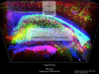
- 1. Tiago Pinheiro May 2015 Supervision: András Simon Macmillan Publishers Ltd: Nature 497: 332–37, copyright 2013
- 2. Key concepts Refractive index Refractive index variation Main obstacle to imaging of intact tissues Refractive index of material needs to be homogeno http://www.microscopyu.com/articles/formulas/images/refractiveindexf
- 3. Key concepts Major types of tissue clearing Refractive index of material needs to be homogeno 1 Dry the sample e.g. ethanol, tetrahydrofuran Refill with high- index solvent that matches the tissue e.g. BABB, DBE, glycerol Fluorophore quenching Sample shrinkage 3 4 Replace the water in the tissue with polar solvents with high refractive index e.g. formamide 2 Increase the refractive index of the tissue by adding water-soluble compounds e.g. glucose, fructose Sample expansion
- 4. More transparency Fluorochrome degradation Less transparency Fluorochrome preservation Usual problem https://www.leica-microsystems.com/science-lab/clarity/clearing-procedures-for-deep-tissue-imaging
- 5. Timeline First commercial light sheet microscope – Lavision BABB clearing - Dodt Deep tissue application of two-photon microscopy - Helmchen FocusClear – Liu First light sheet microscope – Voie Two-photon laser scanning fluorescence microscopy - Denk Superman x-ray vision research – Pittenger JB. Other derivative procedures - clove oil and cedarwood oil Method for reducing refractive index variations in tissue - Spalteholz Publication of 6 anatomy books - Vesallii Dissection of animals – Galen 129 1543 1914 ...1914 1983 1990 1993 2003 2005 2007 2010
- 6. Dissection of animals – Galen 129
- 7. Publication of 6 anatomy books - Vesallii 1543
- 8. Method for reducing refractive index variations in tissue - Spalteholz 1914
- 9. Other derivative procedures - clove oil and cedarwood oil ...1914
- 10. Superman x-ray vision research – Pittenger JB. 1983
- 11. Two-photon laser scanning fluorescence microscopy - Denk 1990
- 12. 1990 First light sheet microscope – Voie 1993 Resolution of 26 um
- 13. FocusClear – Liu 2003 Proprietary and expensive 50 ml – 12.000 kr
- 14. Deep tissue application of two-photon microscopy - Helmchen 2005
- 15. BABB clearing - Dodt 2007 BABB clearing quenches fluorophores
- 16. 1990 First commercial light sheet microscope – Lavision 2010 Rapid zstack acquisition in high resolution
- 17. Timeline First commercial light sheet microscope – Lavision BABB clearing - Dodt Deep tissue application of two-photon microscopy - Helmchen FocusClear – Liu First light sheet microscope – Voie Two-photon laser scanning fluorescence microscopy - Denk Superman x-ray vision research – Pittenger JB. Other derivative procedures - clove oil and cedarwood oil Method for reducing refractive index variations in tissue - Spalteholz Publication of 6 anatomy books - Vesallii Dissection of animals – Galen 129 1543 1914 ...1914 1983 1990 * 1993 2003 2005 2007 2010
- 19. Timeline ...1914 * Developments in technology and tissue clearing techniques converge Scale – Hama 2011 More GFP friendly agents discovered DBE and tetrahydrofuran – Becker CUBIC Whole-body – Tainaka CUBIC Whole-brain – Susaki iDISCO – Renier ClearT – Kuwajima SeeDB – Ke Clarity – Chung 3DISCO – Ertürk 2012 2012 2013 2013 2013 2014 2014 2014
- 20. Scale – Hama 2011 2 Increase the refractive index of the tissue by adding water-soluble compounds e.g. glucose, fructose Sample expansion 3 weeks to 6 months Protein loss Non-proprietary
- 21. More GFP friendly agents discovered DBE and tetrahydrofuran – Becker 2012 Gives rise to 3DISCO later on …
- 22. 3DISCO – Ertürk 2012 1 Dry the sample e.g. ethanol, tetrahydrofuran Refill with high-index solvent that matches the tissue e.g. BABB, DBE, glycerol Non-proprietary 1 day half-time of GFP signal No tissue expansion No fluorescent protein quenching 2-5 days
- 23. Clarity – Chung 2013 Non-proprietary 2 weeks Good antibody labelling No fluorophore quenching Keeps tissue morphology Custom setup, long protocol 4
- 24. SeeDB – Ke 2013 3 days Keeps tissue morphology No fluorescent protein quenching Non-proprietary 1 week 2 Increase the refractive index of the tissue by adding water-soluble compounds e.g. glucose, fructose
- 25. ClearT – Kuwajima 2013 2013 3 Replace the water in the tissue with polar solvents with high refractive index e.g. formamide 1 day - ? No or mild expansion Compatible with lipophilic tracers Non-proprietary Not compatible with fluorescent proteins
- 26. iDISCO – Renier 2014 2014 Same as 3DISCO but for large samples
- 27. CUBIC Whole-brain – Susaki 2014 Mix of compounds joining the best of many techniquesSimple clearing and analysis pipeline Non-proprietary 5 Single-cell resolution, axons and dendrites
- 28. 2014 CUBIC Whole-body – Tainaka 2014 Same as whole-brain CUBIC but applied to other organs… 5
- 29. More applications Viewing neuronal populations and projections; Intact tissue imaging of long-range projections, , local circuit wiring, cellular relationships, subcellular structures, protein complexes, nucleic acids, and neurotransmitters; Quantitation of distances of neural stem cells to blood vessels; …
- 30. “Both complete structural analysis (i.e. not reconstructed across tissue sections) and molecular phenotyping are desired to gain full insights into the relationships and functional mechanisms of biological systems.” “Techniques that focus on molecular labelling require thin tissue sectioning which limits structural reconstruction.” Advantages • No damage or thin sectioning required to visualize whole intact tissue samples • Allows marking and visualization of long-range projections and subcellular structures • Allows multiple rounds of molecular phenotyping • Applicable to multiple tissue types and sizes Disadvantages • Multiple-step process that takes place over several days/weeks • Immunostaining is time-consuming for thicker tissue samples • High start-up and consumable material costs Advantages of tissue clearing to sectioning http://wiki.claritytechniques.org/index.php/Main_Page
Notas del editor
- EYFP (green), parvalbumin-positive neurons (red), and GFAP (blue). Permission from Macmillan Publishers Ltd: Nature 497: 332–37, copyright 2013
- 1-BABB, 3DISCO, iDISCO 2-SeeDB, scale 3-ClearT 4-Clarity – chemical transformation of tissue instead of optical clearing 5-CUBIC
- 1543 – Vesallii was born in Brussels which then was still part of the netherlands. Published books at the age of 28
- 1914 - One option for reducing the refractive index variations in tissue is to remove the water and replace it by an organic compound that has a higher refractive index. Spalteholz described such a treatment with benzyl alcohol and methyl salicylate. He proposed that the final mounting solution must have the same refractive index as the average index of the desiccated material. 1914 - These methods dry the sample with ethanol or tetrahydrofuran and then refill it with a high-index solvent like glycerol, benzyl alcohol-benzyl benzoate (BABB) or dibenzyl ether (DBE). Disadvantages: Immunolabelling is only possible before processing and fluorochromes are not stable. Sample shrinks
- Scale – renders tissue transparent and preserves fluorescent signal. Urea based. Increases refractive index of the aqueous phase by adding water soluble compounds – clear technique type 2.