
Recomendados
Más contenido relacionado
La actualidad más candente
La actualidad más candente (20)
Similar a Electromyography
Similar a Electromyography (20)
Más de TwinkleJoshi4
Más de TwinkleJoshi4 (15)
Último
Último (20)
Electromyography
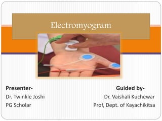
- 1. Presenter- Guided by- Dr. Twinkle Joshi Dr. Vaishali Kuchewar PG Scholar Prof, Dept. of Kayachikitsa Electromyogram
- 2. Introduction Electromyography (EMG) measures muscle response or electrical activity in response to a nerve’s stimulation. Measures electrical activity of muscle during rest, slight or forceful contraction Normally - no electrical signals are produced at rest. Shows ability of the muscle to respond when the nerves are stimulated. Detects neuromuscular abnormalities
- 3. In the test, small needles (electrodes) are inserted through the skin into the muscle. Electrodes detect electrical activity and display on the oscilloscope in the form of waves. Audio-amplifier can be used to hear the activity . Commonly tested nerves- Upper extremities- Radial, Ulnar nerve Lower extremities- Peroneal, Tibial nerve
- 4. Indications Tingling, Numbness Muscle weakness, pain or cramps Brachial plexopathy Cervical spondylosis DMD, BMD Myasthenia gravis Carpal tunnel syndrome or peripheral neuropathies Amyotrophic lateral sclerosis or polio Herniated disk in the spine
- 5. Before the test Avoid caffeinated beverages, such as coffee, tea, etc 2-3 hrs before testing. Stop using lotions or oils on skin Prior notification – pacemaker, blood-thinning medications Wear clothes that permit access to the area to be tested Remove jewelry,pins,glasses, hearing aids, or metal objects Nerve conduction study is done (measures flow of current through a nerve before it reaches the muscle)
- 6. Sit/ lie down locate the muscle to be studied. clean the skin with antiseptic solution 5 or more sterile needles are inserted into muscle ground electrode is kept under arm/leg perform slight/full- strength muscle contraction electrical activity is measured & displayed on oscilloscope. audio amplifier evaluates appearance and sound of electrical potentials Procedure
- 7. Interpretation Amplitude: Height of the wave Conduction velocity (CV): speed at which electrical impulse travels along the nerve. Duration: width of electrical wave. Conduction block: reduction of a signal in an anatomical region, such as wrist due to carpal tunnel syndrome F reflex: impulse travels up to the spine and then back down along the same fiber. It shows conduction along the motor nerve(electrical echo) H reflex: impulse travels to the spinal cord via a sensory nerve, then back along a motor nerve.
- 8. Gives information about both motor and sensory components of PNS. Shows whether the axon or myelin sheath is damaged by a neuropathy. Myelin helps action potentials travel faster, and so in problems of myelin (myelinopathy), conduction velocity is decreased. In axon (axonopathy),fibers that are intact can conduct signals at normal speeds, but there are fewer fibers, which leads to a weaker signal and decreased amplitude.
- 9. Normal EMG: When muscles are at rest, they produce no or very little electrical activity. Activity at rest: In diseases of peripheral nerves, like carpal tunnel syndrome and peripheral neuropathy, muscles show spontaneous activity on their own, which indicates altered nerve supply. It also points to inflammation or a muscle disease. Depicted as fibrillations and positive sharp deflections.
- 10. Abnormal activity during contraction: “Recruitment pattern" As muscle is contracted, nerve fibers signal more and more bits of muscle (called motor units) to join in and help. In peripheral nerve diseases, the nerve is unable to connect to many units. NCS results: Abnormal NCS result occurs due to conduction block, axonopathy (the nerve fiber is damaged), or demyelination (damage to the outer insulating layer of nerves)
- 11. Contra Indications Patients on anticoagulation or antiplatelet therapy. Paraspinal muscles are avoided in patients on anticoagulation due to potential possibility of hematoma formation adjacent to spinal structures. In overlying skin infection Limb affected with lymphedema Pacemakers, internal defibrillators
- 12. Complications Complications rare. Risk of bleeding, infection and nerve injury due to needle electrode Muscle soreness Pain, tenderness, swelling, or pus at the needle insertion sites. Pneumothorax
- 13. Thank you!
- 14. Thought of the day