Stool Examination
•Descargar como PPTX, PDF•
100 recomendaciones•76,731 vistas

Stool Examination

Recomendados
Más contenido relacionado
La actualidad más candente
La actualidad más candente (20)
Similar a Stool Examination
Similar a Stool Examination (20)
Más de Dr. Varughese George
Más de Dr. Varughese George (20)
Último
Último (20)
Stool Examination
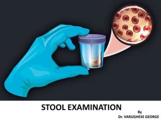
- 1. STOOL EXAMINATION By Dr. VARUGHESE GEORGE
- 2. LEARNING OBJECTIVES • INTRODUCTION • ADVANTAGES OF STOOL EXAMINATION • COLLECTION OF STOOLS SAMPLES • MACROSCOPIC EXAMINATION • PREPARATION OF SLIDES • CONCENTRATION OF STOOL SAMPLES • MICROSCOPIC EXAMINATION • CHEMICAL EXAMINATION
- 3. INTRODUCTION • Normal amount of stools in an adult is 100-200 g/day. • Stools are composed of : – water (up to 75%), – indigestible residue – undigested food – food which is digested but not absorbed – bile, epithelial cells, secretions from digestive tract – inorganic material – Bacteria • Stool analysis determines the various properties of the stool for diagnostic purposes.
- 4. • INTRODUCTION • ADVANTAGES OF STOOL EXAMINATION • COLLECTION OF STOOLS SAMPLES • MACROSCOPIC EXAMINATION • PREPARATION OF SLIDES • CONCENTRATION OF STOOL SAMPLES • MICROSCOPIC EXAMINATION • CHEMICAL EXAMINATION
- 5. ADVANTAGES OF STOOL EXAMINATION Examination of stools aids in investigation of GIT diseases. • Detection of parasites : – detection of worms (adult worms, tapeworms, larvae, ova) – detection of protozoa (trophozoites or cysts). • Evaluation of chronic diarrhea – Random diarrheal sample tested for occult blood, fat, pH, white blood cells, culture, or microscopy. – A 48- or 72-hour sample is examined for weight, fat content, carbohydrate, osmolality or chymotrypsin activity.
- 6. ADVANTAGES OF STOOL EXAMINATION Examination of stools aids in investigation of GIT diseases. • Evaluation of dysentery: – Identification of causative organism is definitive in amebic V/S bacillary dysentery. • Bacteriologic examination: – Infection by bacteria such as Salmonella, Shigella, Vibrio, Yersinia, or Clostridium difficile can be identified by stool culture. – Bacterial toxins released by Clostridium botulinum or Clostridium difficile can also be identified.
- 7. ADVANTAGES OF STOOL EXAMINATION Examination of stools aids in investigation of GIT diseases. • Chemical examination: – to detect occult blood (in ulcerated lesions of gastrointestinal tract, especially occult carcinoma of colon), – excess fat excretion (malabsorption syndrome) – presence or absence of urobilinogen (obstructive jaundice). • Differentiating infection by invasive bacteria (like Salmonella or Shigella) from that due to toxin producing bacteria (like Escherichia coli or Vibrio cholerae): – Increased numbers of polymorphonuclear neutrophils (identified by methylene blue stain) are seen in the former
- 8. ADVANTAGES OF STOOL EXAMINATION Examination of stools aids in investigation of GIT diseases. • Identification of Rotavirus: – common cause of diarrhea in infants & young children. – It can be identified by examination of stool by electron microscopy, latex agglutination, immunofluorescence or ELISA.
- 9. • INTRODUCTION • ADVANTAGES OF STOOL EXAMINATION • COLLECTION OF STOOLS SAMPLES • MACROSCOPIC EXAMINATION • PREPARATION OF SLIDES • CONCENTRATION OF STOOL SAMPLES • MICROSCOPIC EXAMINATION • CHEMICAL EXAMINATION
- 10. COLLECTION OF STOOL SAMPLES • A random specimen of stool (at least 4 ml or 4 cm3) is collected in a clean, dry, container with a tightly fitting lid & transported immediately to the laboratory. • About 20-40 grams of formed stool or 5-6 tablespoons of watery stool should be collected. • Parasites are best detected in warm, freshly passed stools which should be examined as early as possible (preferably within 1 hour of collection). • Stool should not be contaminated with urine, water, soil, or menstrual blood. Urine & water destroy trophozoites. Soil will introduce extraneous organisms & also hinder proper examination.
- 11. COLLECTION OF STOOL SAMPLES • Sample may be refrigerated if delay in examination is anticipated. • A fixative may be used if specimen is to be transported to a distant laboratory. – 10% formalin for preservation of eggs, larvae & cysts. – Polyvinyl alcohol for preservation of trophozoites & cysts & for permanent staining. • 3 separate samples collected at 3-day intervals are recommended to detect all parasite infections. – One negative report for ova and parasites does not exclude the possibility of infection
- 12. • INTRODUCTION • ADVANTAGES OF STOOL EXAMINATION • COLLECTION OF STOOLS SAMPLES • MACROSCOPIC EXAMINATION • PREPARATION OF SLIDES • CONCENTRATION OF STOOL SAMPLES • MICROSCOPIC EXAMINATION • CHEMICAL EXAMINATION
- 13. MACROSCOPIC EXAMINATION Findings :- • Consistency • Color • Odor • Presence of blood • Presence of mucus • Presence of adult worms or segments of tapeworms
- 14. MACROSCOPIC EXAMINATION Consistency of Stool samples • Cysts are likely to be found in formed stools. • Trophozoites are most likely to be found in loose or watery stools or in stools containing blood and mucus. • Trophozoites die soon after being passed and therefore such stools should be examined within 1 hour of passing. • Examination of formed stools can be delayed but should be completed on the same day.
- 15. MACROSCOPIC EXAMINATION Consistency of Stool samples • Dry and hard stools are seen in chronic constipation. • Pasty stools are due to fat contents • Common bile obstruction • Celiac disease • Ribbon like stools are seen in • Spastic bowel • Rectal narrowing • Stricture • Partial GIT obstruction
- 16. MACROSCOPIC EXAMINATION Color/Appearance of Stool samples Color/Appearance Interpretation Brown Normal/Stercobilinogen Black and tarry Bleeding in upper GIT(proximal to cecum) Drugs (iron salts, bismuth salts,charcoal) Red Lower GIT bleeding/tumors, inflammatory process Anal fissure,hemmorhoids,tumors Undigested tomatoes or beetroot. Yellow or yellow green Diarrhoea Clay-colored (gray-white) Biliary obstruction Silvery Carcinoma of ampulla of Vater
- 17. MACROSCOPIC EXAMINATION Color/Appearance of Stool samples Color/Appearance Interpretation Watery Certain strains of Escherichia coli, Rotavirus enteritis, Cryptosporidiosis Rice water Cholera Unformed with blood, mucus, and pus Bacillary dysentery, Ulcerative Colitis, Intestinal tuberculosis, Amoebiasis, Enteritis Unformed, frothy, foul smelling, which float on water Steatorrhoea Pale color stool with greasy appearance Pancreatic deficiency due to malabsorption
- 18. MACROSCOPIC EXAMINATION Odour • Stool odour – Indole and skatole which are formation by bacterial fermentation and putrefaction. • Foul odour – Undigested protein & by excessive intake of carbohydrate. • Sickly odour – Undigested lactose & fatty acids.
- 19. MACROSCOPIC EXAMINATION Presence of mucus in stools • Translucent gelatinous material clinging to surface of stool. • Produced by colonic mucosa in response to parasympathetic stimulation. • Seen in – Severe constipation – Mucous colitis
- 20. MACROSCOPIC EXAMINATION Presence of mucus and blood in stools • Seen in • Bacillary dysentery • Ulcerative Colitis • Intestinal tuberculosis • Amoebiasis • Enteritis Mucus with blood clinging to stool is seen in • Lower GIT malignancy. • Inflammatory lesions of anal canal
- 21. • INTRODUCTION • ADVANTAGES OF STOOL EXAMINATION • COLLECTION OF STOOLS SAMPLES • MACROSCOPIC EXAMINATION • PREPARATION OF SLIDES • CONCENTRATION OF STOOL SAMPLES • MICROSCOPIC EXAMINATION • CHEMICAL EXAMINATION
- 22. PREPARATION OF SLIDES • A drop of normal saline is placed near one end of a glass slide and a drop of Lugol iodine solution is placed near the other end. • A small amount of feces is mixed with a drop each of saline and iodine using a wire loop, and a cover slip is placed over each preparation separately. • If the specimen contains blood or mucus, that portion should be included for examination (trophozoites are more readily found in mucus). • If the stools are liquid, select the portion from the surface for examination.
- 23. PREPARATION OF SLIDES • Saline wet mount is used for demonstration of eggs and larvae of helminths, and trophozoites and cysts of protozoa. • Saline wet mount can also detect red cells and white cells. • The iodine wet mount is useful for identification of protozoal cysts as iodine stains glycogen and nuclei of the cysts. • Trophozoites become non-motile in iodine mounts. • A liquid, diarrheal stool can be examined directly without adding saline.
- 24. • INTRODUCTION • ADVANTAGES OF STOOL EXAMINATION • COLLECTION OF STOOLS SAMPLES • MACROSCOPIC EXAMINATION • PREPARATION OF SLIDES • CONCENTRATION OF STOOL SAMPLES • MICROSCOPIC EXAMINATION • CHEMICAL EXAMINATION
- 25. CONCENTRATION OF STOOL SAMPLES • Useful if very small numbers of parasites are present. • Indicated in cases of negative wet mount examination and there is clinical suspicion of parasitic infection. • Used for detection of ova, cysts, and larvae of parasites. Demerit - Amoebic trophozoites are destroyed in concentrated specimens and therefore cannot be detected.
- 26. CONCENTRATION OF STOOL SAMPLES Concentration techniques are of two main types: • Sedimentation techniques : – Ova and cysts settle at the bottom. – Excessive stool debris may make the detection of parasites difficult. – e.g. Formolethyl acetate sedimentation procedure. • Floatation techniques : – Ova and cysts float on surface. – Some ova and cysts do not float at the top in this procedure. – e.g. Saturated salt floatation technique, zinc sulphate concentration technique.
- 27. CONCENTRATION OF STOOL SAMPLES The most commonly used sedimentation method is formol-ethyl acetate sedimentation method because: i. it can detect eggs and larvae of almost all helminths, and cysts of protozoa. ii. it preserves their morphology well. iii. It is rapid. iv. risk of infection to the laboratory worker is minimal because pathogens are killed by formalin.
- 28. CONCENTRATION OF STOOL SAMPLES • Stool sample suspension is prepared in 10% formalin (10 ml formalin + 1 gram stool sample) & passed through a gauze filter till 7 ml of filtered material is obtained. • Ethyl acetate (3 ml) is added to this filtered material & the mixture is centrifuged for 1 minute. • Eggs, larvae & cysts sediment at the bottom of the centrifuge tube with layers of formalin, stool debris, and ether above. • Stool debris is loosened with an applicator stick & the supernatant is poured off. • One drop of sediment is placed on one end of a glass slide & one drop is placed at the other end. • One of the drops is stained with iodine, cover slips are placed, & the preparation is examined under the microscope. Formol-ethyl acetate sedimentation method
- 29. • INTRODUCTION • ADVANTAGES OF STOOL EXAMINATION • COLLECTION OF STOOLS SAMPLES • MACROSCOPIC EXAMINATION • PREPARATION OF SLIDES • CONCENTRATION OF STOOL SAMPLES • MICROSCOPIC EXAMINATION • CHEMICAL EXAMINATION
- 30. MICROSCOPIC EXAMINATION Findings :- • Leukocytes (WBCs) • Red Blood Cells (RBCs) • Macrophages • Epithelial cells • Bacteria • Ova/ Cysts/ Trophozoites of parasites • Meat/muscle fibres • Fat
- 31. MICROSCOPIC EXAMINATION Leukocytes (WBCs) • Normal stool may contain occasional (0-1) WBCs. • To look for WBCs, the smears should be prepared from areas of mucous or watery stools. RBCs WBCs Wet mount
- 32. MICROSCOPIC EXAMINATION Leukocytes (WBCs) Increased no: of WBCs are stools is associated with • Bacillary dysentery • Chronic ulcerative colitis • Shigellosis • Salmonella infections • Invasive E-Coli infections • Anal/Rectal Fistula • Localised abscess • Amoebiasis & typhoid
- 33. MICROSCOPIC EXAMINATION • Bright red stool is seen in cases of lower GIT bleeding. • Black and tarry blood are seen in cases of – Upper GIT bleeding. – Occult bleeding. • Present in – Dysentery – Hemorrhoids – GIT Malignancies Red Blood Cells (RBCs)
- 34. MICROSCOPIC EXAMINATION Macrophages • seen in – Bacillary dysentery – Ulcerative colitis Wet mount
- 35. MICROSCOPIC EXAMINATION Epithelial cells • Seen in inflammatory conditions of the bowel Epithelial cell
- 36. MICROSCOPIC EXAMINATION Bacteria • Stool cultures are commonly done to identify bacteria associated with enteric infection. • Stool samples are examined to detect toxins & to rule out infection by – Salmonella, Shigella, Campylobacter & predominating numbers of Staphylococcus organisms. – Pseudomonas, Yersinia, Vibrio & Shiga toxin–producing E. coli. – Clostridium difficile causing antibiotic-associated colitis. – Yeast. • At least 3 stool cultures collected on separate days are recommended if the patient’s clinical picture suggests bacterial involvement, despite previous negative cultures.
- 37. MICROSCOPIC EXAMINATION Fat Present in • Malabsorption • Deficiency of pancreatic digestive enzyme • Deficiency of bile
- 38. MICROSCOPIC EXAMINATION Meat/muscle fibres in stools Their presence show impaired intraluminal digestion. • Increased amount of meat fibres are found in • Malabsorption syndrome • Pancreatic functional defect like cystic fibrosis.
- 39. MICROSCOPIC EXAMINATION Ova/ Cysts/ Trophozoites of parasites • Normally there are no parasites/eggs in the stool sample. • Multiple stool samples should be examined to rule out parasitic infestations • At least 3 consecutive days’ stool samples are examined to rule out parasitic infestations
- 40. MICROSCOPIC EXAMINATION OF STOOL SAMPLES
- 41. MICROSCOPIC EXAMINATION - PROTOZOA Entamoeba histolytica • Demonstration of trophozoites of E. histolytica in stool samples is required for diagnosis of amoebic dysentery. • For diagnosis, at least three fresh stool samples should be examined to increase sensitivity. • Trophozoites vary from 15 to 40 μ in diameter. • In saline wet mounts, trophozoites show motility in one direction via pseudopodia, which form rapidly. • Cytoplasm shows outer transparent ectoplasm and inner finely granular endoplasm. • Nucleus is visible in the iodine preparation. • Fine granules of peripheral nuclear chromatin are evenly distributed. Small, single central karyosome. (Motility is lost in iodine mount).
- 42. MICROSCOPIC EXAMINATION - PROTOZOA Entamoeba histolytica Diagnostic feature of E. histolytica trophozoites is the presence of ingested red cells. Trophozoite of Entamoeba histolytica showing ingested red cells (Trichrome stain)
- 43. MICROSCOPIC EXAMINATION - PROTOZOA Entamoeba histolytica trophozoites
- 44. MICROSCOPIC EXAMINATION - PROTOZOA Entamoeba histolytica Uninucleate, binucleate, trinucleate, and quadrinucleate cysts of Entamoeba histolytica • Cysts of E. histolytica are spherical and measure 10-15 μ in diameter. • Nuclei are 1, 2, 3, or 4 and are similar in morphology to trophozoite nucleus (mature cyst contains 4 nuclei). • Nuclear membrane is regular and thin with finely granular peripheral chromatin. • Karyosome is small and central. • Immature cysts may show chromatoid bodies (aggregates of ribosomes) that are oblong structures with rounded ends and glycogen clumps – infective stage.
- 45. MICROSCOPIC EXAMINATION - PROTOZOA Entamoeba histolytica cyst Entamoeba histolytica cyst (arrow)
- 46. MICROSCOPIC EXAMINATION - PROTOZOA Entamoeba histolytica • Plenty of red cells and very few white cells are helpful in differentiating amebic from bacillary dysentery. • Detection of antigen of E. histolytica in stools by commercially available tests based on enzyme immunoassay. • Detection of DNA specific to E. histolytica is possible by polymerase chain reaction-based assays. • Serologic tests like latex agglutination test, indirect hemagglutination test, enzyme immunoassay, and counter immunoelectrophoresis detect antibodies to E. histolytica. • Additionally, endoscopic biopsy of ulcer in the intestine can demonstrate trophozoites of E. histolytica in 50% ofcases. Staining with periodic acid Schiff stain facilitates identification of parasites.
- 47. MICROSCOPIC EXAMINATION - PROTOZOA Differences between amoebic and bacillary dysentery Parameter Amoebic dysentery Bacillary dysentery Cause Entamoeba histolytica Shigella (most common) Onset Gradual Acute Fever/ vomiting Not significant Significant Toxicity Absent Present Abdominal tenderness Localized Generalized Tenesmus Absent Present Frequency 6-8 per day Over 10 per day Odor Offensive Nil Color Dark red Bright red Nature Feces mixed with blood and mucus Blood and mucus with little or no feces Consistency Not adherent Adherent to container Reaction Acid Alkaline
- 48. MICROSCOPIC EXAMINATION - PROTOZOA Differences between amoebic and bacillary dysentery Parameter Amoebic dysentery Bacillary dysentery Microscopic examination of stool • Cellular exudates • Red cells • Pus cells • Macrophages • Eosinophils • Charcot-Leyden crystals • Bacteria • Trophozoites of E. histolytica Scanty Clumps Nil or few Few Present May be present Many, motile Motile trophozoites with ingested red blood cells Abundant Discrete Numerous Many, some with ingested RBCs Absent Absent Few, nonmotile Absent Antigen test for E. histolytica Positive Negative Stool culture Negative Positive for Shigella
- 49. MICROSCOPIC EXAMINATION - PROTOZOA Giardia intestinalis (lamblia) • Cysts are more likely to be found in formed or loose stools. • Cysts of G. lamblia are 8-12 μ in diameter, oval and contain 4 nuclei, axonemes, median bodies and remains of flagella
- 50. MICROSCOPIC EXAMINATION - PROTOZOA Giardia intestinalis (lamblia) • G. lamblia trophozoites are found as clusters in fresh liquid stools, particularly in flakes of mucus. • Trophozoites of G. lamblia are pear-shaped,12-15 μ in diameter, have 4 pairs of flagellae, 2 large and oval nuclei, 2 axonemes (axial filaments of flagella), and 1 or 2 curved median bodies. • Motility is likened to that of “falling leaf”.
- 51. MICROSCOPIC EXAMINATION - PROTOZOA Giardia intestinalis (lamblia) trophozoite
- 52. MICROSCOPIC EXAMINATION - PROTOZOA Giardia intestinalis (lamblia) • Detection of antigen of G. lamblia in stool sample by enzyme immunoassay technique with high sensitivity (90- 99%) and specificity (95-100%). • Direct fluorescent antibody assay is available commercially in a kit form in which cysts are labeled with immunofluorescent antibodies and are detected under fluorescence microscope.
- 53. MICROSCOPIC EXAMINATION - PROTOZOA Coccidia • Demonstration of oocysts of human intestinal coccidia such as Isospora belli, Cryptosporidium parvum & Cyclospora cayetanensis. • These protozoan organisms cause self-limited, mild diarrheal illness. • In immunocompromised patients (such as patients with AIDS) they can induce severe and protracted diarrhea, which may sometimes be life-threatening. • Detection of antigen in stool samples by enzyme immunoassay for detection of specific antigen of C.parvum is available. • Oocysts of Cryptosporidium labelled with fluorescent antibody are readily detected under fluorescence microscope.
- 54. MICROSCOPIC EXAMINATION - PROTOZOA Coccidia - Isospora belli Immature oocyst showing 2 sporoblasts Mature oocyst with 2 sporocysts containing sporozoites • Oocysts are seen in direct wet mounts of stools after formalin-ether concentration technique. • Oocysts are oval and about 32 × 16 μ in size and mature outside the human body • Immature oocysts which are seen in stools contain 2 sporoblasts. • Mature oocysts found outside the human body contains 2 sporocysts, each with 4 sporozoites. • With modified Ziehl-Neelsen stain (on a stool smear prepared from the sediment after formalin-ether concentration), oocysts stain uniform red-pink. • Under ultraviolet light, oocysts show autofluorescence.
- 55. MICROSCOPIC EXAMINATION - PROTOZOA . Under ultraviolet light, oocysts show autofluorescence. With modified Ziehl-Neelsen stain (on a stool smear prepared from the sediment after formalin-ether concentration), oocysts stain uniform red-pink Coccidia - Isospora belli
- 56. MICROSCOPIC EXAMINATION - PROTOZOA • Oocysts of C. parvum are difficult to demonstrate in direct stool wet mounts. • They are demonstrated either by modified Ziehl- Neelsen staining of concentrated stool smear or by immunofluorescence technique. • They are 4-6 μ in size, round to oval, and stain pink-red. Cryptosporidium oocysts appear with a peripheral green fluorescence Cryptosporidium oocysts in stools by modified Ziehl- Neelsen staining Coccidia - Cryptosporidium parvum
- 57. MICROSCOPIC EXAMINATION - PROTOZOA • In direct wet mounts of feces,oocysts measure 8-10 μ in diameter and contain a cluster of refractile globules (morula-like appearance). With modified Ziehl-Neelsen stain, they appear similar to C. parvum, but are larger. Under ultraviolet light (365 nm), oocysts show intense blue autofluorescence. Coccidia - Cyclospora cayetanensis
- 58. MICROSCOPIC EXAMINATION - PROTOZOA • Microsporidia are obligate intracellular protozoa, which cause opportunistic infection in immunocompromised patients leading to persistent diarrhea and weight loss. • Common species causing infection in humans are Enterocytozoon bieneusi, Encephalitozoon intestinalis, and Encephalitozoon hellem. • Spores are very small (1-5 μ), stain red on modified trichrome stain and may show a transverse band. Microsporidia (arrows) in stool stained with modified acid-fast trichrome Microsporidia
- 59. MICROSCOPIC EXAMINATION - HELMINTHS • Diagnosis of A. lumbricoides infection is made by demonstration of eggs on stool examination. • Eggs can be demonstrated in direct wet mount of stools in moderate to heavy infections. • The recommended procedure is formol-ethyl acetate sedimentation technique for concentration of eggs. • In stools, the types of eggs are found: – fertilized (double-shelled or decorticated) – unfertilised (double-shelled or decorticated) Ascaris lumbricoides (Roundworm)
- 60. (A) Adult female and male worms (B) Anterior end of worm. Head-on view, showing one dorsal and two ventral lips with papillae (C) Posterior end of female, showing anal opening, a little above the conical tip (D) Posterior end of male, showing two protruding copulatory spicules(s) (E) Specimen showing Ascaris lumbricoides, male and female. Ascaris lumbricoides (Roundworm)
- 61. MICROSCOPIC EXAMINATION - HELMINTHS Ascaris lumbricoides (Roundworm) Unfertilized eggs: • Single female worms discharge eggs which are slightly larger & elliptical in shape. • Size : 80 μm x 55 μm. • The egg has a thinner shell with an irregular coating of albumin. • The egg is filled with a mass of large disorganized highly refractile granules of various sizes (atrophied ovum). • Eggs do not float in salt solution.
- 62. MICROSCOPIC EXAMINATION - HELMINTHS Fertilized eggs: • Eggs are round, oval, golden brown in color & always bile stained. • Size : 70 μ × 50 μ in size. • Surrounded by thick smooth translucent shell with an outer coarsely mammillated albuminous coat, a thick transparent middle layer & the inner lipoidal vitelline membrane. • The egg contains a single central granular mass (fertilized ovum). • Eggs float in saturated solution of common salt. Ascaris lumbricoides (Roundworm)
- 63. MICROSCOPIC EXAMINATION - HELMINTHS Ascaris lumbricoides (Roundworm) Decorticated eggs : • These eggs do not have the unevenly thick outer albuminous shell & resemble the hookworm eggs.
- 64. MICROSCOPIC EXAMINATION - HELMINTHS Identification of adult worms • Occasionally adult worms are passed spontaneously in the feces and brought to the laboratory for identification. • Adult ascaris worms are cylindrical or round, pinkish, and measure about 15 cm (male) or 30 cm (female) in length. • 0.5 cm in diameter • Tail is either curved (male) or straight (female). • There are three lips at the anterior end (mouth). Ascaris lumbricoides (Roundworm)
- 65. MICROSCOPIC EXAMINATION - HELMINTHS • Hookworms are – Ancylostoma duodenale (old world hookworm) – Necator americanus (new world hookworm). • Diagnosis is based on identification of hookworm eggs on stool • examination. • Technique of formol-ethyl acetate sedimentation is preferred. • Alternatively, direct wet mount of stool sample can demonstrate eggs in moderate to heavy infections. • Eggs of A. duodenale & N. americanus are morphologically similar. Hookworms
- 66. MICROSCOPIC EXAMINATION - HELMINTHS Hookworms • Eggs of A. duodenale & N. americanus are 50-75 μ in length and 40 μ in width, oval, colourless, and have a thin shell. • In fresh stools, eggs show 4-8 gray, granular cells. • If stool > 12 hrs old, eggs will show a rhabditiform larva folded upon itself , which are called embryonated eggs. • If feces > 24 hrs old, free rhabditiform larvae will be seen. • Buccal cavity of hookworm larva is longer which distinguishes itself from • larvae of Strongyloides stercoralis Test for occult blood in stools is positive. • Additional findings in blood - • Blood examination often shows eosinophilia. • Microcytic hypochromic anemia develops due to chronic blood loss. Hookworm egg in fresh stools. Egg in oval shape with a thin shell, has a clear space between wall and developing cleavage, and contains granular cells
- 67. MICROSCOPIC EXAMINATION - HELMINTHS Trichuris trichiura (Whipworm) Diagnosis depends on identification of typical eggs on stool examination. Eggs measure 50 × 25 μ in size, are yellow- brown and barrel-shaped. A rounded, transparent plug is present at both poles Eggs contain central, uniformly granular mass. Eggs are often quantitated to assess the severity of infection.
- 68. MICROSCOPIC EXAMINATION - HELMINTHS Hymenolepsis nana • Eggs are oval and measures 30-50 μ in size. • The inner membrane has 2 poles, from which 4-8 polar filaments spread out between the two membranes. • The oncosphere has 6 hooklets
- 69. MICROSCOPIC EXAMINATION - HELMINTHS Strongyloides stercoralis Diagnosis of S. stercoralis infection depends on demonstration of rhabditiform larvae in fresh stool specimens. Eggs of S.stercoralis are rarely seen in stool samples because they hatch and release rhabditiform larvae in the intestine. Rhabditiform larvae are 200-300 μ in length and 15 μ in width and are actively motile. They have two esophageal swellings, a prominent genital primordium and a short buccal cavity. In old stool samples, rhabditiform larvae of hookworms are seen which resemble those of S. stercoralis; the differentiating feature is the longer buccal cavity of the former.
- 70. MICROSCOPIC EXAMINATION - HELMINTHS Strongyloides stercoralis • Excretion of larvae is often irregular and their number may be few stool examination may yield negative result. • Concentration technique (formol-ethyl acetate) is helpful in suspected cases. • Duodenal fluid can be aspirated for detection of larvae or Entero-test (String test) can be performed. • In disseminated infection, larvae may be detected in sputum. • Enzyme immunoassay test that detects IgG antibodies to S. stercoralis is available & • indicated if organism is not detected in feces, duodenal aspirate, or string test and clinical suspicion is strong. • cannot differentiate between recent and past infection.
- 71. MICROSCOPIC EXAMINATION - HELMINTHS Enterobius vermicularis Special technique for collection and demonstration of pinworm eggs : • Adult female pinworm migrates at night from the intestine (cecum) to the perianal skin folds and deposits eggs which are often not found on routine stool examination. • Pinworm eggs can be collected either by a transparent adhesive tape (“cellophane tape test”) or by anal swab. • Specimen should preferably be collected late into the night or early morning before patient passes urine, feces or takes a bath. • A transparent adhesive tape is folded over the end of a glass slide, spoon handle or a wooden tongue depressor (sticky surface outwards). • Patient’s buttocks are separated & the slide or spoon handle covered with tape is pressed over perianal skin at many sites. • The tape is then spread over a glass slide with adhesive side down & pressed flat onto the slide surface. • Slide covered with tape is then examined under the microscope.
- 72. MICROSCOPIC EXAMINATION - HELMINTHS Enterobius vermicularis • Eggs of E.vermicularis measure 60 μ × 30 μ in size, are oval & flattened on one side. • They are colorless, transparent with a double-lined smooth shell & contain a small granular mass or a larva. • Adult pinworms may be recovered from perianal skin folds (by adhesive tape) or may be found in children’s feces. • They are white, motile & small in size (male: 0.5 cm; female: 1 cm). • Diagnosis depends on demonstration of eggs in samples collected from perianal skin or demonstration of adult worms. Enterobius egg Enterobius eggs collected by transparent tape method
- 73. MICROSCOPIC EXAMINATION - HELMINTHS Enterobius vermicularis Enterobius vermicularis egg
- 74. MICROSCOPIC EXAMINATION - HELMINTHS Taenia solium - Identification of eggs: • Egg measures 30-40 μ in diameter, is round to oval, and has a thick, brown wall with transverse lines. • The egg contains an embryo, which is a round granular mass containing 3 pairs of hooklets and surrounded by a fine membrane.
- 75. MICROSCOPIC EXAMINATION - HELMINTHS Taenia solium - Identification of eggs: • Occasionally, the egg is enclosed in a clear sac. • Eggs are discharged intermittently by the tapeworm and therefore may not be detected easily. • Repeated stool examinations and formol-ether concentration technique are often required for their demonstration.
- 76. MICROSCOPIC EXAMINATION - HELMINTHS Taenia solium - Identification of gravid segments or proglottids: • This allows identification of species. • The segment is flattened between two glass slides and examined under a magnifying glass. • Gravid segment is 13 mm × 8 mm in size, translucent, and pale blue. • It has a central uterine stem with 8-13 lateralbranches. • (Uterine branches are >13 in T. saginata)
- 77. MICROSCOPIC EXAMINATION - HELMINTHS Taenia solium - Identification of scolex (head): • Scolex of a tapeworm is very small (pinhead size) and is rarely seen. • When examined with a magnifying glass, scolex of T. solium shows 4 suckers and a crown of hooklets.
- 78. MICROSCOPIC EXAMINATION - HELMINTHS Taenia saginata - Identification of eggs: • Eggs of T. saginata can be identified in feces and perianal skin. • They are morphologically similar to those of T. solium.
- 79. MICROSCOPIC EXAMINATION - HELMINTHS Taenia saginata - Identification of gravid segments or proglottids: • This allows identification of species. • Segments measure 20 mm × 6 mm in size, and are ivory white. • They contain a central uterine stem > 13 side branches(i.e. 15-20 branches).
- 80. MICROSCOPIC EXAMINATION - HELMINTHS Taenia solium - Identification of scolex (head): • Scolex of T. saginata has 4 suckers but no hooklets. • It measures 2 mm in width.
- 81. MICROSCOPIC EXAMINATION - ARTEFACTS It is also important to identify artefacts during microscopic examination of stool samples which could be confused with ova & cysts of various protozoa & helminths Yeast & fungal elements in stool
- 82. MICROSCOPIC EXAMINATION - ARTEFACTS It is also important to identify artefacts during microscopic examination of stool samples which could be confused with ova & cysts of various protozoa & helminths Pollen grains in stools mistaken for helminth eggs.
- 83. MICROSCOPIC EXAMINATION - ARTEFACTS It is also important to identify artefacts during microscopic examination of stool samples which could be confused with ova & cysts of various protozoa & helminths Plant fibre/Plant cells.
- 84. MICROSCOPIC EXAMINATION - ARTEFACTS It is also important to identify artefacts during microscopic examination of stool samples which could be confused with ova & cysts of various protozoa & helminths Air bubbles
- 85. • INTRODUCTION • ADVANTAGES OF STOOL EXAMINATION • COLLECTION OF STOOLS SAMPLES • MACROSCOPIC EXAMINATION • PREPARATION OF SLIDES • CONCENTRATION OF STOOL SAMPLES • MICROSCOPIC EXAMINATION • CHEMICAL EXAMINATION
- 86. CHEMICAL EXAMINATION Chemical examination of feces is usually carried out for the following tests : • Occult blood • Excess fat excretion (malabsorption) • Urobilinogen • Reducing sugars • stool osmotic gap • stool pH
- 87. CHEMICAL EXAMINATION Test for Occult Blood in Stools • Presence of blood in feces which is not apparent on gross inspection and which can be detected only by chemical tests is called as occult blood. • Causes of occult blood in stools are: i. Intestinal diseases: hookworms, amebiasis,typhoid fever, ulcerative colitis, intussusception,adenoma, cancer of colon or rectum. ii. Gastric and esophageal diseases: peptic ulcer,gastritis, esophageal varices, hiatus hernia. iii. Systemic disorders: bleeding diathesis, uremia. iv. Long distance runners. • Recommended as a screening procedure for detection of asymptomatic colorectal cancer
- 88. CHEMICAL EXAMINATION Test for Occult Blood in Stools - Peroxidase-like activity of Hb • Benzidine and orthotolidine are carcinogenic and are no longer used. • Benzidine test is also highly sensitive and false-positive reactions are common. • Since bleeding from the lesion may be intermittent, repeated testing may be required. Principle: • Hemoglobin has peroxidase-like activity & releases oxygen from hydrogen peroxide. • Oxygen molecule then oxidizes the chemical reagent (benzidine, orthotolidine, aminophenazone, or guaiac) to produce a colored reaction product.
- 89. CHEMICAL EXAMINATION Causes of False +ve Tests 1. Ingestion of peroxidase- containing foods like red meat, fish, poultry, turnips, horseradish, cauliflower, spinach, or cucumber. Diet should be free from peroxidase- containing foods for at least 3 days prior to testing. 2. Drugs like aspirin and other anti- inflammatory drugs, which increase blood loss from GITin normal persons. Causes of False -ve Tests 1. Foods containing large amounts of vitamin C. 2. Conversion of all hemoglobin to acid hematin (which has no peroxidase-like activity) during passage through the gastrointestinal tract. Test for Occult Blood in Stools - Peroxidase-like activity of Hb
- 90. CHEMICAL EXAMINATION Test for Occult Blood in Stools - Immunochemical Tests • These tests specifically detect human hemoglobin. • There is no interference from animal hemoglobin or myoglobin (e.g. meat) or peroxidase-containing vegetables in the diet. • The test consists of mixing the sample with latex particles coated with anti-human haemoglobin antibody & if agglutination occurs, test is positive. • This test can detect 0.6 ml of blood per 100 grams of feces.
- 91. CHEMICAL EXAMINATION Test for Occult Blood in Stools - Apt test • This test devised by Dr. Apt is done to decide whether blood in the vomitus or in the feces of a neonate represents swallowed maternal blood or is the result of bleeding in the GIT. • The baby swallows blood during delivery or during breastfeeding if nipples are cracked. • Apt test is based on the principle that – if blood is of neonatal origin it will contain high proportion of hemoglobin F (Hb F) that is resistant to alkali denaturation. – On the other hand, maternal blood mostly contains adult hemoglobin or Hb A that is less resistant.
- 92. CHEMICAL EXAMINATION Malabsorption of Fat • Dietary fat is absorbed in the small intestine with the help of bile salts and pancreatic lipase. • Fecal fat mainly consists of neutral fats (unsplit fats), fatty acids, and soaps (fatty acid salts). • Normally very little fat is excreted in feces (<7 grams/day in adults). • Excess excretion of fecal fat indicates malabsorption and is known as steatorrhea. • It manifests as bulky, frothy, and foul-smelling stools,which float on the surface of water.
- 93. CHEMICAL EXAMINATION Causes of Malabsorption of Fat i. Deficiency of pancreatic lipase (insufficient lipolysis): chronic pancreatitis, cystic fibrosis. ii. Deficiency of bile salts (insufficient emulsification of fat): biliary obstruction, severe liver disease, bile salt deconjugation due to bacterial overgrowth in the small intestine. iii. Diseases of small intestine: tropical sprue, celiac disease, Whipple’s disease.
- 94. CHEMICAL EXAMINATION Test for Malabsorption of Fat Tests for fecal fat are • qualitative (i.e. direct microscopic examination after fat staining) • quantitative (i.e. estimation of fat by gravimetric or titrimetric analysis).
- 95. CHEMICAL EXAMINATION • A random specimen of stool is collected after putting the patient on a diet of > 80 gm fat per day. • Stool sample is stained with a fat stain (oil red O, Sudan III or Sudan IV) and observed under the microscope for fat globules. • Presence of ≥ 60 fat droplets/HPF indicates steatorrhea. Ingestion of mineral or castor oil and use of rectal suppositories can cause problems in interpretation. Sudan stain on fecal sample: (A) Negative; (B) Positive Microscopic stool examination for fat
- 96. CHEMICAL EXAMINATION Patient should be on a diet of 70-100 gm of fat per day for 6 days before the test. Stool samples are collected over 72 hours and stored in a refrigerator during the collection period. Samples should not be contaminated with urine. Fat quantitation can be done by gravimetric or titrimetric method. In gravimetric method, an accurately weighed sample of feces is emulsified, acidified, and fat is extracted in a solvent; after evaporation of solvent, fat is weighed as a pure compound. Quantitative estimation of fecal fat:
- 97. CHEMICAL EXAMINATION • Titrimetric analysis is the most widely used method. • An accurately weighed stool sample is treated with alcoholic potassium hydroxide to convert fat into soaps. • Soaps are then converted to fatty acids by the addition of hydrochloric acid. • Fatty acids are extracted in a solvent and the solvent is evaporated. • The solution of fat made in neutral alcohol is then titrated against sodium hydroxide. Quantitative estimation of fecal fat:
- 98. CHEMICAL EXAMINATION • Fatty acids comprise about 80% of fecal fat. • Values >7 grams/day are usually abnormal. • Values >14 grams/day are specific for diseases causing fat malabsorption. Quantitative estimation of fecal fat:
- 99. CHEMICAL EXAMINATION • Fecal urobilinogen is determined by Ehrlich’s aldehyde test • Sample should be fresh and kept protected from light. • Normal amount of urobilinogen excreted in feces is 50-300 mg per day. • Increased fecal excretion of urobilinogen is seen in hemolytic anemia. • Urobilinogen is decreased in biliary tract obstruction, severe liver disease, oral antibiotic therapy (disturbance of intestinal bacterial flora) & aplastic anemia (low hemoglobin turnover). • Stools become pale or clay-colored if urobilinogen is reduced or absent. Test for Urobilinogen in Feces
- 100. CHEMICAL EXAMINATION • Deficiency of intestinal enzyme lactase which converts lactose (in milk) to glucose and galactose, is a common cause of malabsorption. • If lactase is deficient, lactose is converted to lactic acid with production of gas which leads to diarrhea, vomiting, and failure to thrive in infants. • Benedict’s test / ClinitestTM tablet test for reducing sugars is used to test freshly collected stool sample for lactose. Test for Reducing Sugars
- 101. CHEMICAL EXAMINATION Trypsin in Stools & Fecal Chymotrypsin • Trypsin is a proteolytic enzyme formed in the small intestine & it is destroyed by bacteria in older children and adults. • Inadequate trypsin secretion can lead to malabsorption & abdominal discomfort. • No trypsin activity is detectable in constipated stools owing to prolonged exposure to intestinal bacteria, which inactivates trypsin. • Chymotrypsin, an intestinal proteolytic enzyme secreted by the pancreas, can be used to assess pancreatic function. • Fecal chymotrypsin is a more reliable measurement of pancreatic function than trypsin.
- 102. CHEMICAL EXAMINATION Stool Electrolytes: Sodium, Chloride, Potassium & Osmolality • Stool electrolyte tests are used to assess electrolyte imbalance in patients with diarrhea. • Stool electrolytes must be evaluated along with the serum and urine electrolytes as well as clinical findings in the • patient. • Stool osmolality is used in conjunction with blood serum osmolality to calculate the osmotic gap and to diagnose intestinal disaccharide deficiency.
- 103. CHEMICAL EXAMINATION Fecal Osmotic Gap • Fecal osmotic gap is calculated from concentration of electrolytes in stool water by the formula 290-2([Na+] + [K+]). (290 is the assumed plasma osmolality). • In osmotic diarrhea, osmotic gap >150 mOsm/kg. • In secretory diarrhea, osmotic gap < 50 mOsm/kg. Fecal pH • Stool pH below 5.6 is characteristic of carbohydrate malabsorption.
- 104. EVALUATION OF CHRONIC DIARRHEA
- 105. • Bulk: 100-200 grams/day • Color: Brown • Water: Up to 75% • pH: 7.0-7.5 • Red blood cells: Absent • White blood cells: Few • Epithelial cells: Present • Crystals: Calcium oxalate, triple phosphate REFERENCE RANGES IN STOOL EXAMINATION
- 106. REFERENCE RANGES IN STOOL EXAMINATION • Fat (Adults): <7 grams/day (gravimetric method), <6 • grams/day (titrimetric method) • Fat droplets: Average 2.5 per high power field in random • sample • Urobilinogen: 50-300 mg/24 hours • Parasites: Nil • Ova, cysts, trophozoites: Nil Critical Values • Stool culture positive for Salmonella, Shigella, Campylobacter, Vibrio, or Yersinia.
Notas del editor
- GIT – Gastrointestinal tract.