1. Measuring the effect on the JNK/AP1 Pathway for Screening
CCR1 Antagonists
Victor M. Suarez1
, GSSRP1
, Yuriko Root1
,
Dr. James Merritt1*
, and Dr. Salvatore Coniglio1*
1
New Jersey Center for Science, Technology, and Mathematics
Introduction
Chemokine receptor CCR1, a protein expressed in
macrophages, promotes chemotactic signal transduction
in response to binding ligands such as CCL3, CCL5, and
CCL6. Evidence supports that CCR1 binding activity
inhibits the ability of macrophages to stimulate invasion of
mouse glioblastoma cell line GL261 and U87. It also
showed that the blockade of CCR1 inhibits the JNK
biochemical pathway, mediating activation of the
transcription factor, AP-1. In this study, a Dual Luciferase
Reporter (DLR) assay was used to test activity of
luciferase bioluminescence in cells transfected with an
AP-1 Firefly Luciferase reporter plasmid. Previous
studies indicate that AP-1-mediated luciferase activity
increases in GL261 cells when co-cultured with microglia
for 24 hours. This project focuses on the addition of the
CCR1 inhibitor JLY-133 and its effect on JNK/AP1
pathway inhibition.
Methods
Process
Results
• CCR1 may be an important target in blocking microglia-
stimulated glioblastoma invasion
• Paracrine signaling between glioblastoma and microglia
involves activation of the JNK/AP1 pathway, which is sensitive
to CCR1 inhibition
• Initial studies suggest this pathway can be used to measure
CCR1 efficacy and perhaps be used as a screening tool for
novel CCR1 antagonists.
Special recognition to my research mentor and advisor, Dr.
Coniglio, for guidance through this on-going project, to
Yuriko Root for her dedication and assistance.
Conclusion
Acknowledgements
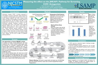
Two approaches to measure JNK/AP1 activity:
1) Cultures of GL261 murine glioblastoma cells were
transfected with a firefly Luciferase reporter gene under
the direction of a promoter containing AP-1 binding
elements (pGL2 backbone). Transfected cells were co-
cultured with murine microglia for 24 hours in DMSO
alone or in the presence of 250 nM JLY133. Cells were
then harvested and luminescence was measured TECAN
M1000 plate reader.
2) For western blotting assays, cell culture was carried
out as described above except cells were lysed in sample
buffer and loaded on 7% SDS-PAGE gels. Anti phospho-
JNK (T183/185) and anti-phospho-cJun primary antibodies
were used to measure levels of JNK pathway activity.
Preparation of Cell Cultures:
Independent cultures were grown
along with one co-culture. Co-
culture showed increase in
malignancy and cell chemotaxis.
CCR1 inhibition blocks cell
malignancy increase.
Dual Luciferase Assay
Process:
Plate and transfect cells with AP-
1 Firefly luciferase plasmid. Then
treat with Luciferase enzyme,
Stop & Glo solution, and LAR II.
Emitted luminescence shows a
decrease in luciferase
expression.
Western Blotting: Cells lysed in sample buffer and loaded onto 7% SDS-PAGE
gels. Post antibody treatment, results were screened using Bio-Rad VersaDoc
Imaging System.
Relative Luciferase Activity
Figure 1: Glioma
when mixed with
Microglia cells
show an intense
increase in
activity. Activity
inhibited when in
the presence of
JnJ and JLY.
Figure 2: Co-
culture of
microglia and
glioma cell lines
show increase in
Luciferase
expression which
is inhibited in the
presence of
JLY133.
*JLY133 acts as the inhibitor of the CCR1 Receptor
*