Presentation1, interpretation of x ray of the abdomen.
•Descargar como PPTX, PDF•
177 recomendaciones•38,461 vistas

Health &medicine

Recomendados
Más contenido relacionado
La actualidad más candente
La actualidad más candente (20)
Similar a Presentation1, interpretation of x ray of the abdomen.
Similar a Presentation1, interpretation of x ray of the abdomen. (20)
Más de Abdellah Nazeer
Más de Abdellah Nazeer (20)
Último
Último (20)
Presentation1, interpretation of x ray of the abdomen.
- 1. Dr/ Abd ALLAH NAZEER. MD. Interpretation of X-Ray of the abdomen.
- 2. Indications for plain abdominal radiograph are 1-Suspected bowel obstruction. 2-Suspected perforation. 3-Suspected foreign body. 4-Moderate to severe undifferentiated abdominal pain with provisional diagnosis of: Toxic megacolon in acute IBD. Bowel ischemia. Metals (mercury). 5-Renal tract calculi follow-up. 6-radio-opaque medical related abdominal ingestions(Iron potassium chloride tables).
- 10. Abdominal X-ray - System and anatomy Introduction Tutorial key points Check the patient details Assess quality Systematically review bowel gas, soft tissues, bones and abnormal calcification Related tutorials Abdominal X-ray - Abnormal bowel gas pattern Abdominal X-ray - Abnormal soft tissues and bones Abdominal X-ray - Abnormal calcification Although anatomy of the abdomen is complicated, many structures are not clearly defined on a radiograph of the abdomen, and therefore cannot be fully assessed. A systematic approach to abdominal X-ray interpretation is therefore relatively straightforward. This involves assessment of the bowel gas pattern, soft tissue structures, and bones. Full assessment includes a check of patient data, image quality, and checking for artifact and abnormal calcification.
- 11. Stomach: If the stomach contains air it may be visible in the left upper quadrant of the abdomen. The lowest part of the stomach crosses the midline.
- 12. Small Bowel. Central position in the abdomen Valvulae conniventes - mucosal folds that cross the full width of the bowel (arrowheads)
- 13. Large Bowel: Peripheral position in the abdomen (the transverse and sigmoid colon occupy very variable positions) Haustra (arrowheads) Contains faeces
- 14. Soft tissue organs visible on abdominal X-rays include the liver, spleen, kidneys, psoas muscles, bladder (within pelvis), and lung bases (within thorax).
- 23. Pathology ABCDE approach: A-Air in a wrong place. B-Bowel loops. C-Calcifications. D-Dense structures like soft tissue and bones densities. E-Everything as foreign bodies.
- 24. Looking for Air in a wrong place. 1-pneumoperitoneum(air at peritoneal cavity). 2-Pneumoretroperitoneal(air at the retroperitoneal space). 3-Pneumatosis intestinalis(Air along the bowel wall). 4-Pneumobilia(air at the biliary tree). 5-Portal venous air(air at the portal vein).
- 25. Pneumoperitoneum is pneumatosis (abnormal presence of air or other gas) in the peritoneal cavity, a potential space within the abdominal cavity. Causes Perforated duodenal ulcer – The most common cause of rupture in the abdomen. Perforated peptic ulcer Bowel obstruction Ruptured diverticulum Penetrating trauma Ruptured inflammatory bowel disease (e.g. megacolon) Necrotizing enterocolitis/Pneumatosis coli. Bowel cancer Ischemic bowel Steroids After laparotomy and laparoscopy Breakdown of a surgical anastomosis Bowel injury after endoscopy Peritoneal dialysis (PD). Vaginal insufflation (air enters via the fallopian tubes). Colonic or peritoneal infection From chest (e.g. bronchopleural fistula).
- 26. Crescent Sign: plain film showing appearance of a sliver of air usually beneath the both hemidiaphragms in pneumoperitoneum. b) Chilaiditi’s Sign: plain film showing interposition of bowel gas between the liver and the right hemidiaphragm.
- 27. Crescent Sign: abdominal radiography showing air beneath both hemidiaphragms, in relation with pneumoperitoneum.
- 28. Crescent Sign: plain film showing appearance of a sliver of air usually beneath the both hemidiaphragms in pneumoperitoneum
- 29. Cupola sign: abdominal radiography showing free intraperitoneal air under the central diaphragmatic tendon.
- 30. Falciform Ligament Sign - abdominal radiography showing the falciform ligament from surrounding air, in pneumoperitoneum.
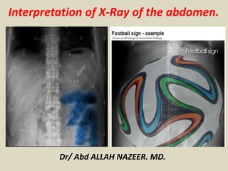
- 32. Football Sign - abdominal radiography showing a large oval radiolucency demarcated by the parietal peritoneum of the abdominal wall.
- 34. Pneumoperitoneum with “football sign.” A, Supine radiograph in a 5-day-old 30-week-gestation premature infant shows a large lucency over the entire abdomen. B, Decubitus view in the same infant confirms the large pneumoperitoneum. Multiple intestinal perforations were found at surgery. C, Another patient with pneumoperitoneum demonstrates the classic football sign on abdominal imaging. Gas outlines the falciform ligament (arrows), and a large lucency overlies the upper abdomen centrally as the gas accumulates anteriorly. At surgery, this patient was found to have a colonic perforation.
- 35. Pyopneumoperitoneum. Massive amount of free air in the abdomen below the diaphragm (white arrows) with air-fluid levels (black arrows) on an upright film.
- 36. Plain X-ray of the chest and upper abdomen displaying obvious Chilaiditi's sign, or presence of gas in the right colic angle between the liver and right.
- 37. Chilaiditi’s syndrome with perforation.
- 39. Rigler sign - abdominal radiography showing free air outlining the small bowel wall, indicating pneumoperitoneum. The Rigler sign, also known as the double wall sign, is seen on a radiograph of the abdomen when the air is present on both sides of the intestine, i.e. when there is air on both the luminal and peritoneal side of the bowel wall.
- 41. Rigler sign.
- 42. Pseudo-Rigler’s Sign - abdominal radiography showing both sides of bowel wall with dilated loops of bowel abut each other(overlapping bowel mimic Rigler sign).
- 43. Pneumoretroperitoneum. Clinical The most common cause of pneumoretroperitoneum is perforation of the second, third, or fourth portion of the duodenum or retroperitoneal colon secondary to trauma, diverticulitis, or ulceration. Post-surgical(Post-urology or adrenal surgery). Radiological findings Pneumoretroperitoneum is most often seen on the right side where the air can outline the right kidney and the undersurface of the liver. In contrast to pneumoperitoneum, air in the retroperitoneum does not move freely with change in position. The gas can extend up into the mediastinum or neck because there is no barrier between them.
- 44. This plain film demonstrates pneumoretroperitoneum with gas outlining the right psoas major (arrows).
- 46. Pneumoretroperitoneum - presence of gas within the retroperitoneal space. Typically the air outlines structures like the kidneys, psoas muscles and retroperitoneal portions of the bowel
- 47. Pneuombilia Clinical Gas in the biliary tree is most commonly secondary to surgical procedures such as choledochoenterostomy or sphincterotmy of the sphincter of Oddi. It may also arise in the setting of trauma, infection by gas producing organisms (i.e. emphysematous cholecystitis), fistulas connecting the biliary system and the intestinal tract (i.e. from dudodenal ulcers, or gallstones), malignant involvement of the ampulla of Vater, or as a congenital anomaly. Radiological findings Ultrasound is the modality of choice for visualizing gas in the biliary tree. Air in the bile ducts generally causes bright linear or globular reflections with shadowing and ring-down artifacts. The gas will move with patient positioning. On plain film, biliary gas may be seen outlining the bile ducts but it is important to be cautious in making this diagnosis as normal periductal fat that surrounds and parallels the course of the bile ducts may give the appearance known as pseudopneumobilia.
- 49. Pneuombilia
- 50. Gas in Portal Vein Clinical With the exception of umbilical vein catheterization in children, gas in the portal veins is a grave prognostic indicator and almost always signals imminent death. There are two major causes of gas in the portal veins. The first is necrosis of the bowel wall from either mechanical obstruction or mesenteric artery occlusion. The break down of the bowel wall allows gas to penetrate the vessel walls and flow to the liver. The second mechanism involves infection of the bowel wall. This may be caused by bowel necrosis with secondary infection with gas producing organisms, or may be due to overwhelming enterocolitis. Radiological findings Gas in the portal veins has a very characteristic appearance. The gas follows the centrifugal flow of the portal veins and thus appears as radiating tubular radiolucencies branching from the porta hepatis. Gas seen in the outermost 2 cm of the liver is indicative of portal vein gas.
- 52. Intramural gas and Portal-venous Gas.
- 53. Gas in the Bowel Wall (Pneumatosis Intestinalis) Clinical Pneumatosis intestinalis can occur as a primary or secondary disorder. Primary pneumatosis intestinalis is less common (15%) and occurs when there is no other underlying respiratory or gastrointestinal abnormality. It is primarily a disease of older adults and is often asymptomatic. Secondary pneumatosis intestinalis is much more common (85%) and occurs int the setting of underlying bowel or pulmonary disease. It may be broken down into three subgroups: GI disease with bowel necrosis (i.e. necrotizing enterocolitis in infants, ischemic necrosis due to mesenteric vascular disease, strangulation, primary infection of the bowel wall); GI disease without bowel necrosis (i.e. pyloroduodenal peptic ulcers, bowel obstruction, IBD, connective tissue disease, endoscopy/colonoscopy, percutaneous jejunostomy tube, steroid therapy, leukemia, intestinal parasites, etc.); obstructive pulmonary disease (i.e. emphysema, bullous disease, chronic bronchitis, asthma). Radiological findings Primary pneumatosis intestinalis will appear as cystic gas in the colon on plain film and CT. Secondary pneumatosis intestinalis will appear as linear gas collections throughout the bowel wall.
- 54. Plain film, from the patient with portal venous gas (arrows) demonstrates massive pneumatosis intestinalis as well.
- 56. Bowel loops pathology. Dilated stomach. Dilated small and large bowel loops. Volvulus. Inflammatory bowel disease. Hernia. Constipation with fecal impaction.
- 58. CT scout image showing massive gastric dilation.
- 60. Double Bubble Sign: abdominal radiography showing two air- filled structures in the upper abdomen, with no air distally. The proximal bubble in the left side filled with air represents the stomach and the second bubble to the right of the midline represents the proximal duodenum.
- 61. Small bowel obstruction/ileus. Dilated small bowel >3cm is considered abnormal Small bowel obstruction and ileus can have similar appearances Causes of small bowel obstruction include: Adhesions from previous abdominal surgery (most common cause) Hernias containing bowel Crohn's disease causing adhesions or inflammatory strictures Neoplasms, benign or malignant Intussusception Volvulus Superior mesenteric artery syndrome, a compression of the duodenum by the superior mesenteric artery and the abdominal aorta Ischemic strictures Foreign bodies (e.g. gallstones in gallstone ileus, swallowed objects) Intestinal atresia.
- 65. Sentinel Loop Sign - abdominal radiography showing dilated loops of small bowel, in a patient with an acute pancreatitis.
- 66. Stepladder Appearance - abdominal radiography showing dilated loops of the small bowel in the left upper quadrant in a mechanical small bowel obstruction.
- 67. String of Beads Sign - abdominal radiography showing linearly arranged small pockets of air in a fluid-dilated small bowel loop.
- 68. Upright abdominal X- ray demonstrating a small bowel obstruction. Note multiple air fluid levels.
- 71. Large bowel obstruction Key points Dilatation of the caecum >9cm is abnormal Dilatation of any other part of the colon >6cm is abnormal Abdominal X-ray may demonstrate the level of obstruction Abdominal X-ray cannot reliably differentiate mechanical obstruction from pseudo-obstruction The most common causes of large bowel obstruction are colorectal carcinoma and diverticular strictures. Less common causes are hernias or volvulus (twisting of the bowel on its mesentery). Adhesions do not commonly cause large bowel obstruction. Radiological appearances of large bowel obstruction differ from those of small bowel obstruction, however, with large bowel obstruction there is often co-existing small bowel dilatation proximally. Dilatation of the caecum >9cm, and >6cm for the rest of the colon is considered abnormal.
- 73. Toxic Megacolon Abdominal X-ray - large bowel obstruction.
- 75. Colon Cutoff Sign: abdominal radiography showing dilated transverse colon to splenic flexure, in this case was associated with pancreatitis.
- 76. Twisting of the bowel - or 'volvulus- is a specific cause of bowel obstruction which can have characteristic appearances on an abdominal X-ray. The two commonest types of bowel twisting are sigmoid volvulus and cecal volvulus. Sigmoid volvulus The sigmoid colon is more prone to twisting than other segments of the large bowel because it is 'mobile' on its own mesentery, which arises from a fixed point in the left iliac fossa (LIF). Twisting at the root of the mesentery results in the formation of an enclosed loop of sigmoid colon which becomes very dilated. If untreated this can lead either to perforation, due to excessive dilatation, or to ischemia due to compromise of the blood supply.
- 78. Coffee-bean Sign - plain film showing dilated sigmoid colon in sigmoid volvulus.
- 80. Cecal volvulus: The caecum is most frequently a retroperitoneal structure, and therefore not susceptible to twisting. However, in up to 20% of individuals there is congenital incomplete peritoneal covering of the caecum with formation of a 'mobile' caecum on a mesentery, such that it no longer lies in the right iliac fossa.
- 82. Bowel wall inflammation Key points Abdominal X-rays sometimes demonstrate signs of bowel inflammation such as mucosal thickening 'thumb-printing' or a featureless colon 'lead pipe' colon. Occasionally, abdominal X-rays show signs of inflammation in patients with inflammatory bowel disease. Abnormalities may relate to either acute or chronic stages of disease.
- 84. Thumbprinting – abdominal radiography showing ‘thumbprinting’(arrows). The normal haustral folds are replaced by wide transverse thickened bands.
- 87. Intra-abdominal calcification is common and the causes may be classified into four broad groups based on morphology: Stones: renal stones, ureteric stones, bladder stones, gallstones pancreatic ductal calcification nodal calcification: most commonly from treated lymphoma, tuberculosis or histoplasmosis Phlebolith, appendicolith, calcified granuloma failed renal transplant encapsulating peritoneal sclerosis Conduit calcification Calcification within the walls of any fluid-filled hollow tube: abdominal aorta, pancreatic duct, ductus deferens, large veins Cystic calcification Calcification in the wall of a mass such as a cyst, pseudocyst or aneurysm. simple serous cysts, Aneurysms, echinococcal cysts, hematoma, 'porcelain' gallbladder, calcified appendiceal mucocele Solid mass calcification mesenteric nodes, adrenal calcifications, uterine fibroids Primary tumours, e.g. ovarian dermoid, metastases, adenoma Spleen (autosplenectomy in sickle cell disease) Renal tuberculosis with autonephrectomy
- 98. A and B, Two patients with pancreatic calculi and chronic pancreatitis in a patient with alcoholism. This is the typical appearance of numerous dense, discrete opacities that cross the midline at the level of L1 to L2 (arrow). The normal pancreas is not visible on abdominal plain films.
- 99. A and B, Numerous phleboliths in two patients. Phleboliths frequently are multiple and bilateral, and they are asymptomatic. They are inconsequential concretions of thrombi attached to the walls of veins. Observe the concentric interior lucency of the phleboliths (arrows). These should not be confused with ureteral stones or calcifications of a pelvic mass The numerous tiny calculi projecting above the pubic symphysis seen in this patient (arrows) are typical of the intraductal calculi often occurring in patients who have chronic inflammation of the prostate.
- 100. Bladder calculi. Three homogeneously dense bladder stones with a continuous rim of calcification typical of concretions. Incidentally noted is a phlebolith with the diagnostic concentric lucency that should not be mistaken for a ureteral stone (arrow).
- 101. Abdominal aorta and iliac arteries calcification. A, Anteroposterior projection. Tubular appearance characteristic of conduit wall calcification. The aortic bifurcation is seen clearly (arrow). B, Lateral view. Notice that the anterior and posterior walls are parallel and the abdominal aorta diameter does not exceed 3.5 cm. Aneurysm should be suspected if the diameter of the abdominal aorta exceeds 3.5 cm. A spondylolytic spondylolisthesis of L5 also is visible (arrow).
- 102. Density(D). Bones Lots of bones are visible on an AXR and it’s important that you can identify each and screen for any pathology (which may be expected or unexpected). In addition, bones on the AXR provide useful landmarks for where you might expect to see a soft tissue structure (e.g. ischial spines are the usual level of the vesicoureteric junction). Bones commonly visible on AXR include: Ribs(Look for increased or decreased density). Lumbar vertebrae( look for alignment, vertebral height and pedicle Sacrum and Coccyx(Look for lytic or sclerotic lesion). Pelvis(look for fracture, lytic or sclerotic lesion). Proximal femurs(Looks for sclerotic, lytic lesion or fractures).
- 103. Scoliosis of the lumbar spine.
- 105. Sclerotic bony metastases (arrows) in a male patient with prostate cancer.
- 106. Extensive lytic and sclerotic bony metastasis with periosteal reaction involving the pubic bones bilaterally and a mainly sclerotic lesion at the left intertrochanteric region likely representing metastatic disease from invasive mucinous adenocarcinoma of the ovaries. Prominent air filled dilated loop of proximal small bowel.
- 107. Other organs and structures Although AXR isn’t well suited to imaging these structures, it’s useful to recognize them to help orientate yourself and spot relevant pathology. Lungs – check the lung bases if visible for pathology (e.g. consolidation) as abdominal pain can sometimes be caused by basal pneumonia Liver – large right upper quadrant (RUQ) structure Gallbladder – rarely seen, look for calcified gallstones and cholecystectomy clips Stomach – left upper quadrant (LUQ) to midline structure, containing a variable amount of air Psoas muscles – lateral edge marked by a relatively straight line either side of the lumbar vertebrae and sacrum Kidneys – often visible, right lower than left due to the liver Spleen – LUQ, superior to left kidney Bladder – variable appearance depending on fullness
- 109. Erect (A) and supine (B) views of the abdominal radiography in a normal child (thin arrows: kidney, thick arrow: psoas shadow).
- 110. Plain X-ray finding of hepatomegaly.
- 111. Splenomegaly. The enlarging spleen (arrows) in the left upper quadrant displaces the splenic flexure of the colon caudally and medially, and the stomach medially.
- 112. Wilms tumour.
- 113. Bowel loops are displaced inferiorly by an enlarged liver. No evidence of obstruction or pneumoperitoneum. No fecal loading. Multiple lytic lesions demonstrated through bony pelvis and proximal femurs. Upper abdominal surgical clips.
- 114. Intussusception with the 'target' and 'meniscus' signs. Supine view shows a round soft-tissue mass in the right upper quadrant. The mass contains a ring-like area of lucency representing the 'target' sign (small arrows). The mass protrudes into the gas-filled transverse colon representing the 'meniscus' sign (long arrows).
- 115. Everything(E) Looks for any abdominal IVC filter, catheter, stent or naso-gastric tube. Looks for metallic F.B. Looks for IUD(intra-abdominal or intra-pelvic). Looks for any surgical stitches for previous surgery. Looks for lung base pathology.
- 122. The IUCD is seen within the anterior lower abdominal cavity, anterior to the upper sacrum, above the presumed site of the uterus.
- 125. Thank You.