SlideShare una empresa de Scribd logo
1 de 61
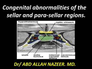
Congenital abnormalities of the
sellar and para-sellar regions.
Dr/ ABD ALLAH NAZEER. MD.
Aim of the lecture.
To show the embryological correlations
of the pituitary gland with adjacent
structures which allow knowledge of
interpretation of the sellar and para-
sellar congenital abnormalities.
Also to show imaging appearance of
the congenital diseases of the sellar
and para-sellar regions. .
Technique:
MRI is the examination of choice for sellar and para-sellar
pathologies due to its superior soft tissue contrast,
multiplanar capability and lack of ionizing radiation. In
addition, MRI also provides useful information about the
relationship of the gland with adjacent anatomical
structures and helps to plan medical or surgical strategy.
MRI techniques in diagnosing pituitary lesions have
witnessed a rapid evolution, ranging from non-contrast MRI
in late 1980s to contrast-enhanced MRI in mid-1990s.
Recently, a variety of advanced MR techniques have been
evolved which are particularly helpful in evaluating specific
cases. These include 3D volumetric analysis of pituitary
volume, high-resolution MR imaging at 3 Tesla (T) for
evaluating pituitary stalk.
The aim of MR imaging is to obtain a high-
spatial-resolution image with a reasonable
signal to noise ratio. It is important to identify
the gland separate from the lesion if possible.
Initially, pre-contrast T1- and T2-weighted spin
echo coronal and sagittal sections are acquired
using a small FOV (20×25 cm), thin slices (2-3
mm), and high-resolution matrix (256×512).
Both the dynamic and routine post-contrast
images and delayed scanning after 30-60
minutes may be combined in one study for
optimum imaging.
Parameters.
TR TE FLIP NXA SLICE MATRIX FOV PHASE OVERSAMPLE GAP
3000-4000 110 130 4 2-3mm 256x512 100-130 R> L 100% 10%
Normal
postnatal
progression of
the pituitary
gland.
Transsphenoidal encephalocele.
MRI and CT images of transsphenoidal encephalocele.
Markedly hypoplastic pituitary with absent bright spot. (A) Sagittal non-
contrast T1- weighted and (B) contrast-enhanced T1-weighted imaging
shows almost complete absence of the gland with an associated shallow
sella (arrowheads). The upper infundibulum is present but the characteristic
high T1-weighted signal is not seen on non contrast imaging.
Anterior pituitary aplasia and posterior pituitary ectopia. No
anterior pituitary gland identified with a thin infundibulum.
Sagittal MRI of the hypothalamic−pituitary axis. The pituitary (lower arrow)
is small and scalloped. There is absence of the normal posterior pituitary and
stalk in and above the pituitary fossa, and an ectopic posterior pituitary
bright spot where the infundibulum normally is (upper arrow).
Ectopic neurohypophysis
with small pituitary gland.
Ectopic posterior pituitary gland and the
pituitary gland is hypoplastic (arrowhead).
Hypothalamic
hamartoma.
Infundibular hypoplasia..
Congenital disorder.
Development of the adenohypophysis.
Rathke,s Cleft Cyst:
CT :
•75% hypodense
•25% iso/hyperdense
•Ca++ rare
•May be difficult to differentiate from other benign cysts or
craniopharyngioma.
Rathke,s cleft cyst.
Sagittal T1W, Fluid suppressed T2W and post contrast
T1W images showing craniopharyngioma in child.
Pars Intermedia Cyst.
Miscellaneous.
Dermoid cyst.
Right para-and supra-sellar dermoid cyst.
Para-sellar teratoma.
THANK YOU.

Más contenido relacionado

La actualidad más candente

Presentation1.pptx, radiological imaging of spinal dysraphism.
Presentation1.pptx, radiological imaging of spinal dysraphism.Presentation1.pptx, radiological imaging of spinal dysraphism.
Presentation1.pptx, radiological imaging of spinal dysraphism.
Abdellah Nazeer
 
Presentation1.pptx sellar and para sellar masses
Presentation1.pptx sellar and para sellar massesPresentation1.pptx sellar and para sellar masses
Presentation1.pptx sellar and para sellar masses
Abdellah Nazeer
 
Fifteen (50) intracranial cystic lesion Dr Ahmed Esawy CT MRI main
Fifteen (50) intracranial cystic lesion Dr Ahmed Esawy CT MRI main Fifteen (50) intracranial cystic lesion Dr Ahmed Esawy CT MRI main
Fifteen (50) intracranial cystic lesion Dr Ahmed Esawy CT MRI main
AHMED ESAWY
 
Presentation1, new mri techniques in the diagnosis and monitoring of multiple...
Presentation1, new mri techniques in the diagnosis and monitoring of multiple...Presentation1, new mri techniques in the diagnosis and monitoring of multiple...
Presentation1, new mri techniques in the diagnosis and monitoring of multiple...
Abdellah Nazeer
 
Presentation1.pptx, perfusiona and specroscopy imaging in brain tumour.
Presentation1.pptx, perfusiona and specroscopy imaging in brain tumour.Presentation1.pptx, perfusiona and specroscopy imaging in brain tumour.
Presentation1.pptx, perfusiona and specroscopy imaging in brain tumour.
Abdellah Nazeer
 
Presentation1.pptx, radiological imaging of cerebral venous thrombosis.
Presentation1.pptx, radiological imaging of cerebral venous thrombosis.Presentation1.pptx, radiological imaging of cerebral venous thrombosis.
Presentation1.pptx, radiological imaging of cerebral venous thrombosis.
Abdellah Nazeer
 
Presentation1.pptx, radiological imaging of spinal cord tumour.
Presentation1.pptx, radiological imaging of spinal cord tumour.Presentation1.pptx, radiological imaging of spinal cord tumour.
Presentation1.pptx, radiological imaging of spinal cord tumour.
Abdellah Nazeer
 

La actualidad más candente (20)

Spinal tumors- Imaging
Spinal tumors- ImagingSpinal tumors- Imaging
Spinal tumors- Imaging
 
Diagnostic Imaging of Central Nervous System Infections
Diagnostic Imaging of Central Nervous System InfectionsDiagnostic Imaging of Central Nervous System Infections
Diagnostic Imaging of Central Nervous System Infections
 
Spots with keys
Spots with keysSpots with keys
Spots with keys
 
Presentation1.pptx, radiological imaging of spinal dysraphism.
Presentation1.pptx, radiological imaging of spinal dysraphism.Presentation1.pptx, radiological imaging of spinal dysraphism.
Presentation1.pptx, radiological imaging of spinal dysraphism.
 
Diagnostic Imaging of Deep Neck Spaces
Diagnostic Imaging of Deep Neck SpacesDiagnostic Imaging of Deep Neck Spaces
Diagnostic Imaging of Deep Neck Spaces
 
Presentation1.pptx sellar and para sellar masses
Presentation1.pptx sellar and para sellar massesPresentation1.pptx sellar and para sellar masses
Presentation1.pptx sellar and para sellar masses
 
Fifteen (50) intracranial cystic lesion Dr Ahmed Esawy CT MRI main
Fifteen (50) intracranial cystic lesion Dr Ahmed Esawy CT MRI main Fifteen (50) intracranial cystic lesion Dr Ahmed Esawy CT MRI main
Fifteen (50) intracranial cystic lesion Dr Ahmed Esawy CT MRI main
 
Radiology spotters
Radiology spottersRadiology spotters
Radiology spotters
 
Diagnostic Imaging of the Pituitary Gland
Diagnostic Imaging of the Pituitary GlandDiagnostic Imaging of the Pituitary Gland
Diagnostic Imaging of the Pituitary Gland
 
Radiology Spotters
Radiology Spotters Radiology Spotters
Radiology Spotters
 
Diagnostic Imaging of Pineal Region Masses
Diagnostic Imaging of Pineal Region MassesDiagnostic Imaging of Pineal Region Masses
Diagnostic Imaging of Pineal Region Masses
 
Presentation1, new mri techniques in the diagnosis and monitoring of multiple...
Presentation1, new mri techniques in the diagnosis and monitoring of multiple...Presentation1, new mri techniques in the diagnosis and monitoring of multiple...
Presentation1, new mri techniques in the diagnosis and monitoring of multiple...
 
Presentation1.pptx, perfusiona and specroscopy imaging in brain tumour.
Presentation1.pptx, perfusiona and specroscopy imaging in brain tumour.Presentation1.pptx, perfusiona and specroscopy imaging in brain tumour.
Presentation1.pptx, perfusiona and specroscopy imaging in brain tumour.
 
radiology Spotters mixed bag
radiology Spotters mixed bagradiology Spotters mixed bag
radiology Spotters mixed bag
 
Presentation1.pptx, radiological imaging of cerebral venous thrombosis.
Presentation1.pptx, radiological imaging of cerebral venous thrombosis.Presentation1.pptx, radiological imaging of cerebral venous thrombosis.
Presentation1.pptx, radiological imaging of cerebral venous thrombosis.
 
Presentation1.pptx, radiological imaging of spinal cord tumour.
Presentation1.pptx, radiological imaging of spinal cord tumour.Presentation1.pptx, radiological imaging of spinal cord tumour.
Presentation1.pptx, radiological imaging of spinal cord tumour.
 
Spots with keys (2)
Spots with keys (2)Spots with keys (2)
Spots with keys (2)
 
Diagnostic Imaging of Intraspinal Masses
Diagnostic Imaging of Intraspinal MassesDiagnostic Imaging of Intraspinal Masses
Diagnostic Imaging of Intraspinal Masses
 
Radiology Spots PPT- 2 by Dr Chandni Wadhwani
Radiology Spots PPT- 2 by Dr Chandni WadhwaniRadiology Spots PPT- 2 by Dr Chandni Wadhwani
Radiology Spots PPT- 2 by Dr Chandni Wadhwani
 
Diagnostic Imaging of Bilateral Abnormalities of the Basal Ganglia & Thalamus
Diagnostic Imaging of Bilateral Abnormalities of the Basal Ganglia & ThalamusDiagnostic Imaging of Bilateral Abnormalities of the Basal Ganglia & Thalamus
Diagnostic Imaging of Bilateral Abnormalities of the Basal Ganglia & Thalamus
 

Destacado

Imaging sectional anatomy of brain part 2
Imaging sectional anatomy of brain   part 2Imaging sectional anatomy of brain   part 2
Imaging sectional anatomy of brain part 2
drnaveent
 
Presentation1.pptx, intra cranial vascular malformation.
Presentation1.pptx, intra cranial vascular malformation.Presentation1.pptx, intra cranial vascular malformation.
Presentation1.pptx, intra cranial vascular malformation.
Abdellah Nazeer
 
Imaging sectional anatomy of brain part 1
Imaging sectional  anatomy  of  brain  part 1Imaging sectional  anatomy  of  brain  part 1
Imaging sectional anatomy of brain part 1
drnaveent
 
Presentation1.pptx, radiological imaging of dementia.
Presentation1.pptx, radiological imaging of dementia.Presentation1.pptx, radiological imaging of dementia.
Presentation1.pptx, radiological imaging of dementia.
Abdellah Nazeer
 
Presentation1.pptx, radiological anatomy of the brain and pituitary gland
Presentation1.pptx, radiological anatomy of the brain and pituitary glandPresentation1.pptx, radiological anatomy of the brain and pituitary gland
Presentation1.pptx, radiological anatomy of the brain and pituitary gland
Abdellah Nazeer
 

Destacado (20)

Case record...Parasellar craniopharyngioma
Case record...Parasellar craniopharyngiomaCase record...Parasellar craniopharyngioma
Case record...Parasellar craniopharyngioma
 
Orbital pathologies.pptx (part 1)
Orbital pathologies.pptx (part 1)Orbital pathologies.pptx (part 1)
Orbital pathologies.pptx (part 1)
 
Suprasellar mass ct scan of a child
Suprasellar mass  ct scan of a child Suprasellar mass  ct scan of a child
Suprasellar mass ct scan of a child
 
Imaging sectional anatomy of brain part 2
Imaging sectional anatomy of brain   part 2Imaging sectional anatomy of brain   part 2
Imaging sectional anatomy of brain part 2
 
Immunogenetics
Immunogenetics Immunogenetics
Immunogenetics
 
Presentation1.pptx, intra cranial vascular malformation.
Presentation1.pptx, intra cranial vascular malformation.Presentation1.pptx, intra cranial vascular malformation.
Presentation1.pptx, intra cranial vascular malformation.
 
20 congenital heart disease Dr. Muhammmad Bin Zulfiqar
20 congenital heart disease Dr. Muhammmad Bin Zulfiqar20 congenital heart disease Dr. Muhammmad Bin Zulfiqar
20 congenital heart disease Dr. Muhammmad Bin Zulfiqar
 
Imaging sectional anatomy of brain part 1
Imaging sectional  anatomy  of  brain  part 1Imaging sectional  anatomy  of  brain  part 1
Imaging sectional anatomy of brain part 1
 
Neurodegenerative disorders MRI approach
Neurodegenerative disorders MRI approachNeurodegenerative disorders MRI approach
Neurodegenerative disorders MRI approach
 
Presentation1.pptx, radiological imaging of dementia.
Presentation1.pptx, radiological imaging of dementia.Presentation1.pptx, radiological imaging of dementia.
Presentation1.pptx, radiological imaging of dementia.
 
craniopharyngioma
 craniopharyngioma craniopharyngioma
craniopharyngioma
 
A pictorial review of “signs in thoracic imaging 02”
A pictorial review of “signs in thoracic imaging 02”A pictorial review of “signs in thoracic imaging 02”
A pictorial review of “signs in thoracic imaging 02”
 
Journal club drug eluting balloon for cad
Journal club   drug eluting balloon for cadJournal club   drug eluting balloon for cad
Journal club drug eluting balloon for cad
 
Imaging in Ocular Pathologies
Imaging in Ocular PathologiesImaging in Ocular Pathologies
Imaging in Ocular Pathologies
 
Eye signs in radiology
Eye signs in radiologyEye signs in radiology
Eye signs in radiology
 
Mri in white matter diseases
Mri in white matter diseasesMri in white matter diseases
Mri in white matter diseases
 
Spinal trauma IMAGING
Spinal trauma  IMAGINGSpinal trauma  IMAGING
Spinal trauma IMAGING
 
Brain anatomy
Brain anatomyBrain anatomy
Brain anatomy
 
Presentation1.pptx, radiological anatomy of the brain and pituitary gland
Presentation1.pptx, radiological anatomy of the brain and pituitary glandPresentation1.pptx, radiological anatomy of the brain and pituitary gland
Presentation1.pptx, radiological anatomy of the brain and pituitary gland
 
CT Abdomen and Pelvis
CT Abdomen and PelvisCT Abdomen and Pelvis
CT Abdomen and Pelvis
 

Similar a Presentation1.pptx, congenital abnormality of the sellar and para sellar regions

Lesiones del cuello en la infancia
Lesiones del cuello en la infanciaLesiones del cuello en la infancia
Lesiones del cuello en la infancia
Lizbet Marrero
 
Presentation1.pptx, radiological imaging of salivary glands diseases.
Presentation1.pptx, radiological imaging of salivary glands diseases.Presentation1.pptx, radiological imaging of salivary glands diseases.
Presentation1.pptx, radiological imaging of salivary glands diseases.
Abdellah Nazeer
 
MR spectroscopy by Dr. Nida Kanwal, Neurosurgery, Liaquat National Hospital
MR spectroscopy by Dr. Nida Kanwal, Neurosurgery, Liaquat National HospitalMR spectroscopy by Dr. Nida Kanwal, Neurosurgery, Liaquat National Hospital
MR spectroscopy by Dr. Nida Kanwal, Neurosurgery, Liaquat National Hospital
noorulainiqbal
 

Similar a Presentation1.pptx, congenital abnormality of the sellar and para sellar regions (20)

Lesiones del cuello en la infancia
Lesiones del cuello en la infanciaLesiones del cuello en la infancia
Lesiones del cuello en la infancia
 
JOURNAL CLUB NEW.ppt
JOURNAL CLUB NEW.pptJOURNAL CLUB NEW.ppt
JOURNAL CLUB NEW.ppt
 
MRI of the Female Pelvis
MRI of the Female PelvisMRI of the Female Pelvis
MRI of the Female Pelvis
 
Presentation1.pptx, radiological imaging of salivary glands diseases.
Presentation1.pptx, radiological imaging of salivary glands diseases.Presentation1.pptx, radiological imaging of salivary glands diseases.
Presentation1.pptx, radiological imaging of salivary glands diseases.
 
MP MRI of prostate by Major Imran from BD.pptx
MP MRI of prostate by Major Imran from BD.pptxMP MRI of prostate by Major Imran from BD.pptx
MP MRI of prostate by Major Imran from BD.pptx
 
recent investigation in epilepsy
recent investigation in epilepsyrecent investigation in epilepsy
recent investigation in epilepsy
 
Neuro ultrasound in carpal tunnel syndrome
Neuro ultrasound in carpal tunnel syndromeNeuro ultrasound in carpal tunnel syndrome
Neuro ultrasound in carpal tunnel syndrome
 
L1-Spinal Cord Sep 2013.ppt
L1-Spinal Cord Sep 2013.pptL1-Spinal Cord Sep 2013.ppt
L1-Spinal Cord Sep 2013.ppt
 
L1-Spinal Cord Sep 2013.ppt
L1-Spinal Cord Sep 2013.pptL1-Spinal Cord Sep 2013.ppt
L1-Spinal Cord Sep 2013.ppt
 
mri vs usg for fetal cns anoalies
mri vs usg for fetal cns anoaliesmri vs usg for fetal cns anoalies
mri vs usg for fetal cns anoalies
 
Mri in urology
Mri in urologyMri in urology
Mri in urology
 
vocal cord paralysis
vocal cord paralysisvocal cord paralysis
vocal cord paralysis
 
RADIOLOGICAL ANATOMY OF THE PITUITARY GLAND AND TECHNIQUES.pptx
RADIOLOGICAL ANATOMY OF THE PITUITARY GLAND AND TECHNIQUES.pptxRADIOLOGICAL ANATOMY OF THE PITUITARY GLAND AND TECHNIQUES.pptx
RADIOLOGICAL ANATOMY OF THE PITUITARY GLAND AND TECHNIQUES.pptx
 
Presentation1, radiological imaging of anal carcinoma.
Presentation1, radiological imaging of anal carcinoma.Presentation1, radiological imaging of anal carcinoma.
Presentation1, radiological imaging of anal carcinoma.
 
Imaging of brachial plexus.Radiology pptx
Imaging of brachial plexus.Radiology pptxImaging of brachial plexus.Radiology pptx
Imaging of brachial plexus.Radiology pptx
 
Advances in neuro-imaging
Advances in neuro-imaging Advances in neuro-imaging
Advances in neuro-imaging
 
Neuroimaging of alzheimer's
Neuroimaging of alzheimer'sNeuroimaging of alzheimer's
Neuroimaging of alzheimer's
 
ANATOMY OF NASOPHARYNX AND STAGING CORRELATION
ANATOMY OF NASOPHARYNX AND STAGING CORRELATION ANATOMY OF NASOPHARYNX AND STAGING CORRELATION
ANATOMY OF NASOPHARYNX AND STAGING CORRELATION
 
MRI Brain
MRI BrainMRI Brain
MRI Brain
 
MR spectroscopy by Dr. Nida Kanwal, Neurosurgery, Liaquat National Hospital
MR spectroscopy by Dr. Nida Kanwal, Neurosurgery, Liaquat National HospitalMR spectroscopy by Dr. Nida Kanwal, Neurosurgery, Liaquat National Hospital
MR spectroscopy by Dr. Nida Kanwal, Neurosurgery, Liaquat National Hospital
 

Más de Abdellah Nazeer

Presentation1, radiological imaging of lateral hindfoot impingement.
Presentation1, radiological imaging of lateral hindfoot impingement.Presentation1, radiological imaging of lateral hindfoot impingement.
Presentation1, radiological imaging of lateral hindfoot impingement.
Abdellah Nazeer
 
Presentation2, radiological anatomy of the liver and spleen.
Presentation2, radiological anatomy of the liver and spleen.Presentation2, radiological anatomy of the liver and spleen.
Presentation2, radiological anatomy of the liver and spleen.
Abdellah Nazeer
 
Presentation1, artifacts and pitfalls of the wrist and elbow joints.
Presentation1, artifacts and pitfalls of the wrist and elbow joints.Presentation1, artifacts and pitfalls of the wrist and elbow joints.
Presentation1, artifacts and pitfalls of the wrist and elbow joints.
Abdellah Nazeer
 
Presentation1, artifact and pitfalls of the knee, hip and ankle joints.
Presentation1, artifact and pitfalls of the knee, hip and ankle joints.Presentation1, artifact and pitfalls of the knee, hip and ankle joints.
Presentation1, artifact and pitfalls of the knee, hip and ankle joints.
Abdellah Nazeer
 
Presentation1, radiological imaging of artifact and pitfalls in shoulder join...
Presentation1, radiological imaging of artifact and pitfalls in shoulder join...Presentation1, radiological imaging of artifact and pitfalls in shoulder join...
Presentation1, radiological imaging of artifact and pitfalls in shoulder join...
Abdellah Nazeer
 
Presentation1, radiological imaging of internal abdominal hernia.
Presentation1, radiological imaging of internal abdominal hernia.Presentation1, radiological imaging of internal abdominal hernia.
Presentation1, radiological imaging of internal abdominal hernia.
Abdellah Nazeer
 
Presentation11, radiological imaging of ovarian torsion.
Presentation11, radiological imaging of ovarian torsion.Presentation11, radiological imaging of ovarian torsion.
Presentation11, radiological imaging of ovarian torsion.
Abdellah Nazeer
 
Presentation1, radiological application of diffusion weighted mri in neck mas...
Presentation1, radiological application of diffusion weighted mri in neck mas...Presentation1, radiological application of diffusion weighted mri in neck mas...
Presentation1, radiological application of diffusion weighted mri in neck mas...
Abdellah Nazeer
 
Presentation1, radiological application of diffusion weighted images in breas...
Presentation1, radiological application of diffusion weighted images in breas...Presentation1, radiological application of diffusion weighted images in breas...
Presentation1, radiological application of diffusion weighted images in breas...
Abdellah Nazeer
 
Presentation1, radiological application of diffusion weighted images in abdom...
Presentation1, radiological application of diffusion weighted images in abdom...Presentation1, radiological application of diffusion weighted images in abdom...
Presentation1, radiological application of diffusion weighted images in abdom...
Abdellah Nazeer
 
Presentation1, radiological application of diffusion weighted imges in neuror...
Presentation1, radiological application of diffusion weighted imges in neuror...Presentation1, radiological application of diffusion weighted imges in neuror...
Presentation1, radiological application of diffusion weighted imges in neuror...
Abdellah Nazeer
 

Más de Abdellah Nazeer (20)

Muculoskeletal Pediatic Imaging..pptx
Muculoskeletal Pediatic Imaging..pptxMuculoskeletal Pediatic Imaging..pptx
Muculoskeletal Pediatic Imaging..pptx
 
Presentation1, Ultrasound of the bowel loops and the lymph nodes..pptx
Presentation1, Ultrasound of the bowel loops and the lymph nodes..pptxPresentation1, Ultrasound of the bowel loops and the lymph nodes..pptx
Presentation1, Ultrasound of the bowel loops and the lymph nodes..pptx
 
Presentation1 Short cases MD..pptx
Presentation1 Short cases MD..pptxPresentation1 Short cases MD..pptx
Presentation1 Short cases MD..pptx
 
Presentation1, MD MCQ Cases..pptx
Presentation1, MD MCQ Cases..pptxPresentation1, MD MCQ Cases..pptx
Presentation1, MD MCQ Cases..pptx
 
Presentation1, Short Cases Quiz..pptx
Presentation1, Short Cases Quiz..pptxPresentation1, Short Cases Quiz..pptx
Presentation1, Short Cases Quiz..pptx
 
Presentation1, radiological imaging of lateral hindfoot impingement.
Presentation1, radiological imaging of lateral hindfoot impingement.Presentation1, radiological imaging of lateral hindfoot impingement.
Presentation1, radiological imaging of lateral hindfoot impingement.
 
Presentation2, radiological anatomy of the liver and spleen.
Presentation2, radiological anatomy of the liver and spleen.Presentation2, radiological anatomy of the liver and spleen.
Presentation2, radiological anatomy of the liver and spleen.
 
Presentation1, artifacts and pitfalls of the wrist and elbow joints.
Presentation1, artifacts and pitfalls of the wrist and elbow joints.Presentation1, artifacts and pitfalls of the wrist and elbow joints.
Presentation1, artifacts and pitfalls of the wrist and elbow joints.
 
Presentation1, artifact and pitfalls of the knee, hip and ankle joints.
Presentation1, artifact and pitfalls of the knee, hip and ankle joints.Presentation1, artifact and pitfalls of the knee, hip and ankle joints.
Presentation1, artifact and pitfalls of the knee, hip and ankle joints.
 
Presentation1, radiological imaging of artifact and pitfalls in shoulder join...
Presentation1, radiological imaging of artifact and pitfalls in shoulder join...Presentation1, radiological imaging of artifact and pitfalls in shoulder join...
Presentation1, radiological imaging of artifact and pitfalls in shoulder join...
 
Presentation1, radiological imaging of internal abdominal hernia.
Presentation1, radiological imaging of internal abdominal hernia.Presentation1, radiological imaging of internal abdominal hernia.
Presentation1, radiological imaging of internal abdominal hernia.
 
Presentation11, radiological imaging of ovarian torsion.
Presentation11, radiological imaging of ovarian torsion.Presentation11, radiological imaging of ovarian torsion.
Presentation11, radiological imaging of ovarian torsion.
 
Presentation1, musculoskeletal anatomy.
Presentation1, musculoskeletal anatomy.Presentation1, musculoskeletal anatomy.
Presentation1, musculoskeletal anatomy.
 
Presentation1, radiological application of diffusion weighted mri in neck mas...
Presentation1, radiological application of diffusion weighted mri in neck mas...Presentation1, radiological application of diffusion weighted mri in neck mas...
Presentation1, radiological application of diffusion weighted mri in neck mas...
 
Presentation1, radiological application of diffusion weighted images in breas...
Presentation1, radiological application of diffusion weighted images in breas...Presentation1, radiological application of diffusion weighted images in breas...
Presentation1, radiological application of diffusion weighted images in breas...
 
Presentation1, radiological application of diffusion weighted images in abdom...
Presentation1, radiological application of diffusion weighted images in abdom...Presentation1, radiological application of diffusion weighted images in abdom...
Presentation1, radiological application of diffusion weighted images in abdom...
 
Presentation1, radiological application of diffusion weighted imges in neuror...
Presentation1, radiological application of diffusion weighted imges in neuror...Presentation1, radiological application of diffusion weighted imges in neuror...
Presentation1, radiological application of diffusion weighted imges in neuror...
 
Presentation1, mr physics.
Presentation1, mr physics.Presentation1, mr physics.
Presentation1, mr physics.
 
Presentation1. ct physics.
Presentation1. ct physics.Presentation1. ct physics.
Presentation1. ct physics.
 
Presentation1, ultrasound physics.
Presentation1, ultrasound physics.Presentation1, ultrasound physics.
Presentation1, ultrasound physics.
 

Último

Call Girls Bhubaneswar Just Call 9907093804 Top Class Call Girl Service Avail...
Call Girls Bhubaneswar Just Call 9907093804 Top Class Call Girl Service Avail...Call Girls Bhubaneswar Just Call 9907093804 Top Class Call Girl Service Avail...
Call Girls Bhubaneswar Just Call 9907093804 Top Class Call Girl Service Avail...
Dipal Arora
 
Call Girls in Gagan Vihar (delhi) call me [🔝 9953056974 🔝] escort service 24X7
Call Girls in Gagan Vihar (delhi) call me [🔝  9953056974 🔝] escort service 24X7Call Girls in Gagan Vihar (delhi) call me [🔝  9953056974 🔝] escort service 24X7
Call Girls in Gagan Vihar (delhi) call me [🔝 9953056974 🔝] escort service 24X7
9953056974 Low Rate Call Girls In Saket, Delhi NCR
 

Último (20)

Call Girls Bhubaneswar Just Call 9907093804 Top Class Call Girl Service Avail...
Call Girls Bhubaneswar Just Call 9907093804 Top Class Call Girl Service Avail...Call Girls Bhubaneswar Just Call 9907093804 Top Class Call Girl Service Avail...
Call Girls Bhubaneswar Just Call 9907093804 Top Class Call Girl Service Avail...
 
Mumbai ] (Call Girls) in Mumbai 10k @ I'm VIP Independent Escorts Girls 98333...
Mumbai ] (Call Girls) in Mumbai 10k @ I'm VIP Independent Escorts Girls 98333...Mumbai ] (Call Girls) in Mumbai 10k @ I'm VIP Independent Escorts Girls 98333...
Mumbai ] (Call Girls) in Mumbai 10k @ I'm VIP Independent Escorts Girls 98333...
 
(👑VVIP ISHAAN ) Russian Call Girls Service Navi Mumbai🖕9920874524🖕Independent...
(👑VVIP ISHAAN ) Russian Call Girls Service Navi Mumbai🖕9920874524🖕Independent...(👑VVIP ISHAAN ) Russian Call Girls Service Navi Mumbai🖕9920874524🖕Independent...
(👑VVIP ISHAAN ) Russian Call Girls Service Navi Mumbai🖕9920874524🖕Independent...
 
Call Girls Jabalpur Just Call 8250077686 Top Class Call Girl Service Available
Call Girls Jabalpur Just Call 8250077686 Top Class Call Girl Service AvailableCall Girls Jabalpur Just Call 8250077686 Top Class Call Girl Service Available
Call Girls Jabalpur Just Call 8250077686 Top Class Call Girl Service Available
 
Night 7k to 12k Navi Mumbai Call Girl Photo 👉 BOOK NOW 9833363713 👈 ♀️ night ...
Night 7k to 12k Navi Mumbai Call Girl Photo 👉 BOOK NOW 9833363713 👈 ♀️ night ...Night 7k to 12k Navi Mumbai Call Girl Photo 👉 BOOK NOW 9833363713 👈 ♀️ night ...
Night 7k to 12k Navi Mumbai Call Girl Photo 👉 BOOK NOW 9833363713 👈 ♀️ night ...
 
Call Girls Guntur Just Call 8250077686 Top Class Call Girl Service Available
Call Girls Guntur  Just Call 8250077686 Top Class Call Girl Service AvailableCall Girls Guntur  Just Call 8250077686 Top Class Call Girl Service Available
Call Girls Guntur Just Call 8250077686 Top Class Call Girl Service Available
 
Top Quality Call Girl Service Kalyanpur 6378878445 Available Call Girls Any Time
Top Quality Call Girl Service Kalyanpur 6378878445 Available Call Girls Any TimeTop Quality Call Girl Service Kalyanpur 6378878445 Available Call Girls Any Time
Top Quality Call Girl Service Kalyanpur 6378878445 Available Call Girls Any Time
 
Call Girls Cuttack Just Call 9907093804 Top Class Call Girl Service Available
Call Girls Cuttack Just Call 9907093804 Top Class Call Girl Service AvailableCall Girls Cuttack Just Call 9907093804 Top Class Call Girl Service Available
Call Girls Cuttack Just Call 9907093804 Top Class Call Girl Service Available
 
VIP Service Call Girls Sindhi Colony 📳 7877925207 For 18+ VIP Call Girl At Th...
VIP Service Call Girls Sindhi Colony 📳 7877925207 For 18+ VIP Call Girl At Th...VIP Service Call Girls Sindhi Colony 📳 7877925207 For 18+ VIP Call Girl At Th...
VIP Service Call Girls Sindhi Colony 📳 7877925207 For 18+ VIP Call Girl At Th...
 
Call Girls Nagpur Just Call 9907093804 Top Class Call Girl Service Available
Call Girls Nagpur Just Call 9907093804 Top Class Call Girl Service AvailableCall Girls Nagpur Just Call 9907093804 Top Class Call Girl Service Available
Call Girls Nagpur Just Call 9907093804 Top Class Call Girl Service Available
 
Call Girls Ludhiana Just Call 9907093804 Top Class Call Girl Service Available
Call Girls Ludhiana Just Call 9907093804 Top Class Call Girl Service AvailableCall Girls Ludhiana Just Call 9907093804 Top Class Call Girl Service Available
Call Girls Ludhiana Just Call 9907093804 Top Class Call Girl Service Available
 
Call Girls Gwalior Just Call 8617370543 Top Class Call Girl Service Available
Call Girls Gwalior Just Call 8617370543 Top Class Call Girl Service AvailableCall Girls Gwalior Just Call 8617370543 Top Class Call Girl Service Available
Call Girls Gwalior Just Call 8617370543 Top Class Call Girl Service Available
 
Call Girls Haridwar Just Call 8250077686 Top Class Call Girl Service Available
Call Girls Haridwar Just Call 8250077686 Top Class Call Girl Service AvailableCall Girls Haridwar Just Call 8250077686 Top Class Call Girl Service Available
Call Girls Haridwar Just Call 8250077686 Top Class Call Girl Service Available
 
Call Girls Tirupati Just Call 8250077686 Top Class Call Girl Service Available
Call Girls Tirupati Just Call 8250077686 Top Class Call Girl Service AvailableCall Girls Tirupati Just Call 8250077686 Top Class Call Girl Service Available
Call Girls Tirupati Just Call 8250077686 Top Class Call Girl Service Available
 
Top Rated Bangalore Call Girls Ramamurthy Nagar ⟟ 9332606886 ⟟ Call Me For G...
Top Rated Bangalore Call Girls Ramamurthy Nagar ⟟  9332606886 ⟟ Call Me For G...Top Rated Bangalore Call Girls Ramamurthy Nagar ⟟  9332606886 ⟟ Call Me For G...
Top Rated Bangalore Call Girls Ramamurthy Nagar ⟟ 9332606886 ⟟ Call Me For G...
 
Call Girls Faridabad Just Call 9907093804 Top Class Call Girl Service Available
Call Girls Faridabad Just Call 9907093804 Top Class Call Girl Service AvailableCall Girls Faridabad Just Call 9907093804 Top Class Call Girl Service Available
Call Girls Faridabad Just Call 9907093804 Top Class Call Girl Service Available
 
Call Girls in Gagan Vihar (delhi) call me [🔝 9953056974 🔝] escort service 24X7
Call Girls in Gagan Vihar (delhi) call me [🔝  9953056974 🔝] escort service 24X7Call Girls in Gagan Vihar (delhi) call me [🔝  9953056974 🔝] escort service 24X7
Call Girls in Gagan Vihar (delhi) call me [🔝 9953056974 🔝] escort service 24X7
 
Premium Call Girls In Jaipur {8445551418} ❤️VVIP SEEMA Call Girl in Jaipur Ra...
Premium Call Girls In Jaipur {8445551418} ❤️VVIP SEEMA Call Girl in Jaipur Ra...Premium Call Girls In Jaipur {8445551418} ❤️VVIP SEEMA Call Girl in Jaipur Ra...
Premium Call Girls In Jaipur {8445551418} ❤️VVIP SEEMA Call Girl in Jaipur Ra...
 
Russian Call Girls Service Jaipur {8445551418} ❤️PALLAVI VIP Jaipur Call Gir...
Russian Call Girls Service  Jaipur {8445551418} ❤️PALLAVI VIP Jaipur Call Gir...Russian Call Girls Service  Jaipur {8445551418} ❤️PALLAVI VIP Jaipur Call Gir...
Russian Call Girls Service Jaipur {8445551418} ❤️PALLAVI VIP Jaipur Call Gir...
 
Call Girls in Delhi Triveni Complex Escort Service(🔝))/WhatsApp 97111⇛47426
Call Girls in Delhi Triveni Complex Escort Service(🔝))/WhatsApp 97111⇛47426Call Girls in Delhi Triveni Complex Escort Service(🔝))/WhatsApp 97111⇛47426
Call Girls in Delhi Triveni Complex Escort Service(🔝))/WhatsApp 97111⇛47426
 

Presentation1.pptx, congenital abnormality of the sellar and para sellar regions

  • 1. Congenital abnormalities of the sellar and para-sellar regions. Dr/ ABD ALLAH NAZEER. MD.
  • 2. Aim of the lecture. To show the embryological correlations of the pituitary gland with adjacent structures which allow knowledge of interpretation of the sellar and para- sellar congenital abnormalities. Also to show imaging appearance of the congenital diseases of the sellar and para-sellar regions. .
  • 3.
  • 4.
  • 5.
  • 6.
  • 7.
  • 8.
  • 9.
  • 10.
  • 11. Technique: MRI is the examination of choice for sellar and para-sellar pathologies due to its superior soft tissue contrast, multiplanar capability and lack of ionizing radiation. In addition, MRI also provides useful information about the relationship of the gland with adjacent anatomical structures and helps to plan medical or surgical strategy. MRI techniques in diagnosing pituitary lesions have witnessed a rapid evolution, ranging from non-contrast MRI in late 1980s to contrast-enhanced MRI in mid-1990s. Recently, a variety of advanced MR techniques have been evolved which are particularly helpful in evaluating specific cases. These include 3D volumetric analysis of pituitary volume, high-resolution MR imaging at 3 Tesla (T) for evaluating pituitary stalk.
  • 12. The aim of MR imaging is to obtain a high- spatial-resolution image with a reasonable signal to noise ratio. It is important to identify the gland separate from the lesion if possible. Initially, pre-contrast T1- and T2-weighted spin echo coronal and sagittal sections are acquired using a small FOV (20×25 cm), thin slices (2-3 mm), and high-resolution matrix (256×512). Both the dynamic and routine post-contrast images and delayed scanning after 30-60 minutes may be combined in one study for optimum imaging.
  • 13. Parameters. TR TE FLIP NXA SLICE MATRIX FOV PHASE OVERSAMPLE GAP 3000-4000 110 130 4 2-3mm 256x512 100-130 R> L 100% 10%
  • 14.
  • 15.
  • 17.
  • 18.
  • 19. Transsphenoidal encephalocele. MRI and CT images of transsphenoidal encephalocele.
  • 20.
  • 21.
  • 22. Markedly hypoplastic pituitary with absent bright spot. (A) Sagittal non- contrast T1- weighted and (B) contrast-enhanced T1-weighted imaging shows almost complete absence of the gland with an associated shallow sella (arrowheads). The upper infundibulum is present but the characteristic high T1-weighted signal is not seen on non contrast imaging.
  • 23. Anterior pituitary aplasia and posterior pituitary ectopia. No anterior pituitary gland identified with a thin infundibulum.
  • 24.
  • 25.
  • 26.
  • 27.
  • 28. Sagittal MRI of the hypothalamic−pituitary axis. The pituitary (lower arrow) is small and scalloped. There is absence of the normal posterior pituitary and stalk in and above the pituitary fossa, and an ectopic posterior pituitary bright spot where the infundibulum normally is (upper arrow). Ectopic neurohypophysis with small pituitary gland.
  • 29. Ectopic posterior pituitary gland and the pituitary gland is hypoplastic (arrowhead).
  • 30.
  • 31.
  • 32.
  • 33.
  • 34.
  • 35.
  • 37.
  • 38.
  • 40. Congenital disorder. Development of the adenohypophysis.
  • 41.
  • 42.
  • 43. Rathke,s Cleft Cyst: CT : •75% hypodense •25% iso/hyperdense •Ca++ rare •May be difficult to differentiate from other benign cysts or craniopharyngioma.
  • 44.
  • 45.
  • 47.
  • 48.
  • 49.
  • 50. Sagittal T1W, Fluid suppressed T2W and post contrast T1W images showing craniopharyngioma in child.
  • 51.
  • 52.
  • 55.
  • 58.
  • 59.