SlideShare una empresa de Scribd logo
1 de 65
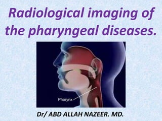
Radiological imaging of
the pharyngeal diseases.
Dr/ ABD ALLAH NAZEER. MD.
Pharynx (oro and hypopharynx).
The hypopharynx is in continuity with the oropharynx and
extends from the level of the hyoid bone to the opening of
the esophagus. It is composed of the inferior aspect of the
middle constrictor and the inferior pharyngeal constrictor
muscles. The hypopharynx sits behind the larynx, and its
lateral-most walls, the pyriform sinuses, are nestled
medial to the thyroid lamina. Immediately posterior to the
hypopharynx is the potential retropharyngeal space; the
prevertebral fascia is posterior to that. Patients with
tumors of the hypopharynx present with progressive
difficulty and pain on swallowing, first to solids and then
to liquids; they often report experiencing referred otalgia.
Anatomy.
Imaging studies:
A. X-rays.
B. CT scans.
C. MRI.
D. PET CT Scan.
Congenital anomalies of oropharynx.
Both pharyngeal atresia (Morris and Reay, 1971) and congenital large
pharynx (Calnan, 1971) have been reported. Patients with congenital
large pharynx and velopharyngeal insufficiency are not helped with
palatal push-back surgery; instead, a posterior pharyngeal flap is
needed. The presence of a subglossopalatal membrane has been
reported in one girl, who developed dyspnea and dysphagia after birth
(Nakajima et al, 1979). A thick, fan-shaped fibrous membrane existed
from the subglossal region to the junction of the cleft of the soft palate
and the hard palate. The literature records a persistent
buccopharyngeal membrane (Chandra et al, 1974) and pharyngeal
membrane (Hoffman, 1979) from the anterior pillar to the base of the
tongue, interfering with speech, posterior pillar mucosal webbing, and
palatal pharyngeal muscle displacement (Warren et al, 1978).
Newcomb (1897) reported 42 cases that included absence of pillars and
tonsils and lymphoid tissue abnormalities.
Pharyngitis is a type of inflammation, most commonly caused by
an upper respiratory tract infection. It may be classified as acute
or chronic. An acute pharyngitis may be catarrhal, purulent or
ulcerative, depending on the virulence of the causative agent and
the immune capacity of the affected individual. Chronic
pharyngitis is the most common otolaringologic disease and may
be catarrhal, hypertrophic or atrophic.
Most acute cases are caused by viral infections (40–80%), with the
remainder caused by bacterial infections, fungal infections, or
irritants such as pollutants or chemical substances.
Tonsillitis is inflammation of the tonsils most commonly caused by
viral or bacterial infection. Symptoms may include sore throat and
fever. When caused by a bacterium belonging to the group A
streptococcus, it is typically referred to as strep throat.
Inflammatory diseases.
Severe acute pharyngitis.
Retropharyngeal Abscess.
CT of retropharyngeal abscess
Retropharyngeal Abscess (CT, )
Retropharyngeal Abscess.
Lateropharyngeal abscess.
Acute bacterial tonsillitis.
Bilateral peritonsillar abscesses (PTA).CT of peritonsillar abscess (late stage).
Tonsillar abscess (CT).
Epiglotitis (Radiography, )
Periodontal abscess (CT).
Benign tumour of the hypo and oropharynx.
Usually uncommon and present as smooth,
well defined, pedunculated and mobile mass.
Papilloma.
Hemangioma.
Pleomorphic adenoma.
Mucous cyst.
Lipoma.
Fibroma.
Leiomyoma.
Vascular tumors.
Malignant tumour of the hypo and oropharynx.
Histological classification:
Squamous cell carcinoma: may be,
well/moderately/ poorly differentiated.
Lymphoepithelioma.
Adenocarcinoma.
Lymphoma, both Hodgkin and non-Hodgkins.
Carcinoma of the hypopharynx occur in order of
frequency:
Pyriform sinus(60%).
Post-cricoid region(30%).
Posterior pharyngeal wall(10%).
Nodular fasciitis caused a benign tumor which is
composed of fibrous tissue. The most commonly
affected site is the upper extremity, which accounts for
half of all cases. Ten to 20% of the lesions occur in the
head and neck area, most of which are subcutaneous.
We report a case of nodular fasciitis, which presented
as an unusually large submucosal tumor of the pharynx.
The tumor was extended from the retropharyngeal to
the parapharyngeal space, and it was measured 8 x 4 x
3 cm in size. Since nodular fasciitis is known for the
spontaneous regression, the tumor was transorally
debulked by the use of YAG laser.
Giant tumor formed by nodular fasciitis of the pharynx.
Nodular fasciitis.
Nodular fasciitis.
Retropharyngeal lipoma.
Neural fibrolipoma in pharyngeal mucosal space.
Hemangioma of the pharynx.
Pleomorphic adenoma of the hypopharynx.
Pleomorphic adenoma.
Pleomorphic adenoma in the parapharyngeal mass.
Hypopharyngeal duct cyst.
Paraganglioma of the hypopharynx.
Vagal schwannoma. Cystic choristoma.
Glomus vagale.Neurofibroma.
Carotid body tumor. True internal carotid artery aneurysm
Internal carotid pseudoaneurysm. Meningioma.
Multiplanar mutisequential MRI of a 24 year-old patient known for neurofibromatosis
type 1. Note the large left sided suprahyoid neurofibroma (arrowheads).
Malignant tumours of the hypo and oropharynx.
Squamous cell carcinomas amount to more than 90% of
malignant tumours of the hypo and oropharynx. As in other
parts of the upper aerodigestive tract, there is a strong and
synergistic association with tobacco smoking and alcohol
abuse. In some regions, particularly the Indian subcontinent,
oral cancer is among the most frequent malignancies, largely
due to tobacco chewing.
The WHO Working Group has made an attempt to unify the
terminology used to define the histological features of
precursor lesions throughout the head and neck region.
Although there has been considerable progress in the
understanding of the genetic and molecular events underlying
the progression of precancerous lesions to invasive carcinomas,
this has yet to be translated into novel therapeutic strategies.
Oropharyngeal squamous cell carcinomas (OSCC) begins
in the oropharynx , the middle part of the throat that
includes the soft palate , the base of the tongue , and
the tonsils . Squamous cell cancers of the tonsils are
more strongly associated with human papillomavirus
infection than are cancers of other regions of the head
and neck.
The hypopharynx includes the pyriform sinuses, the
posterior pharyngeal wall, and the postcricoid area.
Tumors of the hypopharynx frequently have an
advanced stage at diagnosis, and have the most
adverse prognoses of pharyngeal tumors. They tend
to metastasize early due to the extensive lymphatic
network around the larynx .
Parapharyngeal squamous carcinoma.
Squamous cell carcinoma.
Hypopharyngeal squamous cell carcinoma.
Post-cricoid squamous carcinoma.
Right tonsil cancer with spread through constrictor
muscle directly into parapharyngeal fat.
Two cases of left tonsillar carcinoma, which causes effacement
of the fat of the retropharyngeal space on the left.
Metastatic adenopathy from a tonsilar carcinoma.
Adenocarcinoma of left hypopharynx with lymph nodes metastasis.
Tonsillar primary adenocarcinoma with lymph node metastasis
The initial manifestation of certain lymphoid disorders
may be a tumor of the nasopharynx or pharynx. At
the present time there seems to be no accurate way
of predicting the subsequent behavior of the varying
types of lymphoid tumors which appear in this area.
The basis of this report is an analysis of the clinical
and pathological data whose presenting symptoms
was discovered a primary lymphomatous neoplasm
of the pharynx, with no simultaneous evidence of
systemic involvement or of disease elsewhere in the
body.
LYMPHOMAS OF THE PHARYNX:
Non-Hodgkin’s lymphoma.
Diffuse large cell lymphoma.
Mixed cellularity Hodgkin lymphoma in
HIV positive patient.
Follicular lymphoma. B-Cell Lymphoma.
Unilateral mass was found in the tonsil, associated with cervical
lymphadenopathy, in the HL group, nodular sclerosis subtype.
A case of tonsillar B-cell lymphoma with extensive local spread
(arrow) as seen on MIP (A) CT (B) and fused PET/CT (C) Images.
Thank You.

Más contenido relacionado

La actualidad más candente

ANATOMICAL VARIANTS OF CT PNS
ANATOMICAL VARIANTS OF CT PNSANATOMICAL VARIANTS OF CT PNS
ANATOMICAL VARIANTS OF CT PNSsusritha17
 
CT ANATOMY OF PARA NASAL SINUSES
CT ANATOMY OF PARA NASAL SINUSESCT ANATOMY OF PARA NASAL SINUSES
CT ANATOMY OF PARA NASAL SINUSESPrasanna Kumaravel
 
Ascites and Pleural Effusion
 Ascites and Pleural Effusion Ascites and Pleural Effusion
Ascites and Pleural EffusionMedia Genie
 
Larynx anatomy CT and MRI
Larynx anatomy CT and MRILarynx anatomy CT and MRI
Larynx anatomy CT and MRIDr. Mohit Goel
 
Presentation1.pptx, radiological imaging of the nasopharyngeal diseases.
Presentation1.pptx, radiological imaging of the nasopharyngeal diseases.Presentation1.pptx, radiological imaging of the nasopharyngeal diseases.
Presentation1.pptx, radiological imaging of the nasopharyngeal diseases.Abdellah Nazeer
 
Imaging of white matter diseases
Imaging of white matter diseasesImaging of white matter diseases
Imaging of white matter diseasesNavni Garg
 
Radiological approach to neck spaces
Radiological approach to neck spacesRadiological approach to neck spaces
Radiological approach to neck spacesMilan Silwal
 
Presentation1, radiological imaging of endometrial carcinoma.
Presentation1, radiological imaging of endometrial carcinoma.Presentation1, radiological imaging of endometrial carcinoma.
Presentation1, radiological imaging of endometrial carcinoma.Abdellah Nazeer
 
Temporal bone radiology
Temporal bone radiologyTemporal bone radiology
Temporal bone radiologySatish Naga
 
Radiology Spots PPT- 3 by Dr Chandni Wadhwani
 Radiology Spots PPT- 3 by Dr Chandni Wadhwani Radiology Spots PPT- 3 by Dr Chandni Wadhwani
Radiology Spots PPT- 3 by Dr Chandni WadhwaniChandni Wadhwani
 
Presentation1.pptx radiological imaging of mediastinal masses .
Presentation1.pptx radiological imaging of mediastinal masses .Presentation1.pptx radiological imaging of mediastinal masses .
Presentation1.pptx radiological imaging of mediastinal masses .Abdellah Nazeer
 
Imaging modalities of the petrous bone
Imaging modalities of the petrous boneImaging modalities of the petrous bone
Imaging modalities of the petrous boneAbdellah Nazeer
 
Diagnostic Imaging of Orbital Lesions
Diagnostic Imaging of Orbital LesionsDiagnostic Imaging of Orbital Lesions
Diagnostic Imaging of Orbital LesionsMohamed M.A. Zaitoun
 
Benign bone disease spotters
Benign bone disease spottersBenign bone disease spotters
Benign bone disease spottersAGRAWAL14
 
CT anatomy of Neck Spaces RV
CT anatomy of Neck Spaces RVCT anatomy of Neck Spaces RV
CT anatomy of Neck Spaces RVRoshan Valentine
 
Diagnostic Imaging of Cerebellopontine Angle Masses
Diagnostic Imaging of Cerebellopontine Angle MassesDiagnostic Imaging of Cerebellopontine Angle Masses
Diagnostic Imaging of Cerebellopontine Angle MassesMohamed M.A. Zaitoun
 

La actualidad más candente (20)

Imaging in CNS Infections
Imaging in CNS InfectionsImaging in CNS Infections
Imaging in CNS Infections
 
ANATOMICAL VARIANTS OF CT PNS
ANATOMICAL VARIANTS OF CT PNSANATOMICAL VARIANTS OF CT PNS
ANATOMICAL VARIANTS OF CT PNS
 
CT ANATOMY OF PARA NASAL SINUSES
CT ANATOMY OF PARA NASAL SINUSESCT ANATOMY OF PARA NASAL SINUSES
CT ANATOMY OF PARA NASAL SINUSES
 
Ascites and Pleural Effusion
 Ascites and Pleural Effusion Ascites and Pleural Effusion
Ascites and Pleural Effusion
 
Lung cancer radiology
Lung cancer radiologyLung cancer radiology
Lung cancer radiology
 
Larynx anatomy CT and MRI
Larynx anatomy CT and MRILarynx anatomy CT and MRI
Larynx anatomy CT and MRI
 
Presentation1.pptx, radiological imaging of the nasopharyngeal diseases.
Presentation1.pptx, radiological imaging of the nasopharyngeal diseases.Presentation1.pptx, radiological imaging of the nasopharyngeal diseases.
Presentation1.pptx, radiological imaging of the nasopharyngeal diseases.
 
Imaging of white matter diseases
Imaging of white matter diseasesImaging of white matter diseases
Imaging of white matter diseases
 
Radiological approach to neck spaces
Radiological approach to neck spacesRadiological approach to neck spaces
Radiological approach to neck spaces
 
Presentation1, radiological imaging of endometrial carcinoma.
Presentation1, radiological imaging of endometrial carcinoma.Presentation1, radiological imaging of endometrial carcinoma.
Presentation1, radiological imaging of endometrial carcinoma.
 
Temporal bone radiology
Temporal bone radiologyTemporal bone radiology
Temporal bone radiology
 
Radiology Spots PPT- 3 by Dr Chandni Wadhwani
 Radiology Spots PPT- 3 by Dr Chandni Wadhwani Radiology Spots PPT- 3 by Dr Chandni Wadhwani
Radiology Spots PPT- 3 by Dr Chandni Wadhwani
 
SKULL BASE IMAGING
SKULL BASE IMAGINGSKULL BASE IMAGING
SKULL BASE IMAGING
 
Presentation1.pptx radiological imaging of mediastinal masses .
Presentation1.pptx radiological imaging of mediastinal masses .Presentation1.pptx radiological imaging of mediastinal masses .
Presentation1.pptx radiological imaging of mediastinal masses .
 
Imaging modalities of the petrous bone
Imaging modalities of the petrous boneImaging modalities of the petrous bone
Imaging modalities of the petrous bone
 
Diagnostic Imaging of Orbital Lesions
Diagnostic Imaging of Orbital LesionsDiagnostic Imaging of Orbital Lesions
Diagnostic Imaging of Orbital Lesions
 
Ct of the larynx
Ct of the larynxCt of the larynx
Ct of the larynx
 
Benign bone disease spotters
Benign bone disease spottersBenign bone disease spotters
Benign bone disease spotters
 
CT anatomy of Neck Spaces RV
CT anatomy of Neck Spaces RVCT anatomy of Neck Spaces RV
CT anatomy of Neck Spaces RV
 
Diagnostic Imaging of Cerebellopontine Angle Masses
Diagnostic Imaging of Cerebellopontine Angle MassesDiagnostic Imaging of Cerebellopontine Angle Masses
Diagnostic Imaging of Cerebellopontine Angle Masses
 

Similar a Presentation1.pptx, radiological imaging of the pharyngeal diseases

hypopharyngealcancer2-151012175726-lva1-app6892.pptx
hypopharyngealcancer2-151012175726-lva1-app6892.pptxhypopharyngealcancer2-151012175726-lva1-app6892.pptx
hypopharyngealcancer2-151012175726-lva1-app6892.pptxegodoc222
 
Cancer of the hypopharynx
Cancer  of  the hypopharynxCancer  of  the hypopharynx
Cancer of the hypopharynxMohamed Barakat
 
Throat_Pharynx_Carcinoma_Oropharynx_ENT_Lectures.ppt
Throat_Pharynx_Carcinoma_Oropharynx_ENT_Lectures.pptThroat_Pharynx_Carcinoma_Oropharynx_ENT_Lectures.ppt
Throat_Pharynx_Carcinoma_Oropharynx_ENT_Lectures.pptRatna Harika
 
Benign sinonasal masses presentation & management-1
Benign sinonasal masses presentation & management-1Benign sinonasal masses presentation & management-1
Benign sinonasal masses presentation & management-1kamalaiims
 
Cancer of the nasopharynx
Cancer of the nasopharynxCancer of the nasopharynx
Cancer of the nasopharynxIbrahim Barakat
 
Cancer of the nasopharynx
Cancer of the nasopharynxCancer of the nasopharynx
Cancer of the nasopharynxIbrahim Barakat
 
PATHOLOGY OF UPPER RESPIRARTORY
PATHOLOGY OF UPPER RESPIRARTORY PATHOLOGY OF UPPER RESPIRARTORY
PATHOLOGY OF UPPER RESPIRARTORY دكتور مريض
 
CA HYPOPHARYNX.pptx
CA HYPOPHARYNX.pptxCA HYPOPHARYNX.pptx
CA HYPOPHARYNX.pptxDr Monica P
 
Approach to Diseases of Pleura and Mediastinum
Approach to Diseases of Pleura and MediastinumApproach to Diseases of Pleura and Mediastinum
Approach to Diseases of Pleura and MediastinumChetan Ganteppanavar
 
Tumours of nasopharynx (2) itp class dr.davis - 03.06.16
Tumours of nasopharynx (2) itp class   dr.davis - 03.06.16Tumours of nasopharynx (2) itp class   dr.davis - 03.06.16
Tumours of nasopharynx (2) itp class dr.davis - 03.06.16ophthalmgmcri
 

Similar a Presentation1.pptx, radiological imaging of the pharyngeal diseases (20)

hypopharyngealcancer2-151012175726-lva1-app6892.pptx
hypopharyngealcancer2-151012175726-lva1-app6892.pptxhypopharyngealcancer2-151012175726-lva1-app6892.pptx
hypopharyngealcancer2-151012175726-lva1-app6892.pptx
 
Hypopharyngeal cancer
Hypopharyngeal cancer Hypopharyngeal cancer
Hypopharyngeal cancer
 
Pharyngeal tumor
Pharyngeal tumorPharyngeal tumor
Pharyngeal tumor
 
Pharyngeal tumor
Pharyngeal tumorPharyngeal tumor
Pharyngeal tumor
 
Cancer of the hypopharynx
Cancer  of  the hypopharynxCancer  of  the hypopharynx
Cancer of the hypopharynx
 
Throat_Pharynx_Carcinoma_Oropharynx_ENT_Lectures.ppt
Throat_Pharynx_Carcinoma_Oropharynx_ENT_Lectures.pptThroat_Pharynx_Carcinoma_Oropharynx_ENT_Lectures.ppt
Throat_Pharynx_Carcinoma_Oropharynx_ENT_Lectures.ppt
 
Laryngeal carcinoma
Laryngeal carcinomaLaryngeal carcinoma
Laryngeal carcinoma
 
Benign sinonasal masses presentation & management-1
Benign sinonasal masses presentation & management-1Benign sinonasal masses presentation & management-1
Benign sinonasal masses presentation & management-1
 
Cancer of the nasopharynx
Cancer of the nasopharynxCancer of the nasopharynx
Cancer of the nasopharynx
 
Cancer of the nasopharynx
Cancer of the nasopharynxCancer of the nasopharynx
Cancer of the nasopharynx
 
Malignant tumor of neck
Malignant tumor of neckMalignant tumor of neck
Malignant tumor of neck
 
2)acute &chronic pharyngeal abscess
2)acute &chronic pharyngeal abscess2)acute &chronic pharyngeal abscess
2)acute &chronic pharyngeal abscess
 
PATHOLOGY OF UPPER RESPIRARTORY
PATHOLOGY OF UPPER RESPIRARTORY PATHOLOGY OF UPPER RESPIRARTORY
PATHOLOGY OF UPPER RESPIRARTORY
 
CA HYPOPHARYNX.pptx
CA HYPOPHARYNX.pptxCA HYPOPHARYNX.pptx
CA HYPOPHARYNX.pptx
 
ORO PHARYNX.pptx
ORO PHARYNX.pptxORO PHARYNX.pptx
ORO PHARYNX.pptx
 
Approach to Diseases of Pleura and Mediastinum
Approach to Diseases of Pleura and MediastinumApproach to Diseases of Pleura and Mediastinum
Approach to Diseases of Pleura and Mediastinum
 
Pharynx Lecture_3.doc
Pharynx Lecture_3.docPharynx Lecture_3.doc
Pharynx Lecture_3.doc
 
Cancer of larynx
Cancer of larynxCancer of larynx
Cancer of larynx
 
Tumours of nasopharynx (2) itp class dr.davis - 03.06.16
Tumours of nasopharynx (2) itp class   dr.davis - 03.06.16Tumours of nasopharynx (2) itp class   dr.davis - 03.06.16
Tumours of nasopharynx (2) itp class dr.davis - 03.06.16
 
1)metastatic neck disease
1)metastatic neck disease1)metastatic neck disease
1)metastatic neck disease
 

Más de Abdellah Nazeer

Muculoskeletal Pediatic Imaging..pptx
Muculoskeletal Pediatic Imaging..pptxMuculoskeletal Pediatic Imaging..pptx
Muculoskeletal Pediatic Imaging..pptxAbdellah Nazeer
 
Presentation1, Ultrasound of the bowel loops and the lymph nodes..pptx
Presentation1, Ultrasound of the bowel loops and the lymph nodes..pptxPresentation1, Ultrasound of the bowel loops and the lymph nodes..pptx
Presentation1, Ultrasound of the bowel loops and the lymph nodes..pptxAbdellah Nazeer
 
Presentation1 Short cases MD..pptx
Presentation1 Short cases MD..pptxPresentation1 Short cases MD..pptx
Presentation1 Short cases MD..pptxAbdellah Nazeer
 
Presentation1, MD MCQ Cases..pptx
Presentation1, MD MCQ Cases..pptxPresentation1, MD MCQ Cases..pptx
Presentation1, MD MCQ Cases..pptxAbdellah Nazeer
 
Presentation1, Short Cases Quiz..pptx
Presentation1, Short Cases Quiz..pptxPresentation1, Short Cases Quiz..pptx
Presentation1, Short Cases Quiz..pptxAbdellah Nazeer
 
Presentation1, radiological imaging of lateral hindfoot impingement.
Presentation1, radiological imaging of lateral hindfoot impingement.Presentation1, radiological imaging of lateral hindfoot impingement.
Presentation1, radiological imaging of lateral hindfoot impingement.Abdellah Nazeer
 
Presentation2, radiological anatomy of the liver and spleen.
Presentation2, radiological anatomy of the liver and spleen.Presentation2, radiological anatomy of the liver and spleen.
Presentation2, radiological anatomy of the liver and spleen.Abdellah Nazeer
 
Presentation1, artifacts and pitfalls of the wrist and elbow joints.
Presentation1, artifacts and pitfalls of the wrist and elbow joints.Presentation1, artifacts and pitfalls of the wrist and elbow joints.
Presentation1, artifacts and pitfalls of the wrist and elbow joints.Abdellah Nazeer
 
Presentation1, artifact and pitfalls of the knee, hip and ankle joints.
Presentation1, artifact and pitfalls of the knee, hip and ankle joints.Presentation1, artifact and pitfalls of the knee, hip and ankle joints.
Presentation1, artifact and pitfalls of the knee, hip and ankle joints.Abdellah Nazeer
 
Presentation1, radiological imaging of artifact and pitfalls in shoulder join...
Presentation1, radiological imaging of artifact and pitfalls in shoulder join...Presentation1, radiological imaging of artifact and pitfalls in shoulder join...
Presentation1, radiological imaging of artifact and pitfalls in shoulder join...Abdellah Nazeer
 
Presentation1, radiological imaging of internal abdominal hernia.
Presentation1, radiological imaging of internal abdominal hernia.Presentation1, radiological imaging of internal abdominal hernia.
Presentation1, radiological imaging of internal abdominal hernia.Abdellah Nazeer
 
Presentation11, radiological imaging of ovarian torsion.
Presentation11, radiological imaging of ovarian torsion.Presentation11, radiological imaging of ovarian torsion.
Presentation11, radiological imaging of ovarian torsion.Abdellah Nazeer
 
Presentation1, musculoskeletal anatomy.
Presentation1, musculoskeletal anatomy.Presentation1, musculoskeletal anatomy.
Presentation1, musculoskeletal anatomy.Abdellah Nazeer
 
Presentation1, new mri techniques in the diagnosis and monitoring of multiple...
Presentation1, new mri techniques in the diagnosis and monitoring of multiple...Presentation1, new mri techniques in the diagnosis and monitoring of multiple...
Presentation1, new mri techniques in the diagnosis and monitoring of multiple...Abdellah Nazeer
 
Presentation1, radiological application of diffusion weighted mri in neck mas...
Presentation1, radiological application of diffusion weighted mri in neck mas...Presentation1, radiological application of diffusion weighted mri in neck mas...
Presentation1, radiological application of diffusion weighted mri in neck mas...Abdellah Nazeer
 
Presentation1, radiological application of diffusion weighted images in breas...
Presentation1, radiological application of diffusion weighted images in breas...Presentation1, radiological application of diffusion weighted images in breas...
Presentation1, radiological application of diffusion weighted images in breas...Abdellah Nazeer
 
Presentation1, radiological application of diffusion weighted images in abdom...
Presentation1, radiological application of diffusion weighted images in abdom...Presentation1, radiological application of diffusion weighted images in abdom...
Presentation1, radiological application of diffusion weighted images in abdom...Abdellah Nazeer
 
Presentation1, radiological application of diffusion weighted imges in neuror...
Presentation1, radiological application of diffusion weighted imges in neuror...Presentation1, radiological application of diffusion weighted imges in neuror...
Presentation1, radiological application of diffusion weighted imges in neuror...Abdellah Nazeer
 
Presentation1, mr physics.
Presentation1, mr physics.Presentation1, mr physics.
Presentation1, mr physics.Abdellah Nazeer
 
Presentation1. ct physics.
Presentation1. ct physics.Presentation1. ct physics.
Presentation1. ct physics.Abdellah Nazeer
 

Más de Abdellah Nazeer (20)

Muculoskeletal Pediatic Imaging..pptx
Muculoskeletal Pediatic Imaging..pptxMuculoskeletal Pediatic Imaging..pptx
Muculoskeletal Pediatic Imaging..pptx
 
Presentation1, Ultrasound of the bowel loops and the lymph nodes..pptx
Presentation1, Ultrasound of the bowel loops and the lymph nodes..pptxPresentation1, Ultrasound of the bowel loops and the lymph nodes..pptx
Presentation1, Ultrasound of the bowel loops and the lymph nodes..pptx
 
Presentation1 Short cases MD..pptx
Presentation1 Short cases MD..pptxPresentation1 Short cases MD..pptx
Presentation1 Short cases MD..pptx
 
Presentation1, MD MCQ Cases..pptx
Presentation1, MD MCQ Cases..pptxPresentation1, MD MCQ Cases..pptx
Presentation1, MD MCQ Cases..pptx
 
Presentation1, Short Cases Quiz..pptx
Presentation1, Short Cases Quiz..pptxPresentation1, Short Cases Quiz..pptx
Presentation1, Short Cases Quiz..pptx
 
Presentation1, radiological imaging of lateral hindfoot impingement.
Presentation1, radiological imaging of lateral hindfoot impingement.Presentation1, radiological imaging of lateral hindfoot impingement.
Presentation1, radiological imaging of lateral hindfoot impingement.
 
Presentation2, radiological anatomy of the liver and spleen.
Presentation2, radiological anatomy of the liver and spleen.Presentation2, radiological anatomy of the liver and spleen.
Presentation2, radiological anatomy of the liver and spleen.
 
Presentation1, artifacts and pitfalls of the wrist and elbow joints.
Presentation1, artifacts and pitfalls of the wrist and elbow joints.Presentation1, artifacts and pitfalls of the wrist and elbow joints.
Presentation1, artifacts and pitfalls of the wrist and elbow joints.
 
Presentation1, artifact and pitfalls of the knee, hip and ankle joints.
Presentation1, artifact and pitfalls of the knee, hip and ankle joints.Presentation1, artifact and pitfalls of the knee, hip and ankle joints.
Presentation1, artifact and pitfalls of the knee, hip and ankle joints.
 
Presentation1, radiological imaging of artifact and pitfalls in shoulder join...
Presentation1, radiological imaging of artifact and pitfalls in shoulder join...Presentation1, radiological imaging of artifact and pitfalls in shoulder join...
Presentation1, radiological imaging of artifact and pitfalls in shoulder join...
 
Presentation1, radiological imaging of internal abdominal hernia.
Presentation1, radiological imaging of internal abdominal hernia.Presentation1, radiological imaging of internal abdominal hernia.
Presentation1, radiological imaging of internal abdominal hernia.
 
Presentation11, radiological imaging of ovarian torsion.
Presentation11, radiological imaging of ovarian torsion.Presentation11, radiological imaging of ovarian torsion.
Presentation11, radiological imaging of ovarian torsion.
 
Presentation1, musculoskeletal anatomy.
Presentation1, musculoskeletal anatomy.Presentation1, musculoskeletal anatomy.
Presentation1, musculoskeletal anatomy.
 
Presentation1, new mri techniques in the diagnosis and monitoring of multiple...
Presentation1, new mri techniques in the diagnosis and monitoring of multiple...Presentation1, new mri techniques in the diagnosis and monitoring of multiple...
Presentation1, new mri techniques in the diagnosis and monitoring of multiple...
 
Presentation1, radiological application of diffusion weighted mri in neck mas...
Presentation1, radiological application of diffusion weighted mri in neck mas...Presentation1, radiological application of diffusion weighted mri in neck mas...
Presentation1, radiological application of diffusion weighted mri in neck mas...
 
Presentation1, radiological application of diffusion weighted images in breas...
Presentation1, radiological application of diffusion weighted images in breas...Presentation1, radiological application of diffusion weighted images in breas...
Presentation1, radiological application of diffusion weighted images in breas...
 
Presentation1, radiological application of diffusion weighted images in abdom...
Presentation1, radiological application of diffusion weighted images in abdom...Presentation1, radiological application of diffusion weighted images in abdom...
Presentation1, radiological application of diffusion weighted images in abdom...
 
Presentation1, radiological application of diffusion weighted imges in neuror...
Presentation1, radiological application of diffusion weighted imges in neuror...Presentation1, radiological application of diffusion weighted imges in neuror...
Presentation1, radiological application of diffusion weighted imges in neuror...
 
Presentation1, mr physics.
Presentation1, mr physics.Presentation1, mr physics.
Presentation1, mr physics.
 
Presentation1. ct physics.
Presentation1. ct physics.Presentation1. ct physics.
Presentation1. ct physics.
 

Presentation1.pptx, radiological imaging of the pharyngeal diseases

  • 1. Radiological imaging of the pharyngeal diseases. Dr/ ABD ALLAH NAZEER. MD.
  • 2. Pharynx (oro and hypopharynx). The hypopharynx is in continuity with the oropharynx and extends from the level of the hyoid bone to the opening of the esophagus. It is composed of the inferior aspect of the middle constrictor and the inferior pharyngeal constrictor muscles. The hypopharynx sits behind the larynx, and its lateral-most walls, the pyriform sinuses, are nestled medial to the thyroid lamina. Immediately posterior to the hypopharynx is the potential retropharyngeal space; the prevertebral fascia is posterior to that. Patients with tumors of the hypopharynx present with progressive difficulty and pain on swallowing, first to solids and then to liquids; they often report experiencing referred otalgia. Anatomy.
  • 3.
  • 4. Imaging studies: A. X-rays. B. CT scans. C. MRI. D. PET CT Scan.
  • 5. Congenital anomalies of oropharynx. Both pharyngeal atresia (Morris and Reay, 1971) and congenital large pharynx (Calnan, 1971) have been reported. Patients with congenital large pharynx and velopharyngeal insufficiency are not helped with palatal push-back surgery; instead, a posterior pharyngeal flap is needed. The presence of a subglossopalatal membrane has been reported in one girl, who developed dyspnea and dysphagia after birth (Nakajima et al, 1979). A thick, fan-shaped fibrous membrane existed from the subglossal region to the junction of the cleft of the soft palate and the hard palate. The literature records a persistent buccopharyngeal membrane (Chandra et al, 1974) and pharyngeal membrane (Hoffman, 1979) from the anterior pillar to the base of the tongue, interfering with speech, posterior pillar mucosal webbing, and palatal pharyngeal muscle displacement (Warren et al, 1978). Newcomb (1897) reported 42 cases that included absence of pillars and tonsils and lymphoid tissue abnormalities.
  • 6.
  • 7.
  • 8.
  • 9. Pharyngitis is a type of inflammation, most commonly caused by an upper respiratory tract infection. It may be classified as acute or chronic. An acute pharyngitis may be catarrhal, purulent or ulcerative, depending on the virulence of the causative agent and the immune capacity of the affected individual. Chronic pharyngitis is the most common otolaringologic disease and may be catarrhal, hypertrophic or atrophic. Most acute cases are caused by viral infections (40–80%), with the remainder caused by bacterial infections, fungal infections, or irritants such as pollutants or chemical substances. Tonsillitis is inflammation of the tonsils most commonly caused by viral or bacterial infection. Symptoms may include sore throat and fever. When caused by a bacterium belonging to the group A streptococcus, it is typically referred to as strep throat. Inflammatory diseases.
  • 17. Bilateral peritonsillar abscesses (PTA).CT of peritonsillar abscess (late stage).
  • 21. Benign tumour of the hypo and oropharynx. Usually uncommon and present as smooth, well defined, pedunculated and mobile mass. Papilloma. Hemangioma. Pleomorphic adenoma. Mucous cyst. Lipoma. Fibroma. Leiomyoma. Vascular tumors.
  • 22. Malignant tumour of the hypo and oropharynx. Histological classification: Squamous cell carcinoma: may be, well/moderately/ poorly differentiated. Lymphoepithelioma. Adenocarcinoma. Lymphoma, both Hodgkin and non-Hodgkins. Carcinoma of the hypopharynx occur in order of frequency: Pyriform sinus(60%). Post-cricoid region(30%). Posterior pharyngeal wall(10%).
  • 23.
  • 24.
  • 25. Nodular fasciitis caused a benign tumor which is composed of fibrous tissue. The most commonly affected site is the upper extremity, which accounts for half of all cases. Ten to 20% of the lesions occur in the head and neck area, most of which are subcutaneous. We report a case of nodular fasciitis, which presented as an unusually large submucosal tumor of the pharynx. The tumor was extended from the retropharyngeal to the parapharyngeal space, and it was measured 8 x 4 x 3 cm in size. Since nodular fasciitis is known for the spontaneous regression, the tumor was transorally debulked by the use of YAG laser. Giant tumor formed by nodular fasciitis of the pharynx.
  • 30. Neural fibrolipoma in pharyngeal mucosal space.
  • 31. Hemangioma of the pharynx.
  • 32. Pleomorphic adenoma of the hypopharynx.
  • 33. Pleomorphic adenoma of the hypopharynx.
  • 35. Pleomorphic adenoma in the parapharyngeal mass.
  • 36.
  • 38. Paraganglioma of the hypopharynx.
  • 41. Carotid body tumor. True internal carotid artery aneurysm
  • 43. Multiplanar mutisequential MRI of a 24 year-old patient known for neurofibromatosis type 1. Note the large left sided suprahyoid neurofibroma (arrowheads).
  • 44. Malignant tumours of the hypo and oropharynx. Squamous cell carcinomas amount to more than 90% of malignant tumours of the hypo and oropharynx. As in other parts of the upper aerodigestive tract, there is a strong and synergistic association with tobacco smoking and alcohol abuse. In some regions, particularly the Indian subcontinent, oral cancer is among the most frequent malignancies, largely due to tobacco chewing. The WHO Working Group has made an attempt to unify the terminology used to define the histological features of precursor lesions throughout the head and neck region. Although there has been considerable progress in the understanding of the genetic and molecular events underlying the progression of precancerous lesions to invasive carcinomas, this has yet to be translated into novel therapeutic strategies.
  • 45. Oropharyngeal squamous cell carcinomas (OSCC) begins in the oropharynx , the middle part of the throat that includes the soft palate , the base of the tongue , and the tonsils . Squamous cell cancers of the tonsils are more strongly associated with human papillomavirus infection than are cancers of other regions of the head and neck. The hypopharynx includes the pyriform sinuses, the posterior pharyngeal wall, and the postcricoid area. Tumors of the hypopharynx frequently have an advanced stage at diagnosis, and have the most adverse prognoses of pharyngeal tumors. They tend to metastasize early due to the extensive lymphatic network around the larynx .
  • 50.
  • 51.
  • 52. Right tonsil cancer with spread through constrictor muscle directly into parapharyngeal fat.
  • 53. Two cases of left tonsillar carcinoma, which causes effacement of the fat of the retropharyngeal space on the left.
  • 54.
  • 55.
  • 56. Metastatic adenopathy from a tonsilar carcinoma.
  • 57. Adenocarcinoma of left hypopharynx with lymph nodes metastasis.
  • 58. Tonsillar primary adenocarcinoma with lymph node metastasis
  • 59. The initial manifestation of certain lymphoid disorders may be a tumor of the nasopharynx or pharynx. At the present time there seems to be no accurate way of predicting the subsequent behavior of the varying types of lymphoid tumors which appear in this area. The basis of this report is an analysis of the clinical and pathological data whose presenting symptoms was discovered a primary lymphomatous neoplasm of the pharynx, with no simultaneous evidence of systemic involvement or of disease elsewhere in the body. LYMPHOMAS OF THE PHARYNX:
  • 61. Diffuse large cell lymphoma. Mixed cellularity Hodgkin lymphoma in HIV positive patient.
  • 63. Unilateral mass was found in the tonsil, associated with cervical lymphadenopathy, in the HL group, nodular sclerosis subtype.
  • 64. A case of tonsillar B-cell lymphoma with extensive local spread (arrow) as seen on MIP (A) CT (B) and fused PET/CT (C) Images.