V introduction anatomy head and neck for background info
•
0 recomendaciones•135 vistas

Replacing Medical Intitiation. Case: neurosurgicalo abuse with conspiracy of silence by others then direct offenders. Also by the Dutch government there is an active policy to keep abusive practises under the carpet, and cooperating in prevention cases would come up. Exposition with proof will follow.

Recomendados
Más contenido relacionado
La actualidad más candente
La actualidad más candente (20)
Similar a V introduction anatomy head and neck for background info
Similar a V introduction anatomy head and neck for background info (20)
Más de siegfried van hoek
Más de siegfried van hoek (20)
Último
Último (20)
V introduction anatomy head and neck for background info
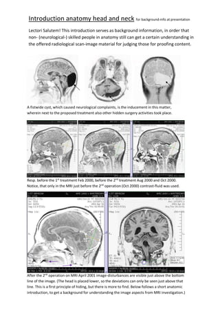
- 1. Introduction anatomy head and neck for background-info at presentation Lectori Salutem! This introduction serves as background information, in order that non- (neurological-) skilled people in anatomy still can get a certain understanding in the offered radiological scan-image material for judging those for proofing content. A fistwide cyst, which caused neurological complaints, is the inducement in this matter, wherein next to the proposed treatment also other hidden surgery activities took place. Resp. before the 1st treatment Feb 2000, before the 2nd treatment Aug 2000 and Oct 2000. Notice, that only in the MRI just before the 2nd operation (Oct 2000) contrast-fluid was used. After the 2nd operation on MRI April 2001 image-disturbances are visible just above the bottom line of the image. (The head is placed lower, so the deviations can only be seen just above that line. This is a first principle of hiding, but there is more to find. Below follows a short anatomic introduction, to get a background for understanding the image aspects from MRI investigation.)
- 2. Vascularisation supply- and drainage arters and veins. Suplly Arteria’s Drainage Veins Aanvoeraders Arteria ’s Afvoeraders Venen Suplly Arteria’s Drainage Veins Real-human anatomic models of resp. supply veins Arteria and (left) the drainage veins Veenas. In the presentation of the case the surgery alterations of the drainage system will be shown more. Nomenclature concerning the drainage veenas: 2 = Vena Sagittalis Superioris Superioris / 8 = Sinus Rectus / 9 = Confluens Sinuüm / (10 = (location) left Vena Transversalis TransVersus).
- 3. SUPPLY: The feeding is separated for both halves of the brains, having both a A.Carotis Interna both coming from the A. Carotis Communis in the neck, and then going both further upwards, supplying simply put each its own side of the brain. There are more other arteries connected to after. Both arteria pairs are connected to the A. Communicantes Posterius. This connection is rather small, but there isn’t noteworthy running a lot of blood through. In Grosso Modo... to put right, there are four major arteries running upward through the neck to feed the entire head (Including the face etc.). DRAINAGE: In Grosso Modo…there is a single central placed drainage for both the brain halves with branching draining channels aside to the left and the right for both halves, with veins on the surface of the brains, the Vena Cerebri Superficialis, and with veins deeper situated in the brain the Vena Cerebri Profundae. The Confluens Sinuum is the junction between these two important drainage branches, which lead to the two Vena Transversalis (L and R) where after blood runs through both their proper Vena Jugelaris (L and R) towards the hart. Schematic image of vascular caring of the brains.
- 4. The schematic imaging in addition on the forgoing vascular schematic-imaging of the drainage veins -marked with a red symbol to compare the difference between this and the actual real situation where veins are missing. The pieces of vein being fold (arrows) indicate a mechanic removal of the left Vein V. Transversus. The circles mark a problem with the Vena Sinus Rectus… (The surgery-report mentions the cutting through an unseen vein inside a kind of Falx Cerebelli.) In the presentation is shown that forgoing (that conscious 2nd operation) the left vein Transversus (L) still was sufficiently actively present even with a cyst measured at about its largest size... The image on top to the left is a first indication of a vein-deviation compensating lack of functioning of the removed veins Vena Sinus Rectus and Vena Traversalis (L). (Also, body- foreign material hast been left behind in the head, but also in the neck and in some molars.
- 5. Brain-fluid circulation in Ventricles and Cisterna’s and Brain-membranes. The brains are surrounded with mesodermal integuments: the brain membranes (meninges), which are built up in three layers. The tough outer layer (protecting) Dura Mater (1), the Arachnoid membrane (2) with its arachnoid space, and at the most inside the soft protecting Pia Mater (3). Through the Pia Mater run veins down shielded in the brain substance. The Arachnoid membrane and the Pia Mater together form the Leptomeninx. The Aranoidea is laying close to inside surface of the dura, separated by a small capillary gap (Cavum Duralis), and is connected by trabecula (14) and septs, which form a close network and likewise a system of connected vessels (-the Cyst is that too-) inside the Arachnoid space. (That space inside also is called Subarachnoid space (13).) From that arachnoid space the Granulates of Pachioni (15) penetrate the veins. They are the most present around the Vena Sinus Sagittalis Superioris (16, being the primary vein of drainage) and near the Lacunae Lateralis (17). They reach seldom the Spiral nerves. At elderly people by age these flakes grew inside the veins of Diploë (18). At those flakes the neural fluid moves into the venous blood.
- 6. (Left image) In between the supply and drain system is the phase of liquor spaces. There are 4 inner and 4 outer liquor spaces, and they are connected near the 4th ventricle (in between the brain trunk and the little brain). The inner spaces are called ventricles and are indicated here with Roman ciphers. The outer liquor space is limited by the arachnoid (and sub-arachnoid) space where the cyst is located). The biggest outer liquor space is the Cisterna Cerebella Medullaris (another shorter name for is Cisterna Magna), which is located just under the little brain. (Through the Arachnoid wall and Pia Mater a hole had to be made towards the Cisterna Magna to stop the cyst from expanding.) Close to the intermediary brain are the Cisterna Interpeduncular and the Cisterna Chiasmatas located. And finally, the Cisterna Ambiens (permeated with wide meshed connective tissue) is limited by the surface of the little brain and the Tectum Mesencephalic. By the expansion of the cyst, which puts the liquor circulation in disadvantage, also the risk of a stroke rose, which in 2016 did happen finally (officially the reason for that stroke is unknown). (Right Image) The schematic view at the forgoing page of the Liquor-circulation (which runs through till the Cisterna Terminalis (just below vertebral L5) is supplemented with the image to the right. At that image (R) we see a schematic view of a piece the vertebral column in exploded view. Anatomy: at the back side of the vertebral surrounded by the protecting pedicle is situated in between the spinal cord, which is surrounded with that same system of membranes as the brains are, again the Leptomeninges (the Pia Mater and the Arachnoid membrane against it with the ArachnoideTrabbeculae inside), with a global cross-section diameter of + 1mm), and finally the protecting membrane Pia Dura. Inside the Spinal Cord starting from the brains are running downwards 32 pairs of neural pathways. (From each vertebral there are two branches coming off leading to neural paths for the control of the body resp. at the left and right side.)
- 7. Falx -Cerebelli and Falx -Cerebri, Tentorium, and enclosed vascularization. Left image: (0= opening for the Brain Stem, 4=Falx Cerebri, 7= Dura with Sinus Rectus, circle = cyst). The cyst is situated at the left side of the brain. The Falx Cerebelli is one of the sides of the cyst as well the layers of the meninges for the rest of the cyst being enclosed in the arachnoid space. The V. Sinus rectus is a unanimous vein in the human body situated in the intersection of the Tentorium and the Falx Cerebri and Falx Cerebelli. On both images we see how both halves of the hemispheres in their caring-system are separated by the Falx Cerebri ((4) for the big brains and the Falx Cerebelli for the little brain; and further the big brains are separated from the little brains by the Tentorium spreading from the right to the left in a kind of tent-shape. Under the scull around the brains are the meninges situated with enclosed the Veena’s (causing the name sinus) V. Sinus Rectus (7), the V. Sinus Superioris (4) and the V. Sinus transversus (L + R). The blood going back from the brains are first led through the tough (!) blood vessels inside the tough membrane (Dura Mater), and then goes further down (as also the other blood vessels in the head) through the veins of the neck (V. Jugulars) towards the right atrium of the heart. All major brain veenas are situated near the (sub-) arachnoid space. The Sinus Sagittalis Suprioris is running under the cranial vault along the attachment of the brain sickle (the Falx Cerebri part) and from the brains smaller veins are leading to that. From the Confluence Sinuum (cross-section where also V. Sinus Sagittalis superioris and V. Transversalis (L + R) and Vena Sinus Rectus connect), also, the insignificant Sinus Occiptalis (most adults like me do not even have this vein) is running downwards after connecting back branching smaller veins in the back side of the head and then splitting near the Foramen Magnum to end in both the V. Jugelaris. It is incorrect to state that this V Occipitalis would have been hit and causing a thrombosis of the left V. Transversalis, because that Vena Occipitalis is missing, and the bloodstream drainage does not go up. X
- 8. Anatomy of the neck and findings of foreign body material. To locate the exact spot of (clandestine) foreign body material in anatomy was easier to do, after it was discovered on MRI. In total there are 7 neck vertebras, starting with the Atlas. The Atlas carries the head at the occipitalis bone and forms a joint with the 2nd vertebral Axis. That Axis has a rather big protrusion sticking backwards (Processus Spinoza). Counting downwards the metal object is situated near the 3rd vertebral. Professor Seibel named the presence of an extinction artefact. The two scans tot he left show that there are two separate inorganic objects to be found, whereby the lower implant is placed as a kind of ring around the location of the Spinal canal. At right: blood traces from just under the skin in the backside of the neck going till the vertebral canal indicate a result of surgical activity, whereby apparently a (clandestine) inorganic object was placed near C3. The reason to operate at the height of C3-C4 is that the muscles are in such way situated around, so that they are easily put aside for committing surgery behind towards the vertebral body /- arch.
- 9. A metal Michel Clip size 14x3 mm2 would have sank down from into the head towards the neck in six weeks time. Technically then it would be inside the leptomeninges with a an available space of 1mm, and it then would be pressing against the spinal cord causing neural damage. On the right photo that Michel Clip shown however is even passing beyond the area of the C3 vertebral arch with the bottom leg. Technically this is all together inpossible (!), because when that clip really sagged down from the head, then it is locked inside the leptomeninges (Pia Mater+ arachnoid membrane=1 mm cross-section space), but on the image to the right above it is even reaching beyond the vertebral arch, meaning it is outside the spinal canal and then it is also not inside the leptomeninges inside the spinal canal, but if it sagged down from the head then it can only arrive inside the leptomeninges (near vertebral C3). The leptomeninges (cross section 0,1 cm) with the ArachnoidTrabbeculae inside is hidering sagging. RESUMEÉ: In this short theoretic exposure about the overall anatomy and caring of the brains various sources of theoretical material were used, whereafter a few personal scanselection were linked to that. After a short explanation about the causality for the initial agreed medical treatment the intruction started with the vasulair care system of the brain (, for convienence also a few scan selections regarding the damaging of some venas were added directly after for anatomy comparing); followed by a short theory about the ‘ Brain fluid circulation in the Ventrickles (Cisterna’s) and the system of meninges. en Vlienzenstelsel’ en ‘Falx -Cerebelli en -Cerebri, Tetorium en ingesloten Vene Sinus Rectus; with at end still some information about the anatomy of the neck added with some scanselections regarding the presence of foreign body material inside the neck. Already in this introduction in preparation of the case (scan) presentation, allready it became clear that that there is something at hand. In the presentation more related scan selection will be shown for actually proofing a medical crime.