Sprint Final 09 B
•Descargar como PPT, PDF•
2 recomendaciones•2,517 vistas

The document describes the intracellular organization of a liver cell (hepatocyte). It includes a table showing the relative volumes occupied by major intracellular compartments, including the cytosol (54% of cell volume), mitochondria (22%), rough endoplasmic reticulum (9%), and nucleus (6%). It notes that the endoplasmic reticulum forms a single large compartment, while the Golgi apparatus is organized into discrete stacked cisternae.

Recomendados
Más contenido relacionado
La actualidad más candente
La actualidad más candente (20)
Similar a Sprint Final 09 B
Similar a Sprint Final 09 B (20)
Más de Alfonso Enrique Islas Rodríguez
Más de Alfonso Enrique Islas Rodríguez (20)
Último
Último (20)
Sprint Final 09 B
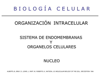
- 1. B I O L O G Í A C E L U L A R ALBERTS, B., BRAY, D., LEWIS, J., RAFF, M., ROBERTS, K., WATSON, J.D. MOLECULAR BIOLOGY OF THE CELL. 3RD EDITION. 1994 ORGANIZACIÓN INTRACELULAR SISTEMA DE ENDOMEMBRANAS Y ORGANELOS CELULARES NUCLEO
- 2. Table 12-1. The Relative Volumes Occupied by the Major Intracellular Compartments in a Liver Cell (Hepatocyte) ________________________________________________________________________ Intracellular Compartment Percent of Total Cell Volume Approximate Number per Cell* ________________________________________________________________________ Cytosol 54 1 Mitochondria 22 1700 Rough ER cisternae 9 1 Smooth ER cisternae plus Golgi cisternae 6 Nucleus 6 1 Peroxisomes 1 400 Lysosomes 1 300 Endosomes 1 200 ________________________________________________________________________ *. All the cisternae of the rough and smooth endoplasmic reticulum are thought to be joined to form a single large compartment. The Golgi apparatus, in contrast, is organized into a number of discrete sets of stacked cisternae in each cell, and the extent of interconnection between these sets has not been clearly established.
- 10. B I O L O G Í A C E L U L A R ALBERTS, B., BRAY, D., LEWIS, J., RAFF, M., ROBERTS, K., WATSON, J.D. MOLECULAR BIOLOGY OF THE CELL. 3RD EDITION. 1994 ORGANIZACIÓN INTRACELULAR SISTEMA DE ENDOMEMBRANAS Y ORGANELOS CELULARES NUCLEO
- 30. B I O L O G Í A C E L U L A R ALBERTS, B., BRAY, D., LEWIS, J., RAFF, M., ROBERTS, K., WATSON, J.D. MOLECULAR BIOLOGY OF THE CELL. 3RD EDITION. 1994 ORGANIZACIÓN INTRACELULAR SISTEMA DE ENDOMEMBRANAS Y ORGANELOS CELULARES RETÍCULO ENDOPLÁSMICO
- 31. B I O L O G Í A C E L U L A R ALBERTS, B., BRAY, D., LEWIS, J., RAFF, M., ROBERTS, K., WATSON, J.D. MOLECULAR BIOLOGY OF THE CELL. 3RD EDITION. 1994 ORGANIZACIÓN INTRACELULAR SISTEMA DE ENDOMEMBRANAS Y ORGANELOS CELULARES COMPLEJO DE GOLGI
- 32. Es tructura y Función de Los Organelos
- 34. Los Organelos son diminutas estructuras que están dentro de las células. Llevan a cabo funciones delicadas. En las células eucarioticas existen docenas de tipos diferentes de organelos. A continuación enfocaremos nuestra atención solo en los mas importantes respecto a su papel al nivel molecular, por ejemplo la mitocondria y su importancia en la obtención de energía útil para la célula. Otro ejemplo la membrana que permite el paso selectivo de sustancias de afuera hacia adentro y de dentro hscia afuera de la célula.
- 35. NUCLEO
- 36. RETICULO RUGOSO
- 38. RETICULO LISO
- 39. MITOCONDRIA
- 43. CITOESQUELETO B I O L O G Í A C E L U L A R ALBERTS, B., BRAY, D., LEWIS, J., RAFF, M., ROBERTS, K., WATSON, J.D. MOLECULAR BIOLOGY OF THE CELL. 3RD EDITION. 1994
- 44. The cytoskeleton. A cell in culture has been fixed and stained with Coomassie blue, a general stain for proteins. Note the variety of filamentous structures that extend throughout the cell. (Courtesy of Colin Smith.)
- 46. A centrosome with attached microtubules. As indicated, the slow-growing minus end of each microtubule is embedded in the centrosome matrix ( light green ) that surrounds a pair of structures called centrioles. By nucleating the growth of new microtubules, this matrix helps to determine the number of microtubules in a cell.
- 47. Growth and shrinkage in a microtubule array. The array of microtubules anchored in a centrosome is continually changing, as new microtubules grow ( red arrows ) and old microtubules shrink ( blue arrows ).
- 48. Fish pigment cells. These giant cells, which are responsible for changes in skin coloration in several species of fish, contain large pigment granules ( brown ), which can change their location in the cell in response to a neuronal or hormonal stimulus. (A) Schematic view of a pigment cell, showing the dispersal and aggregation of pigment granules, which occur along microtubules. (B) Scanning electron micrograph of a pigment cell following a brief exposure to detergent. The plasma membrane and soluble contents of the cytoplasm have been removed, exposing the array of microtubules and associated pigment granules. (C and D) Bright-field images of the same cell in a scale of an African cichlid fish, showing its pigment granules either dispersed throughout the cytoplasm or aggregated in the center of the cell. (E) An immunofluorescence picture of another cell from the same fish stained with antibodies to tubulin, showing large bundles of parallel microtubules extending from the centrosome to the periphery of the cell. (B, from M.A. McNiven and K.R. Porter, J. Cell Biol. 103:1547-1555, 1986, by copyright permission of the Rockefeller University Press; C, D, and E, courtesy of Leah Haimo.)
- 49. An experiment showing that a microtubule array can find the center of a cell. After the arm of a fish pigment cell is cut off with a needle, the microtubules in the detached cell fragment reorient with their minus ends near the center of the fragment.
- 50. The motor proteins that move along microtubules. Kinesins move toward the plus end of a microtubule, whereas dyneins move toward the minus end. As indicated, both types of microtubule motor proteins exist in many forms, each of which is thought to transport a different cargo.
- 51. The placement of organelles by microtubules. (A) Schematic diagram of a cell showing the typical arrangement of microtubules ( green ), endoplasmic reticulum ( blue ), and Golgi apparatus ( yellow ). The nucleus is shown in brown and the centrosome in light green . (B) Cell stained with antibodies to endoplasmic reticulum ( upper panel ) or to microtubules ( lower panel ). Motor proteins pull the endoplasmic reticulum along microtubules, stretching it like a net from its attachments to the nuclear envelope. (C) Cell stained with antibodies to the Golgi apparatus ( upper panel ) or to microtubules ( lower panel ). In this case motor proteins move the Golgi apparatus inward to its position near the centro-some. (B, courtesy of Mark Terasaki and Lan Bo Chen; C, courtesy of Viki Allan and Thomas Kreis.)
- 52. Actin filaments often shape the plasma membrane of animal cells. Three examples of plasma membrane changes caused by the cortical network of actin filaments. (A) Thin, spiky protrusions such as microspikes form on the surface of cells by the assembly of supporting bundles of actin filaments anchored in the cell cortex. (B) Sheetlike extensions, called lamellipodia, also form on the surface, in this case supported by a flattened web of actin filaments rather than discrete bundles. (C) Invaginations of the cell surface, as occur during cell division, are produced by a contractile
- 53. The intermediate filaments in the cytoplasm of a tissue culture cell. Rat kangaroo epithelial cells (Ptk2 cells) in interphase were labeled with antibodies to one class of intermediate filaments (called keratin filaments) and examined by fluorescence microscopy. (Courtesy of Mary Osborn.)
- 54. A current model of intermediate filament construction. The monomer shown in (A) pairs with an identical monomer to form a dimer (B) in which the conserved central rod domains are aligned in parallel and wound together into a coiled-coil. Two dimers then line up side by side to form an antiparallel tetramer of four polypeptide chains (C). Within each tetramer the dimers are staggered with respect to one another, thereby allowing it to associate with another tetramer, as shown in (D). In the final 10-nm ropelike intermediate filament, tetramers are packed together in a helical array (E). An electron micrograph of the final filament is shown upper left. (Diagram based on data from Murray Stewart; micrograph courtesy of Roy Quinlan.)
- 55. The domain organization of intermediate filament protein monomers. Most intermediate filament proteins share a similar rod domain that is usually about 310 amino acids long and forms an extended a helix. The amino-terminal and carboxyl-terminal domains are non-a-helical and vary greatly in size and sequence in different intermediate filaments.
- 56. Migratory cells from fish epidermis. (A) Light micrographs of a keratocyte in culture taken at 15-second intervals. The cell shown is migrating at about 15 micrometer/second. (B) Keratocyte seen by scanning electron microscopy, showing its highly flattened leading edge, with the body of the cell, containing the nucleus, trailing at the rear. (C) Distribution of cytoskeletal filaments in this unusual type of cell. Actin filaments ( red ) fill the flattened leading margin of the cell and are responsible for its migration. Microtubules ( green ) and intermediate filaments ( blue ) are restricted to the region close to the nucleus. (Micrographs courtesy of Juliet Lee.)
- 57. The polarization of a cytotoxic T cell after target-cell recognition. (A) Changes in the cytoskeleton of a cytotoxic T cell after it makes contact with a target cell. (B) Immunofluorescence micrograph in which both the T cell ( top ) and its target cell ( bottom ) have been stained with an antibody against microtubules. The centrosome and the micro-tubules radiating from it in the T cell are oriented toward the point of cell-cell contact. In contrast, the microtubule array in the target cell is not polarized. (B, reproduced from B. Geiger, D. Rosen, and G. Berke, J. Cell Biol. 95:137-143, 1982, by copyright permission of the Rockefeller University Press.)
- 58. An immuno-fluorescence micrograph of glial filaments in cultured astrocytes. The bundles of intermediate filaments ( green ) are stained with antibodies to glial fibrillary acidic protein. Nuclei are stained with a blue DNA-binding dye. (Courtesy of Nancy L. Kedersha.)
- 59. Electron micrographs of two types of intermediate filaments in cells of the nervous system. (A) Freeze-etch image of neurofilaments in a nerve cell axon, showing the extensive cross-linking through protein cross-bridges - an arrangement believed to provide great tensile strength in this long cell process. The cross-links are formed by the long, nonhelical extensions at the carboxyl terminus of the largest neurofilament protein. (B) Freeze-etch image of glial filaments in glial cells illustrating that these filaments are smooth and have few cross-bridges. (C) Conventional electron micrograph of a cross-section of an axon showing the regular side-to-side spacing of the neurofilaments, which greatly outnumber the microtubules. (A and B, courtesy of Nobutaka Hirokawa; C, courtesy of John Hopkins.)
- 60. Keratin filaments join cells together in cell sheets. Immunofluorescence micrograph of the network of keratin filaments in a sheet of epithelial cells in culture. The filaments in each cell are indirectly connected to those of its neighbors by desmosomes. (Courtesy of Michael Klymkowsky.)
- 61. The nuclear lamina. (A) Schematic drawing showing the nuclear lamina in cross-section in the region of a nuclear pore. The lamina is associated with both the chromatin and the inner nuclear membrane. (B) Electron micrograph of a portion of the nuclear lamina in a frog oocyte prepared by freeze-drying and metal shadowing. The lamina is formed from a square lattice of intermediate filaments composed of nuclear lamins (not always as highly organized as that shown here). (C) Electron micrograph of metal-shadowed isolated lamin dimers (marked L). They have an overall form similar to muscle myosin (marked M), with a rodlike tail and two globular heads, but they are much smaller molecules. The globular heads are formed from the two large carboxyl-terminal domains. (B and C, courtesy of Ueli Aebi.)
- 62. Blistering of the skin caused by a mutant keratin gene. A mutant gene encoding a truncated keratin protein (lacking both the amino- and carboxyl-terminal domains) was expressed in a transgenic mouse. The defective protein assembles with the normal keratins and thereby disrupts the keratin filament network in the basal cells. Light micrographs of normal (A) and mutant (B) skin show that the blistering results from the rupturing of cells in the basal layer of the mutant epidermis. The sketch in (C) of three cells observed by electron microscopy in the basal layer of the mutant epidermis shows that the cells rupture between the nucleus and the hemidesmosomes, which connect the keratin filaments to the underlying basal lamina. (From P.A. Coulombe, M.E. Hutton, R. Vassar, and E. Fuchs, J. Cell Biol. 115:1661-1674, 1991, by copyright permission of the Rockefeller University Press.)
- 63. Mechanical properties of actin, tubulin, and vimentin polymers. Networks composed of either microtubules or actin filaments or vimentin filaments, all at equal concentration, were exposed to a shear force in a viscometer and the resulting degree of stretch measured. The results show that microtubule networks are easily deformed but that they rupture (indicated by red starburst ) and begin to flow without limit when stretched beyond 50% of their original length. Actin filament networks are much more rigid, but they also rupture easily. Vimentin networks, by contrast, are easily deformed, but unlike microtubule networks, they withstand large stresses and strains without rupture. Vimentin filaments are therefore well suited to maintain cell integrity. (Adapted from P. Jamney et al. , J. Cell Biol. 113:155-160, 1991.)
- 65. Microtubules. (A) Electron micrograph of a microtubule seen in cross-section, with its ring of 13 distinct subunits, each of which corresponds to a separate tubulin molecule (an a/b heterodimer). (B) Cryoelectron micrograph of a microtubule assembled in vitro. (C and D) Schematic diagrams of a microtubule, showing how the tubulin molecules pack together to form the cylindrical wall. (C) The 13 molecules in cross-section. (D) A side view of a short section of a microtubule, with the tubulin molecules aligned into long parallel rows, or protofilaments. Each of the 13 protofilaments is composed of a series of tubulin molecules, each an a/b heterodimer. Note that a microtubule is a polar structure, with a different end of the tubulin molecule (a or b) facing each end of the microtubule. (A, courtesy of Richard Linck; B, courtesy of Richard Wade; D, drawn from data supplied by Joe Howard.)
- 66. Chemical structures of colchicine and taxol. A third drug, colcemid, is a close relative of colchicine in which the group shown in yellow is replaced by -CH 3 . Its binding to tubulin, unlike that of colchicine, is readily reversible.
- 67. Polymerization of pure tubulin. A mixture of tubulin, buffer, and GTP is warmed to 37°C at time zero. The amount of microtubule polymer, measured by light-scattering, follows a sigmoidal curve. During the lag phase individual tubulin molecules associate to form metastable aggregates, some of which go on to nucleate microtubules. The lag phase reflects a kinetic barrier to this nucleation process. During the rapid elongation phase, subunits add to the free ends of existing micro-tubules. During the plateau phase, polymerization and depolymerization are balanced because the amount of free tubulin has dropped to the point where a critical concentration has been reached. For simplicity, subunits are shown coming on and off the microtubule at only one end.
- 68. Electron micrograph showing preferential polymerization of tubulin onto the plus ends of microtubules. A stable bundle of microtubules obtained from the core of a cilium (discussed later) was incubated with tubulin subunits under polymerizing conditions. Microtubules grow fastest from the plus end of the microtubule bundle (the end above the bundle in this figure). (Courtesy of Gary Borisy.)
- 69. Microtubule polarity as revealed by the hook-decoration method. All the microtubules in this electron micrograph (seen in cross-section) have the same orientation. The hooks formed by the added tubulin curve clockwise, which indicates that the microtubules are being viewed as though looking along each filament from its plus end toward its minus end. Microtubule polarity can also be determined by decoration with dynein molecules (not shown). (Courtesy of Ursula Euteneuer.)
- 70. The interphase array of microtubules in a cultured fibroblast. The microtubules ( green ) are stained with an antibody to tubulin; the cell nucleus ( blue ) is stained with a fluorescent DNA-binding dye. (Courtesy of Nancy L. Kedersha.)
- 71. Microtubules growing out from the centrosome after the removal of colcemid. Immunofluorescence micrographs showing the arrangement of microtubules in cultured cells as revealed by staining with anti-tubulin antibodies. A normal tissue-culture cell is shown in (A). The cells shown in (B) were treated with colcemid for 1 hour to depolymerize their microtubules and were then allowed to recover; microtubules appear first in a starlike aster and then elongate toward the periphery of the cell. (A, courtesy of Eric Karsenti and Marc Kirschner; B, from M. Osborn and K. Weber, Proc. Natl. Acad. Sci. USA 73:867-871, 1976.)
- 72. The centrosome matrix. (A) Electron micrograph of a centrosome in a purified preparation. The matrix surrounds a barrel-shaped centriole, and it appears as a fibrous material that contains fine granules. (B) Light micrograph of a dividing human cell in culture stained with an antibody to b-tubulin ( green ) and with an antibody to g-tubulin ( red ), a protein that is located in the centro-some in cells from a wide variety of organisms. The superimposition of the red and green staining causes the g-tubulin-containing regions at the spindle poles to be yellow. (A, courtesy of Stephen Fuller; B, courtesy of M. Katherine Jung and Berl R. Oakley.)
- 73. A microtubule-organizing center in a fungal cell. Electron micrograph of the spindle pole body in yeast. (Courtesy of John Kilmartin.)
- 74. Microtubule dynamics in a living cell. A fibroblast was injected with tubulin that had been covalently linked to rhodamine, so that approximately 1 tubulin subunit in 10 in the cell was labeled with a fluorescent dye. The fluorescence at an edge of the cell was then observed using an extremely sensitive electronic imaging device. Below are tracings of the micrographs that show selected microtubules more clearly. Note, for example, that microtubule #1 first grows and then shrinks rapidly, whereas microtubule #4 grows continuously. (From P.J. Sammak and G.G. Borisy, Nature 332:724-736, 1988. © 1988 Macmillan Journals Ltd.)
- 75. The dynamic instability of microtubule growth. Fluctuations in length of a single microtubule in a solution of pure tubulin as seen by video-enhanced dark-field microscopy. Images of the same microtubule were recorded at intervals of 1 to 2 minutes and displayed in sequential order on a monitor screen. The two ends go through cycles of elongation and shortening independently, with the plus end showing the greatest fluctuations. (From T. Horio and H. Hotani, Nature 321:605-607, 1986. © 1986 Macmillan Journals Ltd.)
- 76. GTP hydrolysis after polymerization destabilizes microtubules. Analysis of the growth and shrinkage of microtubules in vitro suggests the following model for dynamic instability. (A) Addition of tubulin heterodimers carrying GTP to the end of a protofilament causes it to grow in a linear conformation that can readily pack into the cylindrical wall of the microtubule, thereby becoming stabilized. Hydrolysis of GTP after assembly changes the conformation of the subunits and tends to force the protofilament into a curved shape that is less able to pack into the microtubule wall. (B) In an intact microtubule, protofilaments made from GDP-containing subunits are forced into a linear conformation by the many lateral bonds within the microtubule wall, especially in the stable cap of GTP-containing subunits. Loss of the GTP cap, however, allows the GDP-containing protofilaments to relax to their more curved conformation. This leads to progressive disruption of the microtubule and the eventual disassembly of protofilaments into free tubulin dimers.
- 77. The selective stabilization of microtubules can polarize a cell. A newly formed microtubule will persist only if both of its ends are protected from depolymerizing. In cells the minus ends of microtubules are generally protected by the organizing centers from which these filaments grow. The plus ends are initially free but can be stabilized by other proteins. Here, for example, a nonpolarized cell is depicted in (A) with new micro-tubules growing and shrinking from a centrosome in all directions randomly. The array of microtubules then encounters hypothetical structures in a specific region of the cell cortex that can cap (stabilize) the free plus end of the microtubules (B). The selective stabilization of those microtubules that happen by chance to encounter these structures will lead to a rapid redistribution of the arrays and convert the cell to a polarized form (C and D).
- 78. A microtubule-associated protein. (A) Electron micrograph showing the regularly spaced side arms formed on a microtubule by a large microtubule-associated protein (known as MAP-2) isolated from vertebrate brain. Portions of the protein project away from the microtubule, as shown schematically in (B). (Electron micrograph courtesy of William Voter and Harold Erickson.)
- 79. An example of the cytoplasmic compartmentalization of nerve cells. This micrograph shows the distribution of tau protein ( green ) and MAP-2 ( orange ) in a hippo-campal neuron in culture. Whereas tau is confined to the axon, MAP-2 is confined to the cell body and dendrites. The antibody used to detect tau binds only to dephosphor-ylated tau, which is confined to the axon; other data show that phosphor-ylated tau is present in dendrites. (Courtesy of James W. Mandell and Gary A. Banker.)
- 80. Kinesins and cytoplasmic dyneins are microtubule motor proteins that generally move in opposite directions along a microtubule (A). These proteins (drawn here to scale) are complexes composed of two identical heavy chains plus several smaller light chains. Each heavy chain forms a globular head region that attaches the protein to microtubules in an ATP-dependent fashion. (B and C) Freeze-etch electron micrographs of a kinesin molecule (B) and a molecule of cyto-plasmic dynein (C). Whereas both kinesin and cytoplasmic dynein are two-headed molecules, ciliary dynein (D) has three heads (see Figure16-44). (Freeze-etch electron micrographs prepared by John Heuser.)
- 81. Vesicle transport in two directions. Kinesin and cytoplasmic dynein carry their cargo in opposite directions along microtubules, as illustrated in a fibroblast (A) and in the axon of a neuron (B).
- 83. C O M U N I C A C I Ó N Y C I C L O C E L U L A R B I O L O G Í A C E L U L A R ALBERTS, B., BRAY, D., LEWIS, J., RAFF, M., ROBERTS, K., WATSON, J.D. MOLECULAR BIOLOGY OF THE CELL. 3RD EDITION. 1994
- 84. Figure 17-2. The events of cell division as seen under a microscope. The easily visible processes of nuclear division (mitosis) and cell fission (cytokinesis), which are together called the M phase, typically occupy only a small fraction of the cell cycle. The other, much longer part of the cycle is known as interphase. During M phase an abrupt change in the biochemical state of the cell occurs at the transition from metaphase to anaphase; a cell can pause in metaphase before this transition point, but once the point is passed, the cell will carry on smoothly to the end of mitosis and through cytokinesis into interphase.
- 85. Figure 17-3. The four successive phases of a standard eucaryotic cell cycle. During interphase the cell grows continuously; during M phase it divides. DNA replication is confined to the part of interphase known as S phase. G 1 phase is the gap between M phase and S phase; G 2 is the gap between S phase and M phase.
- 86. Figure 17-4. The standard cell cycle compared with the early embryonic cell cycle. In the early embryonic cycle no growth occurs, so that each of the two daughter cells of each division is half the size of the parent cell. The cycle time is extraordinarily short, and S phases and M phases alternate without any intervening G 1 or G 2 phases.
- 87. Labeling of S-phase cells by autoradiography. The tissue has been exposed for a short period to 3 H-thymidine. Silver grains ( black dots ) in the photographic emulsion over a nucleus indicate that the cell incorporated 3 H-thymidine into its DNA, and thus was in S phase, sometime during the labeling period. In this specimen, showing sensory epithelium from the inner ear, the presence of an S-phase cell is evidence of cell proliferation occurring in response to damage. (Courtesy of Mark Warchol and Jeffrey Corwin.)
- 88. Analysis of DNA content with a fluorescence-activated cell sorter. The graph shows typical results obtained for a growing cell population when the DNA content of its individual cells is determined. The fluorescence-activated cell sorter (see Figure4-31) is used here simply to make measurements on the individual cells, rather than to sort them. The cells are stained with a dye that becomes fluorescent when it binds to DNA, so that the amount of fluorescence is directly proportional to the amount of DNA in each cell. The cells fall into three categories: those that have an unreplicated complement of DNA (one arbitrary unit) and are therefore in G1 phase, those that have a fully replicated complement of DNA (two arbitrary units) and are in G2 or M phase, and those that have an intermediate amount of DNA and are in S phase. The distribution of cells in the case illustrated indicates that there are greater numbers of cells in G1 than in G2 + M, implying that G1 is longer than G2 + M in this population.
- 89. The control of the cell cycle. The essential processes, such as DNA replication and mitosis and cytokinesis, are triggered by a central cell-cycle control system. By analogy with a washing machine, the control system is drawn as an indicator that rotates clockwise, triggering essential processes when it reaches specific points on the outer dial.
- 90. Two alternative conceptions of cell-cycle control. The green slabs represent essential processes of the cell cycle, such as DNA replication, chromosome condensation, spindle formation, and so on. In (A) the performance of one process is the trigger for the next, as in a chain of falling dominoes. In (B) the processes are not directly coupled in this way but are caused to occur in succession by a control device that operates independently of them. The mechanism shown in (B) corresponds more closely to the real cell cycle, where a cyclic control device, which we shall call the cell-cycle control system, triggers the downstream processes of the cycle.
- 91. Checkpoints and inputs of regulatory information to the cell-cycle control system. Feedback from downstream processes and signals from the environment can prevent the control system from passing through certain specific checkpoints. The most prominent checkpoints are where the control system activates the triggers shown in yellow boxes.
- 92. Two key components of the cell-cycle control system. A complex of cyclin with Cdk acts as a protein kinase to trigger downstream processes. Without cyclin, Cdk is inactive.
- 93. The core of the cell-cycle control system. Cdk is thought to associate successively with different cyclins to trigger the different downstream processes of the cycle. Cdk activity is terminated by cyclin degradation.
- 94. A typical neuron of a vertebrate. The arrows indicate the direction in which signals are conveyed. The neuron shown is from the retina of a monkey. The longest and largest neurons in a human extend for about 1 million µm and have an axon diameter of 15 µm. (Drawing of neuron from B.B. Boycott in Essays on the Nervous System [R. Bellairs and E.G. Gray, eds.]. Oxford, UK: Clarendon Press, 1974.)
- 95. The complex organization of nerve cell connections. This semischematic drawing depicts a section through a small part of a mammalian brain - the olfactory bulb of a dog, stained by the Golgi technique. The black objects are neurons; the thin lines are axons and dendrites, through which the various sets of neurons are interconnected according to precise rules. (From C. Golgi, Riv. sper. freniat. Reggio-Emilia 1:405-425, 1875; reproduced in M. Jacobson, Developmental Neurobiology, 3rd ed. New York: Plenum, 1992.)
- 97. Comparison of the layering of neurons in the cortex of normal and reeler mice. In the reeler mutant an abnormality of cell migration causes an approximate inversion of the normal relationship between neuronal birthday and position. The misplaced neurons nevertheless differentiate according to their birthdays and make the connections appropriate to their birthdays.
- 98. Formation of axon and dendrites in culture. A young neuron has been isolated from the brain of a mammal and put to develop in culture, where it sends out processes. One of these processes, the future axon, has begun to grow out faster than the rest (the future dendrites) and has bifurcated. (A) A phase-contrast picture; (B) the pattern of staining with fluorescent phalloidin, which binds to filamentous actin. Actin is concentrated in the growth cones at the tips of the processes that are actively extending and at some other sites of lamellipodial activity. (Courtesy of Kimberly Goslin, from Z.W. Hall, An Introduction to Molecular Neurobiology. Sunderland, MA: Sinauer, 1992.)
- 99. NGF effects on neurite outgrowth. Dark-field photomicrographs of a sympathetic ganglion cultured for 48 hours with ( above ) or without ( below ) NGF. Neurites grow out from the sympathetic neurons only if NGF is present in the medium. Each culture also contains Schwann (glial) cells that have migrated out of the ganglion; these are not affected by NGF. Neuronal survival and maintenance of growth cones for neurite extension represent two distinct effects of NGF. The effect on growth cones is local, direct, rapid, and independent of communication with the cell body; when NGF is removed, the deprived growth cones halt their movements within a minute or two. The effect of NGF on cell survival is less immediate and is associated with uptake of NGF by endocytosis and its intracellular transport back to the cell body. (Courtesy of Naomi Kleitman.)
- 100. Connections between eye and brain in a Xenopus tadpole. In this specimen a tracer molecule has been injected into one eye (dark object at left ), taken up by the neurons there, and carried along their axons, revealing the paths they take to the optic tectum in the brain. (Courtesy of Jeremy Taylor.)
- 104. G-protein coupled receptors (GPCRs) are one of the largest gene families of signaling proteins. Residing in the plasma membrane with seven transmembrane domains, GPCRs respond to extracellular stimuli that include catecholamine neurotransmitters, neuropeptides, larger protein hormones, lipids, nucleotides and other biological molecules. When a GPCR binds its extracellular ligand, it interacts with a G-protein to transduce a signal across the membrane into the cellular interior. G-proteins are a heterotrimeric complex containing a Ga subunit with GTPase activity, as well as b and g subunits. Ga can exist in an active state and an inactive state. Ga in the off state has GDP bound and does not activate downstream signaling molecules. When a GPCR is activated by ligand, it stimulates Ga subunits to bind GTP instead of GDP and become active, dissociating from the receptor and from the b/g subunits to activate downstream signaling factors like the enzyme adenylyl cyclase that synthesizes cyclic-AMP (cAMP) from ATP. Ga turns itself back off again with its intrinsic GTPase activity, hydrolyzing GTP to GDP to become inactive again. Activated G proteins interact with downstream signaling factors to alter the production of second messenger signaling molecules like inositolphosphates, calcium and cAMP. GPCRs that activate the Gi class of Ga subunits inhibit cAMP production and GPCRs that activate the Gs class of Ga subunits activate cAMP production. cAMP in turn activates the cAMP-dependent protein kinase, protein kinase A (PKA). PKA is a tetramer composed of catalytic subunits and regulatory subunits that repress the catalytic units when they are bound together. The regulatory subunits bind cAMP when it is present and release the catalytic units, releasing their inhibition of the catalytic subunits as well. The catalytic subunits of PKA when they are released and active phosphorylate target substrate proteins on serine and threonine residues, altering the activity of the modified protein and creating a cellular response to the extracellular stimulus acting on the GPCR.The PKA activation pathway is an example of a signal transduction cascade, in which tying several signaling events together amplifies the original signal in the cell. For each GPCR molecule that is activated, many G-proteins can be activated, and each active G protein can synthesize many cAMP molecules, continuing the cascade to PKA and further downstream. One of the roles of PKA that was first studied was the regulation of glycogen metabolism. When epinephrine binds to its receptor in liver, it activates cAMP production and PKA activation. One substrate of PKA is phosphorylase kinase, which is activated phosphorylation and is part of the signaling cascade that mobilizes glycogen into glucose to provide energy that fuels the response to epinephrine. Another substrate of PKA is glycogen synthase, which is turned off by PKA phosphorylation. PKA also participates in many other signaling pathways, integrating the key signaling intermediary cAMP with a wide range of biological responses
- 105. Interleukin 2 (IL-2) is a potent cytokine that can lead to cellular activation and proliferation. IL-2 Receptors are found on activated B-Cells, LPS treated Monocytes, and many T cells. The receptor is formed from three chains alpha (CD25), beta (CD122), and gamma (CD132). Primary signaling is through the JAK/Stat pathway and MAPKs.
- 107. The T Cell Receptor plays a key role in the immune system. The specificity of the receptor is governed by the binding site formed from the mature alpha and beta chains (shown here) or gamma and delta chains in gamma/delta T Cells. It is the ability of this receptor to bind a complex of foreign peptide in the groove of an MHC molecule that leads to T cell activation. Upon activation the T cell can assist in activating other cells or carry out cytolytic attacks depending on the particular T cell type. The CD3 complex and CD4 (Th cells) or CD8 (Tc cells) work to transmit the activation signal to the T cell's transcriptional machinary upon engagement of the receptor.